Abstract
Transitioning from pluripotency to differentiated cell fates is fundamental to both embryonic development and adult tissue homeostasis. Improving our understanding of this transition would facilitate our ability to manipulate pluripotent cells into tissues for therapeutic use. Here, we show that membrane voltage (Vm) regulates the exit from pluripotency and the onset of germ layer differentiation in the embryo, a process that affects both gastrulation and left-right patterning. By examining candidate genes of congenital heart disease and heterotaxy, we identify KCNH6, a member of the ether-a-go-go class of potassium channels that hyperpolarizes the Vm and thus limits the activation of voltage gated calcium channels, lowering intracellular calcium. In pluripotent embryonic cells, depletion of kcnh6 leads to membrane depolarization, elevation of intracellular calcium levels, and the maintenance of a pluripotent state at the expense of differentiation into ectodermal and myogenic lineages. Using high-resolution temporal transcriptome analysis, we identify the gene regulatory networks downstream of membrane depolarization and calcium signaling and discover that inhibition of the mTOR pathway transitions the pluripotent cell to a differentiated fate. By manipulating Vm using a suite of tools, we establish a bioelectric pathway that regulates pluripotency in vertebrates, including human embryonic stem cells.
Subject terms: Pluripotency, Embryology, Mesoderm, Ion channel signalling
The plasma membrane’s electrical potential is maintained by ion channels, though the impact of this potential on cell fate has not been clearly elucidated. Here they show that changes in membrane potential can affect calcium levels and mTOR in pluripotent stem cells, altering their transition from pluripotency to differentiation.
Introduction
Action potentials are fundamental to the function of excitable cells, including neurons, cardiomyocytes, and pancreatic cells. They are produced through tightly orchestrated changes in the membrane potential (Vm). However, most animal cells, excitable or not, have a resting state Vm (resting membrane potential) that depends on (a) the permeability of the plasma membrane for each ion (p in the Goldman-Hodgkin-Katz (GHK) equation, Fig. 1a, indicating the number of active ion channels), and (b) the driving force for each ion across the plasma membrane, determined by its electrochemical gradient (e.g., [K]i vs. [K]o in GHK equation). Although molecules that influence membrane potential have established roles in excitable tissues, their functions in embryonic or adult “non-excitable” tissues are emerging. For example, ion channels and pumps appear to be crucial for the formation of the left-right (LR) body axis1. While the vertebrate body plan may appear symmetrical across the LR axis, some of our internal organs, including the heart and gut, require asymmetry across the LR axis for proper formation or function. Chemical inhibition or overexpression of ion channels or pumps disrupt the proper alignment of internal organs along the left-right axis and affect global LR patterning2–7. Notably, using voltage sensitive dyes, Vm appears to vary across the developing embryo suggesting it could play instructive roles3,6,8. There is now a growing field that has implicated Vm in various embryonic contexts including Drosophila wing patterning8,9, craniofacial morphogenesis10,11, and chondrogenesis12,13 as well as the differentiation of excitable tissues such as muscle cells and neurons14. A challenge in the field is connecting changes in Vm to voltage responsive effectors that lead to the gene expression changes that pattern the embryo.
Fig. 1. Membrane potential is important for gastrulation and LR patterning.
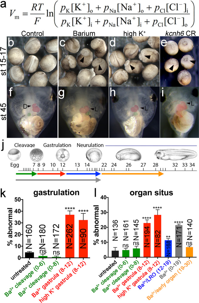
a GHK equation for Vm; R = gas constant, T = temperature, F = Faraday’s constant, p = permeability for each ion, [X]o = ion concentration outside of cell, [X]i = ion concentration inside the cell. b–i Effects of depolarizing treatments (barium chloride and high K+) and kcnh6 depletion on gastrulation (b–e; stage 15–17 embryos; arrowheads indicate incomplete blastopore closure) and organ situs (f–i; stage 45 tadpoles; ventral views; arrowheads indicate normal (D) and inverse (L) heart looping). j Different stages of barium chloride application (color key in j for bar graphs k and l; green = cleavage stages (stages 0–6 or 0–8); red = gastrulation (stages 8–12); blue = LRO signaling (stages 12–19); orange = early organogenesis (stages 19–30); gray = cleavage through LRO signaling (stages 0–19). Xenopus illustrations © Natalya Zahn (2022) from Xenbase (www.xenbase.org RRID:SCR_003280). k, l Percentages of embryos with incomplete blastopore closure at stage 15 (k) and abnormal organ situs at stage 45 (l) after treatment with barium or high K+ at different stages (see j for color code); p-values are in k: (Ba2+ gastrula vs untreated) = 2.28e–015, (high K+ gastrula vs untreated) = 1.36e–006; in l: (Ba2+ gastrula vs untreated) = 5.85e–007, (high K+ gastrula vs untreated) = 2.08e–008, (Ba2+/LRO vs untreated) = 8.51e–003, and (Ba2+ 0–19 vs untreated) = 1.2e–006. All graphs depict mean ± SEM and report total embryo numbers (N) collected over 3 independent experiments for high K+, and over 2 independent experiments for the 47 h barium time course. Key for asterisks: *p ≤ 0.05, **p ≤ 0.01, ****p ≤ 0.0001, ns nonsignificant with p > 0.05. Source data are provided as a Source Data file.
In order to respond, voltage sensitive effector molecules depend on the magnitude in the change of Vm. Quantitative Vm measurements in early embryos are rare but were performed in the 1960s and 1970s from 1-cell stage embryos through blastula stages in Triturus and Xenopus embryos15,16. The blastula embryo has completed a series of rapid cell divisions (cleavages), has established germ-layer cell fates (ectoderm, mesoderm, and endoderm), and is poised to begin gastrulation, the process by which cell movements transform the embryo to acquire the adult body plan. Notably, while the Vm at early cleavage stages is depolarized (=more positive Vm) (Vm@2-cell = −19 ± 10 mV), it becomes progressively hyperpolarized (=more negative) towards blastula stages (−50 mV)16. The implications of this progressive Vm polarization during early development are unclear, as is a mechanism by which Vm could transduce a signal within embryonic cells or act complementary to signals transduced biochemically (i.e., ligand-receptor).
Here we show that the Vm established in the blastula is essential for LR patterning and the exit from pluripotency. Depolarization of Vm using a variety of approaches leads to loss of ectodermal and paraxial mesodermal cell fates due to a persistence of pluripotency in these tissues. Membrane depolarization leads to the opening of voltage-gated calcium channels elevating intracellular calcium and maintaining pluripotency. Using RNA-seq, we find that mTOR signaling is downstream of Vm and modulates the transition from pluripotency to differentiated cell fate. These results define an electrochemical signaling pathway that acts complementary to biochemical (ligand-receptor) signaling pathways that transition pluripotent embryonic stem cells to differentiated cell fates.
Results
Vm of the blastula regulates LR patterning and gastrulation
To address the question of when Vm is critical for embryonic development, we employed barium ions to block K+ channels at different time points of embryonic development, since K+ conductance is paramount for determining Vm. Because K+ conductance drives the membrane voltage to a negative (hyperpolarized) potential, blocking K+ channels depolarizes cells. In line with the previous electrophysiological evidence demonstrating that embryos first become polarized at the blastula stage16, we found that Barium treatment affected embryonic development primarily when embryos were treated from blastula stages through gastrulation rather than at earlier cleavage stages (Fig. 1b, c, f, g, j–l)17. Embryonic development was affected in two ways: (1) 37 ± 3% (SEM) of the embryos failed to complete gastrulation (compared to just 4.7 ± 1% in control embryos) (Fig. 1b, c, j, k), and (2) 23 ± 2% that completed gastrulation exhibited misplacement of their organs relative to the left-right axis (compared to just 4 ± 2% in control embryos; Fig. 1f, g, j, l); these included abnormal heart looping to the left, an L-loop (vs a normal D-loop to the right), inverse gut rotation and misplacement of the gall bladder on the left (vs a normal positioning of the gall bladder on the right side of the body axis) (Fig. 1f, g). Because Barium can affect more than just K+ channels, we tested an alternative strategy for achieving membrane depolarization, namely manipulating Vm by increasing extracellular potassium ([K]o in GHK eq. Fig. 1a). Increasing the extracellular potassium reduces the chemical driving force for potassium to leave the cell and decreases the potassium current which depolarizes the embryo (more positive Vm). Incubating embryos in high K+ at blastula/gastrula stages was sufficient to cause a) gastrulation failure in 32 ± 6% of embryos (compared to just 6 ± 1% in controls Fig. 1b, d, k) and b) defective organ situs at later stages in 28 ± 3% of embryos (compared to just 4 ± 2% in controls; Fig. 1f, h, l). Thus, our results suggest that establishing proper Vm at blastula stages is essential for both gastrulation and LR patterning, providing context to previous work showing that Vm varies during embryonic development by becoming steadily more polarized from egg to blastula.
KCNH6 is essential for LR patterning and gastrulation
Recent studies in patients with congenital heart disease identified a number of variants in KCNH ether-a-go-go (EAG) potassium channels (Table 1)18 as candidate disease genes. Many of these patients had heterotaxy, a disorder of LR development that can have a significant impact on the structure and function of the heart that can be life-threatening. While multiple ions can affect membrane potential, the flow of potassium down its electrochemical gradient (K+in>>K+out) has the largest impact on Vm because in most cell types cell membranes are most permeable to potassium. Since KCNH6 was the most common family member in a total of five patients with heterotaxy (Table 1), we began our studies by examining the CHD/Htx candidate gene, KCNH6.
Table 1.
KCNH gene variants identified in patients with CHD
| Blinded ID | Gene | Phenotype | Allele type | Class | AA change |
|---|---|---|---|---|---|
| 1-09347 | KCNH1 | CTD/Htx | LOF het | splice | . |
| 1-01004 | KCNH1 | CTD/Htx | LOF het | frameshift_deletion | p.G149fs |
| 1-05070 | KCNH3 | LVO | LOF het | stopgain | p.R139X |
| 1-07611 | KCNH3 | CTD | CmpHet | misD/misD | A911V/ S1021P |
| 1-01856 | KCNH3 | LVO | CmpHet | misD misD | A357T/ F542L |
| 1-05499 | KCNH3 | CTD | de novo | misD | p.E859D |
| 1-02696 | KCNH5 | Htx/TGA | de novo | misD | p.N817S |
| 1-05146 | KCNH6 | Htx | LOF het | stopgain | p.S858X |
| 1-07078 | KCNH6 | CTD | LOF het | stopgain | p.E587X |
| 1-02620 | KCNH6 | other | LOF het | stopgain | p.Q487X |
| 1-06077 | KCNH6 | Htx | LOF het | frameshift_insertion | p.S671fs |
| 1-02515 | KCNH6 | Htx | de novo | misD | p.T274M |
| 1-01783 | KCNH7 | LVO | LOF het | stopgain | p.Y1162X |
| 1-12888 | KCNH7 | CTD | LOF het | stopgain | p.E944X |
| 1-06600 | KCNH8 | Htx/TGA | LOF het | frameshift_deletion | p.D782fs |
| 1-06579 | KCNH8 | other | LOF het | stopgain | p.E576X |
CTD conotruncal defect, Htx heterotaxy, LVO left ventricular outflow tract obstruction, TGA transposition of the great arteries, LOF loss of function, CmpHet compound heterozygote, misD damaging missense mutation according to previously published criteria.
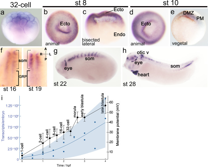
In Xenopus, we found kcnh6 to be expressed in the prospective ectoderm and dorsal/paraxial mesoderm at gastrulation onset, suggesting that it could play a role during gastrulation (Fig. 2a–h). Additionally, high temporal resolution RNA-seq shows that the increase in kcnh6 transcripts parallels the trend of Vm polarization in the frog embryo (Fig. 2i)16,19. We thus tested a role for kcnh6 in early embryonic development, and in determining Vm specifically. Two F0 CRISPRs independently targeting two different exons in kcnh6 as well as a translation blocking morpholino oligo (MO) recapitulated the morphological defects observed in embryos depolarized with barium and high K+, including: (a) inability to complete gastrulation (Fig. 3a) (CRex3: 27 ± 4%; CRex4: 29 ± 5% and MO:31 ± 3% vs only 4 ± 1% in controls), and (b) abnormal organ situs (Supplementary Fig. 1a–h) (CRex3: 43 ± 12%; CRex4: 19 ± 1% and MO:91 ± 7% vs only 5 ± 3% in controls). Importantly, we established specificity of our depletion studies by the following criteria: (1) phenocopy with two non-overlapping sgRNAs via F0 CRISPR and one translation blocking MO (Fig. 3a and Supplementary Fig. 1a–h), (2) rescue of the MO phenotype with human KCNH6 mRNA (Fig. 3a), and (3) detection of gene editing of the kcnh6 locus by PCR amplification and Inference of CRISPR Edits (Supplementary Fig. 2)20. Moreover, treatment of blastula/gastrula embryos (stage 8 to stage 12) with Ergtoxin, a scorpion peptide that specifically acts as a pore blocker of the KCNH channel family21, also led to identical gastrulation and LR defects (Fig. 3a and Supplementary Fig. 1f–h). These results indicate that Kcnh channels, and specifically Kcnh6, contribute to gastrulation and LR development, consistent with their identification in patients with Htx/CHD.
Fig. 2. kcnh6 expression analysis for kcnh6 during Xenopus development.
a–h kcnh6 transcripts were detected by a full-length antisense probe via WMISH. Embryos and tissues displayed are in the following orientations: a animal pole to the top, b animal pole view c bisected with animal pole to the top, d animal pole view, e vegetal view and dorsal to the top, f gastrocoel roof plate with anterior to the top and vegetal view, g, h lateral view with anterior to the left and dorsal to the top; Ecto prospective ectoderm, Endo prospective endoderm, DMZ dorsal marginal zone (mesoderm), PM paraxial mesoderm, som somites, GRP gastrocoel roof plate. Representative images from N = 60 embryos (per developmental stage) over 3 independent experiments. i kcnh6 transcripts19 (blue) and Vm16 (black) plotted during early Xenopus development. Blue dots represent mean transcript levels by temporal resolution RNA-seq, while blue shaded region marks Gaussian process 95% confidence interval of the data; n = 2 biological replicate time courses in original study. Membrane potential is presented as mean ± SD from at least n = 14 embryos in original study.
Fig. 3. Membrane potential is important for gastrulation and regulates calcium levels at gastrulation onset.
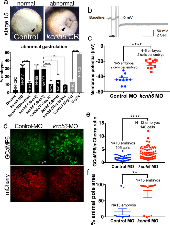
a Percentages of embryos with abnormal gastrulation after depletion of kcnh6 (MO, CRISPR) or Kcnh channel blockade with Ergtoxin, and rescue of kcnh6 depletion with medium conditions that hyperpolarize the Vm (low K+, val valinomycin, sodium substitution with choline; treatments performed stages 8–12). Above: examples of embryos scored for the graph; posterior views (dorsal to the top) of stage 15 embryos after successful (control) or unsuccessful (kcnh6 CRISPR = CR) gastrulation; arrowhead points to blastopore closure. Graph reports mean ± SEM; total embryo numbers (N) in the graph are from 3 independent experiments (except for Ergtoxin: 2 independent experiments with devitellinized embryos); p-values are (kcnh6MO vs Control MO) = 8.66e–010, (kcnh6MO+mRNA vs kcnh6MO) = 2.59e-004, (kcnh6CRex4 vs Control) = 7.19e-018, (kcnh6CRex3 vs Control) = 5.79e-022, (kcnh6CR+low K+ vs kcnh6CR) = 1.64e-005, (kcnh6CR+val vs kcnh6CR+DMSO) = 1.22e-002, (kcnh6CR+choline vs kcnh6CR) = 2.34e-006, (ErgTx vs Control) = 6.57e-010; two-sided Fisher’s exact test. b Representative intracellular recording in the prospective ectoderm of a control stage 10 embryo; Vm is measured relative to the medium (baseline); the dip in membrane potential indicates the electrode breaking into the cell. c The Vm as measured by intercellular recordings in the prospective ectoderm of stage 10 Control MO and kcnh6 MO-injected embryos; graph reports mean ± SEM; p-value (kcnh6MO vs Control MO) is 1.07e-006 (unpaired two-tailed student’s t-test); each data point represents one cell; data from 10 cells/5 embryos/3 independent experiments. d Live animal pole images of GCaMP6/mCherry at stage 10. e Quantification of GCaMP6 fluorescence intensity normalized to mCherry in mCherry+ cells; graph shows mean ± SEM; data points represent single cells; data from N cells (in graph)/10 embryos/3 independent experiments; p = 1.14e-006; unpaired two-tailed student’s t-test. f Maximum area undergoing simultaneous Ca2+ transients within a 20 s time lapse recording as a percentage of total animal pole area; the animal poles of 13 Control MO and 15 Kcnh6 MO embryos were recorded over 3 independent experiments; p = 1.04e-003; unpaired two-tailed student’s t-test. Key for asterisks: *p ≤ 0.05, **p ≤ 0.01, ***p ≤ 0.001, ****p ≤ 0.0001, ns nonsignificant with p > 0.05. Source data are provided as a Source Data file.
Vm, rather than KCNH6 per se, is essential for gastrulation
Depletion/inhibition of potassium channels or elevation of extracellular K+ should lead to membrane depolarization. Thus, we reasoned that the inverse condition, namely hyperpolarizing by reducing extracellular K+, should rescue kcnh6-depleted embryos. Lowering extracellular K+ ([K]o in GHK eq. Fig. 1a) increases the outward driving force for flow of K+, provided that other K+ channels are present. Indeed, lowering extracellular K+ rescues the gastrulation defect in kcnh6-depleted embryos (CRISPR and MO) (Fig. 3a and Supplementary Fig. 3a). To test the significance of K+ conductance independently of a specific K+ channel, we employed valinomycin, a K+ selective ionophore that inserts itself into the plasma membrane and mimics the ability of a K+ channel to passively conduct K+ down its electrochemical gradient. Application of valinomycin also rescued the gastrulation defect in kcnh6-depleted embryos (30 ± 1% gastrulation defect in kcnh6 CR/DMSO- vs 16 ± 7% in kcnh6 CR/Valinomycin-treated embryos) emphasizing the importance of K+ flux rather than a specific need for Kcnh6 itself or an alternative role of Kcnh6 in cell signaling (Fig. 3a and Supplementary Fig. 3a). Finally, to differentiate between membrane potential and an alternative role for K+ flux across the plasma membrane (e.g., cell volume regulation), as well as to unlink membrane potential from a specific conductance (i.e., potassium), we hyperpolarized by reducing extracellular Na+ and replacing it with equimolar choline. Choline has equivalent cationic charge to Na+ but cannot pass through channels and therefore does not influence the Vm. Importantly, replacement of Na+ with equimolar choline does not affect the osmotic properties of the medium, ensuring that external Na+ depletion will only act on Vm and not on cell volume22. To determine the amount of sodium to replace with choline, we measured Vm using intracellular electrodes in the animal pole in the context of high K+ and rescue by choline substitution of sodium. Embryos exposed to high K+ were depolarized (Vm = −20 ± 4 mV) while embryos treated with high K+ and ½ sodium replacement with choline were repolarized to roughly the normal membrane potential (Vm = −41 ± 4 mV) (Supplementary Fig. 3b). Therefore, we tested ½ sodium replacement with choline on embryos and assayed for gastrulation which led to a remarkable rescue in kcnh6-depleted embryos (Fig. 3a). These data suggest that Vm, which is determined by the conductance of K+ through Kcnh6 and influenced by other K+ and Na+ channels, is key to gastrulation.
Finally, we sought to measure the change in Vm when kcnh6 is depleted. Using intracellular electrodes in the animal pole of kcnh6 MO vs control MO-injected embryos at gastrulation onset, we recorded a Vm of −24 ± 1.9 mV in kcnh6 MO vs −44 ± 2.1 mV in control MO embryos (Fig. 3b, c). Thus, Kcnh6 contributes ~20 mV to the cell’s negative resting potential, and embryos lacking kcnh6 are abnormally depolarized compared to their control counterparts.
Depolarized Vm increases calcium levels
We then asked how Vm is transduced into a signal that affects embryonic development. There are a limited number of voltage responsive elements in a cell. We reasoned that depolarization (Vm = −24 mV) in kcnh6-depleted embryos could aberrantly activate voltage-gated Ca2+ channels (VGCCs), which facilitate inward Ca2+ flux23. L-type VGCCs are present in the prospective ectoderm and dorsal mesoderm and can induce potent intracellular Ca2+ increases that can alter germ-layer patterning24,25, yet upstream regulators of these calcium channels remain elusive. Interestingly, intracellular Ca2+ is elevated after fertilization and during early cleavage stages but declines as the embryo approaches gastrulation26 concomitant with the onset of membrane polarization. We argued that, if VGCCs are aberrantly activated due to an abnormally depolarized Vm, we should be able to detect changes in intracellular Ca2+ levels. To assess this, we microinjected the calcium indicator GCaMP627 mRNA together with mCherry mRNA (to enable ratiometric analysis) into control MO or kcnh6 MO embryos and performed calcium imaging in animal cells of early gastrula embryos. Within the animal pole of stage 10 control MO-injected embryos, we observed multiple intracellular calcium increases, signified by a pulse-like appearance of GCaMP6 fluorescence in isolated cells, which then propagated to adjacent cells (Supplementary Movie 1). These increases are well documented in Xenopus stage 8–12 gastrulae, i.e., last a few seconds, in which they spread to adjacent cells and then extinguish, are VGCC dependent and may contribute to neural induction25,28. We confirmed the existence of Ca2+ transients at stage 10 by performing 20 s time lapse recordings, and additionally observed that they are of low intensity and typically do not simultaneously affect more than 16 ± 10% of the total animal pole area (Supplementary Movie 1 and Fig. 3d–f). Interestingly, the same transients were dramatically increased in stage 10 kcnh6 MO embryos both in intensity and area (Supplementary Movie 2 and Fig. 3d–f), affecting 71 ± 11% of the animal pole on average, with most embryos displaying simultaneous calcium increases in >90% of the animal pole. Thus, kcnh6 contributes to a hyperpolarized Vm and is key for suppressing calcium levels at gastrulation onset, a signal that may facilitate correct gastrulation.
Depolarization affects paraxial mesoderm and ectoderm
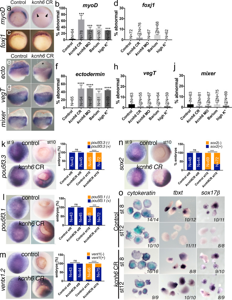
For gastrulation to proceed normally, two steps are critical: first, the germ layers of the blastula embryo (ectoderm, mesoderm, and endoderm) must be patterned correctly and second, the embryo must undergo the cellular rearrangements that drive morphogenesis. Calcium has been previously identified to play a role in morphogenesis cell behaviors during gastrulation29,30. Alternatively, calcium may play a role in patterning the mesoderm that also drives gastrulation cell movements. Patterning precedes morphogenesis, and morphogenesis can fail as a result of abnormal patterning. We, therefore, first examined if patterning is disrupted in Vm-depolarized embryos via marker gene expression. Since the mesoderm is critical for gastrulation movements, we began with this germ layer. Markers of the dorsal (gsc, nodal3) and ventral mesoderm (vent2) appeared unaffected in kcnh6-depleted, barium and high K+ depolarized embryos (Supplementary Fig. 4e–j); however, the paraxial mesoderm fates appeared lost as marked by myoD, myf5, and tbxt (brachyury, xbra) (Fig. 4a–d and Supplementary Fig. 4a–d). In fact, absent patterning of paraxial mesoderm by myoD persisted into the Left-Right Organizer (Supplementary Fig. 5), a transient structure formed at the end of gastrulation where cilia driven flow is thought to pattern the LR axis31,32. In the LRO, dand5 (coco) is normally expressed in the paraxial mesoderm symmetrically until cilia driven flow suppresses dand5 expression on the left33,34. However, consistent with a mispatterning of the paraxial mesoderm in the LRO, dand5 was also absent even before the occurrence of cilia driven flow (Supplementary Fig. 1i, j). A disruption in the LRO is further supported by defective pitx2c expression in the left lateral plate mesoderm at later stages (Supplementary Fig. 1k, l). Therefore, in kcnh6-depleted embryos, the paraxial LRO is mispatterned, and a defect in this tissue can be detected already at the onset of gastrulation.
Fig. 4. Membrane potential affects early gastrula patterning and pluripotency.
a–j WMISH for germ-layer markers in early gastrula embryos (stage 10; a, c: vegetal views with dorsal to the top; e, g, and i: lateral view of bisected embryos, dorsal to right). Markers are for paraxial mesoderm (myoD), superficial dorsal mesoderm (foxj1), ectoderm (ectodermin = ecto), endoderm (vegT, mixer). Graphs (b, d) and (f–j) depict percentages of embryos with absent or strongly reduced expression of these markers; presented are mean ± SEM; total embryo numbers (N) are from 3 independent experiments; p-values (vs Control) are in b: (CR vs Control) = 1.61e-006, (MO vs Control) = 4.99e-003, (Ba2+ vs Control) = 4.45e-003, (high K+ vs Control) = 7.56e-004, and in f: (CR vs Control) = 6.81e-013, (MO vs Control) = 2.7e-013, (Ba2+ vs Control) = 1.58e-009, (high K+ vs Control) = 6.61e-012; ns (nonsignificant) for p > 0.05; two-sided Fisher’s exact test. k–n WMISH for Xenopus pluripotency genes pou5f3.3, pou5f3.1, ventx1.2, and sox2 at stages 9 and 10 (animal pole views). Graphs (k–n) depict mean percentages of embryos with present (+/blue) or absent (−/orange) gene expression; total embryo numbers (N) are from 3 independent experiments; p-values (vs Control) are in k: p = 2.01e-008, in l: p = 1.52e-010, in m: p = 6.73e-018 and in n: p = 1.85e-006; ns (nonsignificant) for p > 0.05. Key for asterisks in all graphs: *p ≤ 0.05, ***p ≤ 0.001, ****p ≤ 0.0001, ns nonsignificant. o Differentiation potential of animal caps excised at stage 8 or 12 and treated with no activin (differentiation into epidermis marked by cytokeratin), low activin (differentiation into mesoderm marked by tbxt) and high activin (differentiation into endoderm marked by sox17β). In each image, the numbers on the bottom right report caps with the indicated phenotype vs total number of caps analyzed over 2 independent experiments. Source data are provided as a Source Data file.
When certain biochemical signaling factors are depleted, loss of one-cell fate (e.g., paraxial mesoderm) is often concomitant with gain of another cell fate35–37. Since the dorsal or ventral mesoderm appeared unaffected (Supplementary Fig. 4e–j), we considered that the ectoderm or endoderm might be expanded into the mesodermal area of embryos with abnormally depolarized Vm38,39. Interestingly, while the endoderm (vegT) and its border to the mesoderm (mixer) seemed unaffected (Fig. 4g–j), ectodermal fates (ectodermin and foxI1a) were lost similar to the paraxial mesoderm (Fig. 4e, f and Supplementary Fig. 4k, l. In fact, depletion of ectodermin (trim33) leads to developmental arrest midway through gastrulation39, which corresponds to the arrested phenotype in a portion of depolarized embryos. These results indicate that Vm has an effect on germ-layer differentiation, and specifically paraxial mesoderm and ectoderm, at gastrulation onset (Fig. 4a–j).
Cacna1c responds to Vm depolarization
In depolarized embryos, we have established (1) changes in cell fate and (2) elevated intracellular calcium levels, so we next tested if these aberrant cell fates are dependent on voltage-gated calcium channels. To determine the specific embryonic VGCCs downstream of Vm, we reviewed our available high temporal resolution RNA-Seq data19. Xenopus contains detectable transcripts of L- and T-type VGCCs between the 1-cell and gastrula stages, while other VGCC types (N-, R-, and P/Q) are less abundant. L-type VGCCs become activated at Vm > −40 mV (and then inactivated at Vm > 10 mV) and have been previously implicated in gastrula patterning23,24,29 while T-type channels become inactivated at Vm > −60 mV and would be inactive both at physiological Vm (~−50 mV) and at more depolarized potentials. Therefore, we tested the L-type VGCC blocker nifedipine. This significantly ameliorated both ectodermin (ectoderm) and myf5 (paraxial mesoderm) expression losses in kcn6 knockdown embryos (Fig. 5a–d). Specifically, myf5 was lost only in 16 ± 1% and 18 ± 5% of nifedipine-treated kcnh6 CR and MO embryos (vs 38 ± 5% and 35 ± 5% in kcnh6CR or MO embryos treated only with DMSO, Fig. 5b, d). Similarly, absent ectodermin was only observed in 16 ± 7% and 17 ± 3% of nifedipine-treated kcnh6 CR and MO embryos (vs 48 ± 2% and 51 ± 6% in kcnh6 CR or MO embryos treated with DMSO, Fig. 5a, c). Of the VGCCs identified in our RNA-seq data at blastula/gastrula stages, two genes cacna1c (Cav1.2; L-type) and cacna1g (Cav3.1; T-type) encode alpha (pore-forming) channel subunits, which are indispensable for channel function. Co-depletion of cacna1c in kcnh6-depleted embryos rescued expression of ectodermin and myf5, while co-depleting cacna1g (Fig. 5a–d) resulted in no rescue. Thus, Kcnh6, which sets a negative Vm, is essential to limit the activation of L-type VGCCs and specifically Cacna1c, a critical step for ectodermal and paraxial mesodermal differentiation.
Fig. 5. The role of VGCCs in germlayer differentiation.
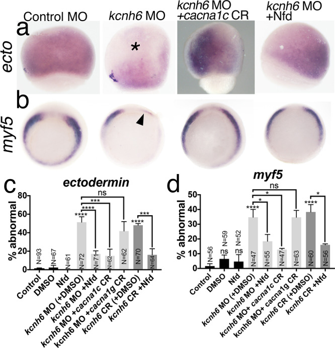
a WMISH for ectodermin; lateral views with the animal pole to the top; asterisk marks the animal pole with loss of ectodermin expression; embryos with absent expression are quantified in c; CR CRISPR, Nfd nifedipine. b WMISH for myf5: vegetal views with dorsal to the top; arrowhead marks loss of expression; embryos with abnormal expression are quantified in d. c, d Percentages of embryos with abnormal ectodermin (c) and myf5 (d) expression. Graphs depict mean ± SEM; p-values are in c: (kcnh6MO+DMSO vs DMSO) = 2.88e-006, (kcnh6MO+Nfd vs kcnh6MO+DMSO) = 4.59e-002, (kcnh6MO+cacna1cCR vs kcnh6MO) = 1.53e-002, (kcnh6MO+cacna1gCR vs kcnh6MO) > 9.99e-001 (ns), (kcnh6CR+DMSO vs DMSO) = 3.50e-007, (kcnh6CR+Nfd vs kcnh6CR+DMSO) = 1.20e-002; in d: p-values are (kcnh6MO+DMSO vs DMSO) = 4.07e-015, (kcnh6MO+Nfd vs kcnh6MO+DMSO) = 8.92e-005, (kcnh6MO+cacna1cCR vs kcnh6MO) = 4.78e-005, (kcnh6MO+cacna1gCR vs kcnh6MO) = 2.24e-001 (ns), (kcnh6CR+DMSO) = 2.04e-014, (kcnh6CR+Nfd) = 1.45e-003; two-sided Fisher’s exact test; total embryo numbers (N) in the graphs are from at least 2 independent experiments; Key for asterisks: *p ≤ 0.05, ***p ≤ 0.001, ****p ≤ 0.0001, ns nonsignificant for p > 0.05. Source data are provided as a Source Data file.
Vm depolarization maintains pluripotency in gastrula embryos
The loss of some cell fates (ectoderm and paraxial mesoderm) without a concomitant expansion of other cell fates was puzzling given that most biochemical signaling factors (Wnt, BMP, Nodal) generally balance different cell fates in the early embryo. We speculated that these unspecified cells may simply lack the ability to assume any cell fate because they remain pluripotent abnormally. To test this hypothesis, we examined markers of pluripotency OCT4, NANOG, and SOX2. In Xenopus, there are three OCT4 homologs (pou5f3.1, 2, and 3, formerly oct91, oct25, and oct60)40, and the ventx1.2/2.2 factors, which have overlapping functions in maintaining differentiation competence and are thought to be structurally and functionally equivalent to mammalian Nanog41. Sox2, a core pluripotency factor in mammals, is highly conserved in amphibians and also expressed at high levels prior to lineage commitment throughout the Xenopus blastula42,43. We examined the prospective ectoderm of embryos, which is best characterized in its pluripotent properties, and confirmed that pou5f3.1, pou5f3.3, sox2, and ventx1.2 are robustly expressed at stage 9 prior to lineage commitment, but their transcripts are sharply reduced by stage 10 in control embryos (Fig. 4k–n). In contrast, kcnh6 CR embryos retain robust expression of these factors well beyond stage 9 and into stage 10, a prolonged expression compared to wildtype embryos (Fig. 4k–n). This, in turn, is not due to a general delay in development, since kcnh6 CR embryos were staged according to the physical progression of gastrulation, i.e. presence of blastopore lip. Moreover, in kcnh6-depleted late gastrula embryos, abnormal maintenance of pou5f3.3 and ventx1.2 can be abolished by incubating the embryos in L-type VGCC blocker nifedipine (Supplementary Fig. 6b–d). These results suggest that kcnh6 is upstream of Vm and VGCCs in promoting the exit from pluripotency, which takes place as gastrulation proceeds.
Based on this result, we sought to test the pluripotency of these kcnh6-depleted embryos. In the blastula (stage 9), the prospective ectoderm or “animal cap” contains cells that when explanted will differentiate into epidermis (Fig. 4o). Importantly, when stage 8–9 explanted animal cap cells are treated with activin, they can be differentiated into mesodermal and endodermal cell fates indicating that they are pluripotent (Fig. 4o), an assay used for decades to test the activity of a host of differentiation factors. However, towards the end of gastrulation at stage 12, these animal cap cells are no longer pluripotent and when explanted will only differentiate into epidermis, even when stimulated with activin43,44 (Fig. 4o). Using this animal cap assay, we sought to test the role of kcnh6 in determining pluripotency. We explanted stage 9 and stage 12 animal caps and assayed differentiation under three conditions: (1) no activin to examine spontaneous differentiation into epidermis (marked by cytokeratin), (2) low activin to stimulate differentiation into mesoderm (tbxt), and (3) high activin to stimulate differentiation into endoderm (sox17β). Both control and kcnh6 CR animal caps explanted from stage 9 embryos were capable of differentiating into all three germ layers, indicating full differentiation potential even when kcnh6 is depleted (Fig. 4o). On the other hand, as expected, animal caps explanted from stage 12 control embryos differentiated into epidermal fate but not into meso- or endoderm despite activin administration (Fig. 4o). Strikingly, stage 12 animal caps explanted concurrently from kcnh6-depleted embryos were able to differentiate into cell fates of all three germ layers with activin administration (Fig. 4o). From these and the previous experiments, we conclude that Vm polarization via kcnh6 enables the exit from pluripotency.
Vm limits mTOR to allow for exit from pluripotency
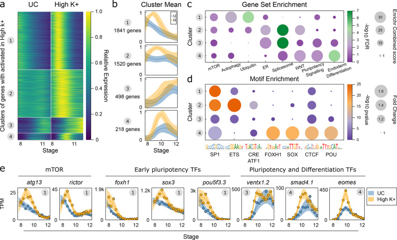
Our findings indicate that a polarized Vm limits voltage-gated calcium channels and intracellular calcium, a process that reduces the expression of pluripotency genes as germ-layer differentiation initiates. A critical question is what are the signaling pathways invoked when Vm is depolarized or intracellular calcium is elevated. To address this question in an unbiased manner, we temporally profiled gene expression via RNA-Seq in control and high K+ depolarized embryos by collecting embryos every 30 min from pre- to post-gastrula stages (stages 8–12; Supplementary Fig. 7a–d). We identified genes exhibiting temporal differential expression employing a Gaussian Process framework19; this determines genes whose expression trajectory differs between control and high K+ embryos over the time course. We found 4043 genes on average activated in high K+ over the time course, and 1101 genes on average repressed (Supplementary Fig. 7e). We further used k-means clustering to subdivide these into 8 clusters, 4 activated (A1–4, Fig. 6a, b) and 4 repressed (R1-4, Supplementary Fig. 7f, g). In both cases, the clustering segregated genes showing dysregulated gene expression prior to gastrulation (Clusters 1, 2) and during gastrulation (Clusters 3,4) (Fig. 6a, Supplementary Fig. 7f, g). To assess the composition of these clusters, we performed gene set enrichment using Enrichr45. Comparing activated clusters to repressed clusters over seven different annotated gene set libraries, we found 1279 terms significantly associated with at least one of the 4 activated clusters, but only 44 terms significantly associated with repressed clusters. Therefore, given the total number of genes and associated terms, we focused our attention on the analysis of the activated genes.
Fig. 6. High-resolution temporal RNA-seq identifies mTOR and Ca2+ Gene Regulatory Network.
a, b Summary of activated gene clusters by a heatmap and b cluster average. Data is Gaussian process median for each gene normalized by maximal value, shaded region in b is ±1 SD for each cluster. UC untreated control; High K+ (=depolarizing conditions). c Bubble plot for selection of gene set enrichment terms, calculated with Enrichr, see Methods for definition of terms, and Supplementary Data 1 for full set of enrichments. Bubble size reflects Enrichr Combined score and color indicates −log10 FDR. d Bubble plot enrichment of TF motifs in 500 bp upstream of cluster promoters, see also Supplementary Fig. 7g. Bubble size reflects fold change over background and color is −log10 Hypergeometric right tail p-value for enrichment. e Expression of exemplar genes in control and high K+. Central line and shaded region are transformed Gaussian process median and 95% CI. Circle in top right hand corner gives cluster number. UC (=untreated control), High K+ (=depolarizing conditions). Data analysis performed from N = 13 samples/each 10 embryos over one biological replicate time course.
We found a hierarchy of gene set enrichments from early to late in our time course, reflecting the changing response in the transcriptome (Fig. 6c). Notably, we found enrichment for the mTOR signaling pathway activated early with members including rictor, depdc5, pik3cb, stk11, atg13 in cluster A1 (Fig. 6e) and akt1s1, gsk3b, lamtor1/2 in cluster A2. The enrichment for mTOR members continues to span all activated clusters (FDR < 3 × 10⁻⁵ over all activated clusters combined). This enrichment is accompanied by pathways associated with mTOR, including autophagy, ubiquitin transferase activity, and ER response (Fig. 6c). The intermediate clusters A2 and A3 show the most prominent ER response enrichment together with significant upregulation of spliceosome machinery (including 12 snRNPs and 5 SRSF family members). In contrast, in later clusters, we find enrichments for terms explaining the sustained pluripotency and germ-layer defects, including pluripotency (pou5f3.1, pou5f3.2, ventx1.2) and WNT signaling (fzd7, wnt8a, tcf7l1) (Fig. 6c, e). Key members of the Xenopus pluripotency network are also found activated early, in cluster A1 including foxh1, sox3, and pou5f3.3 (Fig. 6c, e).
To build the underlying gene regulatory networks, we examined transcription factor motif enrichment in the promoters of each of these activated gene clusters. Mirroring the gene set enrichments, we found motif enrichments segregated between early (A1,2) and later (A3,4) gene clusters (Fig. 6d, Supplementary Fig. 7h). We find strong enrichment for SP1, ETS, YY1, RFX, and CRE/ATF1 motifs driving gene expression changes in clusters A1 and A2. Interestingly, both ETS and CRE motifs are bound by factors responsive to calcium. Calcium induced phosphorylation of ETS1 inhibits binding activity46, and CRE elements are bound by calcium responsive family members, CREB1, CREM, and ATF147–49. In our data, each of these factors are expressed at high levels at stage 8, and then are gradually downregulated to a minimum at stage 12. Of note, their mRNAs are not upregulated in high K+ conditions (Supplementary Fig. 7i). This suggests that the activity of these factors is post-translationally modified in depolarized embryos experiencing high Ca2+ to drive gene expression changes. In the case of ETS1, this factor may act to repress gene expression in normal germ-layer resolution and this repression is removed in high Ca2+ embryos. Supportive of these factors driving gene expression, we find a large intersection between activated gene clusters A1 and A2 and genes found in proximity to publicly available ETS1, CREB1 and CREM binding sites (Supplementary Fig. 7j); in the case of activated cluster A2 this remarkable enrichment accounts for 794/1520 (52.2%) of genes found in proximity to one of ETS1, CREB1, and CREM (FDR < 10−26,10−23,10−35, respectively, Fisher Exact Test). Therefore, we find that initial transcriptional responses captured by activated gene clusters A1 and A2 appear to be largely driven directly by calcium response. This includes the activation of mTOR and pluripotency genes in high K+ conditions; mTOR genes show an enrichment in CRE sites in their promoters across all clusters (p < 0.0072, Odds Ratio 2.46, Fisher’s Exact Test), pluripotency genes show enrichment for CRE sites in cluster A1 and A2 (p < 0.017, Odds Ratio 3.08, Fisher’s Exact Test), and ETS sites in cluster A2 (p < 0.021, Odds Ratio 5.15, Fisher’s Exact Test).
Turning to the genes activated later, particularly, those associated with pluripotency and germ-layer commitment in cluster A4, we find comprehensive enrichment of FOXH1, SOX and POU motifs in their promoters (Fig. 6d). These motifs correspond precisely with the early pluripotency TFs whose transcripts are activated in cluster A1 (Fig. 6e). Together, our high-resolution temporal profiling of the transcriptome in control and high K+ conditions reveals a cascade of transcriptional activation. We propose a model where a depolarized membrane opens VGCCs and elevates intracellular calcium leading to the expression of transcripts (including mTOR and pluripotency factors) whose promoters are enriched with calcium responsive motifs. This is followed by the activation of transcripts involved in pluripotency and germ-layer commitment, driven by the pluripotency factors activated in the early wave of gene expression.
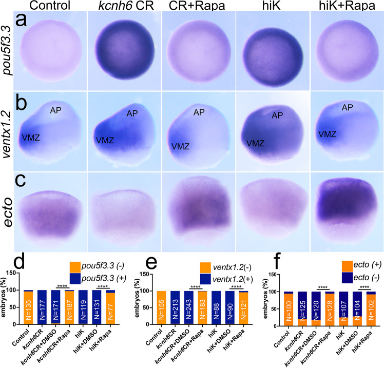
Our transcriptome analysis not only revealed a potential gene regulatory network but pointed towards a role for mTOR. mTOR is critical for multiple cellular processes including autophagy, nutrient sensing, and an emerging role in pluripotency50–56. Because the expression of mTOR pathway members was increased in depolarizing conditions and pathways associated with mTOR, we reasoned that mTOR signaling was upregulated and maintained pluripotency in these depolarized embryos. To test this hypothesis, we applied the mTORC1 inhibitor, rapamycin, to depolarized gastrulating embryos to see if this could abolish the aberrant expression of pluripotency markers pou5f3.3 and ventx1.2 in the animal pole and activate germ-layer differentiation. Rapamycin dramatically lowered expression of pou5f3.3 and ventx1.2 in the animal pole in kcnh6 CR and high K+ treated embryos compared to those embryos treated with vehicle alone and appeared comparable to untreated control embryos (Fig. 7a, b, d, e). Conversely, the expression of the ectodermal marker, ectodermin, which was reduced in depolarizing conditions (kcnh6 depletion or exposure to high K+), was recovered with rapamycin treatment (Fig. 7c, f). Therefore, a polarized Vm at gastrulation onset is critical for limiting mTOR in order to suppress pluripotency genes and enter differentiation.
Fig. 7. Vm polarization limits mTOR which promotes pluripotency.
a–c WMISH for pluripotency markers pou5f3.3, ventx1.2, and differentiation marker ectodermin in stage 10 embryos, depolarized by kcnh6 depletion (kcnh6CR) or high K+ (hiK), and treated with vehicle (DMSO) or rapamycin (Rapa) as indicated; views are a animal pole; b, c lateral view with dorsal to the right; AP animal pole, VMZ ventral marginal zone. d–f Quantification of stage 10 embryos with present (+) or absent (−) expression of markers pou5f3.3 (d), ventx1.2 (e), and ectodemin (f) in the animal pole area. p-values in d are: (kcnh6CR+Rapa vs kcnh6CR+DMSO) = 3.34e-087 and (hiK+Rapa vs hiK+DMSO) = 2.89e-047, in e: (kcnh6CR+Rapa vs kcnh6CR+DMSO) = 1.60e-110 and (hiK+Rapa vs hiK+DMSO) = 1.93e-050, and in f: (kcnh6CR+Rapa vs kcnh6CR+DMSO) = 3.38e-040 and (hiK+Rapa vs hiK+DMSO) = 4.89e-02; two-sided Fisher’s exact test; total embryo numbers (N) in the graphs were collected over 3 independent experiments for pou5f3.3 and ventx1.2 and over 4 independent experiments for ectodermin; key for asterisks: ****p ≤ 0.0001. Source data are provided as a Source Data file.
Finally, we tested whether our findings would also apply to human embryonic stem cells (hESCs). At stage 9, Xenopus animal cap cells are pluripotent in that they can, under appropriate conditions, form derivatives of any of the three germ layers (Fig. 4o). However, they contrast with hESCs in their limited capacity for self-renewal as the brisk pace of embryonic development proceeds. Therefore, we turned our attention to hESCs to test our findings in the context of a self-renewing pluripotent state and to determine their relevance to human development. hESCs are already highly pluripotent, and we wondered if depolarization would lead to elevations in the pluripotency markers OCT4 and SOX2. Indeed, hESCs grown for two days with Ergtoxin to specifically block KCNH channels showed a modest but significant elevation of these markers over their already high levels in the pluripotency state as indicated by immunostaining and qPCR against these markers (Fig. 8a, b, Supplementary Fig. 8a, b). While not as specific as Ergtoxin for KCNH channels, Barium showed similar trends but did not rise to statistical significance. qPCR for the markers OCT4, SOX2, and NANOG also revealed upregulation of these genes at the transcript level, with OCT4 and SOX2 upregulated by day 2 and all three genes upregulated on day 5 (Supplementary Fig. 8a, b).
Fig. 8. Potassium channels affect pluripotency in hESCs.
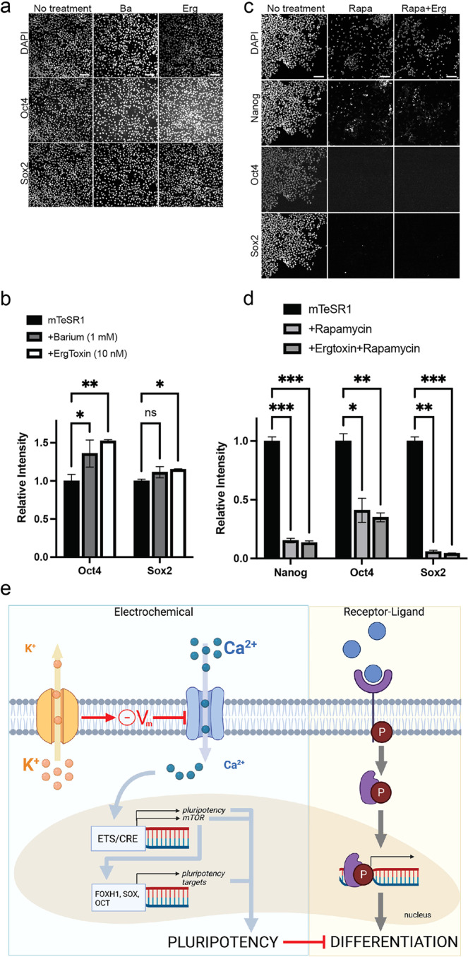
a Images showing untreated hESCs grown in mTeSR1 media or cells treated with 1 mM Barium or 25 nM Ergtoxin and immunostained for pluripotency factors. Scale bar = 100 μm. b Quantification of the results in a; AU arbitrary units. p-values are Oct4: (control vs Ba) = 0.03, (control vs Erg) = 4.5e-04; Sox2 (control vs Ba) = 0.06, (control vs Erg) = 4.4e-04. c Images showing untreated hESCs grown in MEF-CM media or cells treated with 100 nM rapamycin with or without 25 nM Ergtoxin. Scale bar = 100 μm. d Quantification of the results in c; AU arbitrary units. p-values are Nanog: (control vs Rapa) = 4.5e-04, (control vs Rapa+Erg) = 4.3e-04; Oct4: (control vs Rapa) = 0.002, (control vs Rapa+Erg) = 0.002; Sox2 (control vs Rapa) = 7.7e-04, (control vs Rapa+Erg) = 7.6e-04. Graphs (b) and (d) present mean ± SEM; key for asterisks: *p ≤ 0.05, **p ≤ 0.01; ***p ≤ 0.001; ns nonsignificant for p < 0.05; data were derived from 400 cells/3 independent replicates over at least 2 independent experiments. Source data are provided as a Source Data file. e Model for the onset of embryonic differentiation depicting classical biochemical signaling (right) that is complemented by regulation via membrane potential (left). In the electrophysiological pathway, potassium channels set the membrane potential, which limits activation of voltage-gated calcium channels and suppresses intracellular Ca2+ levels. Both pathways result in changes in gene expression, mediated by intracellular signal transducers (right), or by factors that require calcium (left). While biochemical pathways are essential to induce expression of differentiation factors, the electrophysiological pathway affects cell fate indirectly by controlling the timing of downregulation of pluripotency genes. Adapted from “Transporters”, by BioRender.com (2022). Retrieved from https://app.biorender.com/biorender-templates.
We also tested whether blocking K+ channels with Ergtoxin affected the kinetics of differentiation. BMP4 induces differentiation to either mesodermal or extraembryonic fates in a dose-dependent manner57–59. Ergtoxin caused a significant delay in downregulation of pluripotency markers such as SOX2 at 12 h with a similar trend in NANOG (Supplementary Fig. 8c, d). As in Xenopus, the timing of differentiation of human embryonic stem cells appears significantly affected under depolarizing conditions.
To test whether the role of mTOR signaling downstream of membrane depolarization is conserved, we treated hESCs with rapamycin with or without Ergtoxin. Treatment with rapamycin led to reduction of pluripotency markers in a dose-dependent manner with near complete loss by 5 days (Fig. 8c). In the presence of rapamycin, Ergtoxin had no effect on pluripotency markers (Fig. 8d), indicating that mTOR is downstream of membrane depolarization in hESCs as in Xenopus. Although rapamycin did reduce the final cell number per well, it inhibited the effect of Ergtoxin on pluripotency marker expression independently of the density at which cells were seeded (Supplementary Fig. 8e) and of the final cell number in the well (Supplementary Fig. 8f). Taken together, our data support that the polarization of membrane potential via KCNH channels promotes the exit from pluripotency and the activation of differentiated cell fates in Xenopus and human cells.
Discussion
We propose a model in which membrane voltage regulates intracellular calcium during a critical stage of embryonic development, at which point cells need to extinguish pluripotency factors in order to activate a program of cellular differentiation (Fig. 8e). The K+ and Ca2+ channel network upstream of pluripotency factor expression potentially represents an extremely robust control mechanism over the first stages of organism development: ion channels are effective at low expression levels, modular and thus partially redundant with respect to each other (i.e., subunits of different channels can heterodimerize to form a channel if subunits of the same channel are unavailable), and their function depends on the existence of a simple ion concentration gradient across the plasma membrane. The basic elements of this regulation, K+ and Ca2+, are readily available extracellularly, conferring this system with some independence from protein-dependent cell signaling. Ultimately, ion channel networks may represent an additional mechanism to regulate cell fate during development, which is complementary to the established paradigm of gene expression regulation by secreted factors and ligand-receptor signaling (Fig. 8e).
Our work and the work by others supports the notion that membrane potential modulates development in a variety of contexts including LR patterning2–6, chondrocyte differentiation in chick limbs13 as well as morphogenesis of the developing Drosophila wing8,9. Additionally, our work adds to the molecular mechanisms that have associated membrane potential with stem cells differentation12,14,60–62. Of note, previous work has suggested that K+ itself may have signaling properties that affect the directed differentiation of hESCs63. However, our study indicates that it is Vm and not K+ itself that is critical. This is consistent with the notion that, because K+ is so abundant inside the cell, it is unlikely to act as a signaling moiety, unlike Ca2+.
While elevations in intracellular calcium do have physiological functions in the prospective mesoderm and ectoderm of the gastrula, both in patterning24,25 and morphogenesis30, we show that regulation of voltage-gated calcium channels by Vm is critical for the exit from pluripotency. Based on this, we suggest that low intracellular calcium reduces the expression of mTOR and pluripotency factors, which is conducive to differentiation onset. While we have not eliminated other voltage responsive signaling, our results indicate that calcium is critical in this context.
Regulation by Vm and Ca2+ presents interesting implications for developmental timing, as they appear to be key in specifying the exact time frame of pluripotency exit. Indeed, previous work has also implicated mTOR in developmental timing of the pluripotent state64, and our work demonstrates the upstream regulation of mTOR in this context. The requirement of Vm for this developmental period is also underscored by measurements in developing Xenopus16 and Triturus15 showing a clear hyperpolarization of Vm towards the onset of germ-layer differentiation that is concurrent with a reduction in calcium levels26. Therefore, it may be that membrane polarization may act as a timing mechanism to allow embryonic cells to be synchronized before the onset of differentiation. To do so, the embryo employs ion channels, mTOR, and an array of pluripotency transcription factors. Then secreted factors can act on these pluripotent cells to activate different programs of differentiation across the embryo (Fig. 8e). Of note, tissues with abnormal ion homeostasis at gastrulation onset do have differentiation potential and may eventually differentiate at later stages, but the significant delay in exiting pluripotency dramatically affects gastrulation morphogenesis itself as well as LR patterning.
Importantly, genetic data from Htx/CHD patients identified the KCNH family and especially KCNH6. Recent work has identified ion channels as an interesting intersection between CHD and autism65. While ion channels in neuronal development are well established14, our work adds to the role of ion channels in early embryonic development that includes pluripotency, LR patterning, and congenital heart disease.
Finally, the induction and maintenance of pluripotency clearly depends on a set of transcription factors, and our work connects Vm and intracellular calcium as upstream of this pluripotency program. This finding may be applicable to multiple contexts in which stem cells play a role in embryonic development or adult tissue homeostasis. Our work demonstrates the importance of Vm in vivo during early embryonic development as well as in vitro in human stem cells. Importantly, this pathway is readily manipulated by a wide range of highly specific channel inhibitors or simple changes in extracellular ionic concentrations. Therefore, we define multiple tools for pluripotency manipulations in embryos, organoids, and adult tissues where stem cells play a critical role.
Methods
Xenopus husbandry
In this study, we used Xenopus tropicalis from the N (Nigerian) strain. Adult X. tropicalis were raised and housed according to our established protocols which were approved by the Yale Institutional Animal Care and Use Committee. We induced ovulation, performed IVF, and raised embryos in 1/9× MR as previously described66. We staged X. tropicalis embryos according to Nieuwkoop and Faber67. In Xenopus, sex is determined in a 50:50 ratio genetically, and our experiments were all performed in embryos under 72 h of age before the sex of the animal can be determined morphologically.
Morpholino oligonucleotides, mRNA, and CRISPRs
All injections of Xenopus embryos were performed at the one-cell stage using a fine glass needle and Picospritzer system, as previously described68. A kcnh6 translation blocking (kcnh6 MO, 5’-GGTCCTCGAAGTTTAGGATAAACAT-3’) and a scrambled morpholino oligonucleotide were obtained from Gene Tools LLC and injected at 10 ng to deplete kcnh6 or as a control, respectively. CRISPR sgRNAs for kcnh6 targeted either exon 3 or exon 4 based on the v7.1 gene model of the X. tropicalis genome (CRex3: 5’-GGAATAAGGGGTGAAGACAGCGG-3’ and CRex4: 5’-AGGGCGCTCTACATTTCCAATGG-3’). CRISPR sgRNAs for cacna1c (5’-GCAGACGGGGGCAGCGCCATTGG-3’) and cacna1g (5’-GGTTAATGGCTCTCAGCGGGCGG-3’) were designed from the v7.1 model of the Xenopus tropicalis genome. For F0 CRISPR knockdown, embryos were injected with 1.5 ng Cas9 Protein (PNA-Bio) and 400 pg of targeting sgRNA and raised to desired stages as previously described69. For pitx2 and coco expression analyses, the dose of kcnh6 sgRNA was halved to a subphenotypic dose of 200 pg to obtain embryos without gross morphological gastrulation defects. Full-length human KCNH6 (NM_030779.3 cloned in pCS107), GCaMP6 (subcloned in pCSDest) and mCherry cDNAs (Addgene #34935; in pCS2 + ), were used to generate capped mRNAs in vitro by first linearizing with appropriate restriction enzymes and then transcribing with the mMessage machine kit (Ambion). mRNAs were injected at 3 pg (human KCNH6), 150 pg (GCaMP6), and 150 pg (mCherry) per embryo. Embryos were raised at 21 °C to allow time for sufficient expression levels at blastula/gastrula stages.
Inference of CRISPR edits (ICE) analysis
Genomic DNA from CRISPR and control embryos were obtained by lysing individual, stage 45 tadpoles in 50 mM NaOH as previously described69 and amplifying PCR fragments around the CRISPR target site that encompass ~200 bp upstream and 500 bp downstream of the site. The following primers were used for CRISPRs targeting exons 3 and 4 of the kcnh6 locus, respectively: CRex3-F: 5’-CAGGACTGATGAAAGCAAGC-3’ and CRex3-R: 5’-GCTTATCCATAGCTGTAACAACG-3’; CRex4-F: 5’-GAGACAGTAGGCTGTTCC-3’ and CRex4-R: 5’-CCACAAGCAGTTTCACTACC-3’. PCR fragments were Sanger sequenced using the same forward primers, and sequencing traces were uploaded for analysis with the Synthego ICE analysis web tool to assess editing outcomes.
Organ situs
Stage 45 Xenopus embryos were paralyzed with benzocaine or tricaine and scored with a light stereomicroscope. Cardiac looping was determined by position of the outflow tract; D-loop: rightward, L-loop: leftward; A-loop: midline. Normal intestinal looping was scored as counter-clockwise rotation of the gut, while abnormal intestinal looping was scored as completely inverse gut rotation (clockwise) or complete lack of looping (unlooped). While a completely inverted gut rotation is clearly an abnormality of LR patterning, an unlooped gut is less clear so we only considered an unlooped gut as abnormal situs when combined with abnormal placement (left-sided or midline) of the gall bladder. To quantify total abnormal organ situs, each tadpole was counted only once, regardless of whether multiple organs were affected.
Whole mount in situ hybridization
Digoxigenin-labeled antisense probes for pitx2 (TNeu083k20), dand5/coco (TEgg007d24), myoD (Tneu017H11), myf5 (TGas127b01), tbxt (TNeu024F07), foxj1 (Tneu058M03), ectodermin (TNeu104j16), foxI1a (Tgas002H16), mixer (TGas105b05), vegT (TGas066f22), gsc (TNeu077f20), xnr3 (Tgas011k18), vent2 (BG885317), oct25 (TGas051h05), oct60 (IMAGE: 7526158), oct91 (IMAGE: 7575764), vent1 (BG487195), sox2 (Tgas061h22), cytokeratin (IMAGE:6991625), and sox17β (BG886038) were in vitro transcribed using T7 High Yield RNA Synthesis Kit (E2040S) from New England Biolabs. In order to generate a full-length antisense probe for X. tropicalis kcnh6, kcnh6 cDNA was cloned from stage 45 tadpole whole mRNA using primers xtkcnh6-F: 5’- ATGTTTATCCTAAACTTCGAGGACC-3’ and xtkcnh6-R: 5’-CTAACTTCCTGGAAGACCTGGG-3’ (XM_012952904.1). We note that kcnh6 had been misannotated as kcnh2 in the v7.1 model of the X. tropicalis genome (We used NCBI Annotation XP_012808358.2 to identify KCNH6). Embryos were collected at the desired stages, fixed in MEMFA for 1–2 h at room temperature (RT) and dehydrated in 100% ethanol. GRPs were dissected post fixation and prior to dehydration to detect dand5. To detect putative gene expression in the prospective endoderm (mixer, vegT, kcnh6) gastrula stage embryos were bisected to facilitate better probe access. Briefly, whole mount in situ hybridization of digoxigenin-labeled antisense probes was performed overnight, the labeled embryos were then washed, incubated with anti-digoxigenin-AP Fab fragments (Roche 11093274910), and signal was detected using BM-purple (Roche 11442074001), as previously described in detail68.
Medium conditions and treatments of embryos
Normal embryonic medium is 1/9x modified Ringer’s (MR) containing 11 mM NaCl, 0.2 mM KCl, 0.2 mM CaCl2, 0.1 mM Mg2Cl and 0.55 mM HEPES. To allow Ergtoxin to penetrate the embryos, we manually removed the vitelline envelope of stage 8 embryos and incubated embryos in 1/9× MR containing 50 nM Ergtoxin (Alomone STE-450) until stage 12. Embryos were then transferred back into 1/9× MR lacking Ergtoxin to develop until stage 45 in order to score organ situs. Barium chloride was applied into the medium at 20 mM and embryos were thoroughly rinsed in 1/9× MR after each incubation period for further development in Ba2+-free medium. For extracellular K+ manipulations, the KCl concentration in 1/9x MR was modified from 0.2 mM (normal) to 20 mM (high) or 0 mM (low). The ionophore valinomycin (ACROS) and L-type VGCC blocker Nifedipine (ACROS) were diluted in DMSO as stock solutions and applied to embryos in 1/9× MR at 2 and 10 μM, respectively. Treatments performed during gastrulation were applied from stage 8 through stage 12, and embryos were then rinsed thoroughly and returned into 1/9× MR. For rapamycin, we created a standard stock solution of 50 mg/ml in DMSO. The stock solution was diluted 1:2500 in the appropriate embryonic media (final 20 μg/ml). Embryos were treated at stage 7 and then fixed at stage 11 for in situ hybridization.
GRP immunofluorescence
Embryos were fixed at stage 17 in 4% paraformaldehyde-PBS for 2 h at RT, washed in PBS, and then dissected to obtain GRPs. GRPs were permeabilized for 30 min at RT using 0.1% Triton-PBS (PBST), then blocked in 1% BSA-PBST for 1 h at RT and incubated in primary antibodies diluted in 1% BSA-PBST overnight at 4 °C (anti-myoD 1:100; LsBio C143580-100 or anti-acetylated tubulin 1:2000 Sigma T-6793). GRPs were then washed in PBST for 30 min and then incubated with secondary antibodies in 1% BSA-PBST for 1 h at RT. Phalloidin (1:50; Molecular Probes) and Hoechst 33342 (1:1000; Molecular Probes) were diluted into the secondary antibody solution. Images were acquired using a ZEISS 710 laser scanning confocal microscope.
Intracellular Vm recordings
For recordings, devitellinized, stage 10 kcnh6 or control MO-injected embryos were mounted into non-toxic clay with their animal pole exposed and covered with 1/9x MR. To investigate the resting potential, animal pole cells were impaled with a high-impedance (~70 MΩ), sharp microelectrode filled with 3 M KCl for intracellular recordings. The recordings were made using an Axon 200B amplifier and digitized using a Digidata 1320 digitizer. Jclamp software for Windows was used in current clamp mode. All electrodes were zeroed just before entry into the cells.
For the series of intracellular recordings in high K+ and choline treated embryos, stage 8–9 embryos were impaled similarly with an electrode of ~40 MΩ. These recordings were made using a HEKA EPC10 amplifier. We used HEKA PatchMaster v2x67software for Windows. All electrodes were zeroed just before entry into the cells.
Calcium imaging
GCaMP6 and mCherry mRNAs were mixed and injected into embryos at the one-cell stage. Half of these embryos were then injected with kcnh6 MO and the other half with control MO, still at the one-cell stage. Embryos were transferred at stage 10 into the round wells of a press-to-seal silicone isolator (Sigma S3685) mounted between two cover slips in 2% Methylcellulose-1/9x MR. GcaMP6 and mCherry fluorescence was then captured for 20 s (1 frame per second) via time lapse in the whole animal pole of each embryo with a ×20 objective of an LSM710 confocal microscope using identical acquisition settings across Control MO and kcnh6 MO embryos. Time lapse recordings were conducted randomly and in an unbiased manner in regard to presence and intensity of calcium transients. However, all embryos did display transient increases in GcaMP6 fluorescence, varying in intensity and spreading to multiple cells. The frames of each recording were sorted to identify the calcium transient peak (in area), and GcAMP6 fluorescence intensity was quantified at peak as a ratio to mCherry in mCherry+ cells. The maximum Ca2+ transient area was calculated by demarcating in Fuji the GCaMP6(+) vs GcaMP6(–) area of the animal pole at transient peak. To avoid mosaicism artifacts, only embryos with even, non-mosaic mCherry expression across the entire animal pole were considered. To avoid embryonic stage dependent fluctuations in Ca2+ transient size, we verified each embryo for stage by progression of blastopore closure and alternated recordings of control and kcnh6 MO embryos. Of note, there were no notable differences in mCherry expression between Control MO and kcnh6 MO embryos.
Animal cap pluripotency assays
After manually removing the vitelline envelope of stage 9 or 12 embryos, animal caps were excised and placed on agarose coated dishes in 1/9× MR solution. Caps were then directly placed into agarose coated wells of a 96-well plate in 1/3× MR containing 0.1% BSA and cultured without activin to allow for differentiation into epidermis, with low (20 ng/ml) activin to induce mesoderm, or high activin (200 ng/ml) to induce endoderm, as previously described43. Explants were raised at 25 °C until reaching the equivalent of stage 18 (monitored in whole embryos of the same batch), then fixed in 4% paraformaldehyde, washed in PBS, bleached to eliminate pigmentation (0.5× SSC, 5% formamide, 1.2% H2O2), and then processed by in situ hybridization as described above.
RNA-seq
For RNA-Seq, embryos were kept at 25 °C either in 1/9× MR or in 10 mM KCl solution, and 10 embryos were harvested per time point and condition every 30 min starting at stage 8 and concluding at stage 13. Samples were immediately frozen and kept at −80 °C until homogenized in 100 μl Trizol spiked with ERCC RNA Spike-In Mix. 10 μl ERCC RNA Spike-In Mix (Thermo Fisher Scientific) were first diluted into a final volume of 870 μl DEPC water and then further diluted 1:10 into Trizol, which was used to homogenize the samples. Total RNA was purified from the embryo Trizol homogenates according to the manufacturer’s recommendations. After isopropanol precipitation, RNAs were resuspended in DEPC water and any contaminating genomic DNA was removed by overnight precipitation in 5 M LiCl at 4 °C. RNA was subsequently pelleted and washed twice with 70% ethanol. All RNAs were resuspended in DEPC water (2 μl/embryo), and finally, RNA quality was verified by Bioanalyzer. All libraries were sequenced with 100 bp paired-ends on an Illumina NovaSeq6000.
Quantification RNA-seq
Stranded paired-end 100 bp RNA-seq reads were aligned to the Xt9.1 genome combined with ERCC spikes using STAR70 and quantified as transcripts per million (TPM) for each isoform with RSEM71 using the RSEM-STAR pipeline, with additional options “--seed 1618 --calc-pme --calc-ci --estimate-rspd --paired-end”. Using the ERCC spikes we identified a batch-dependent GC bias where AT-rich transcripts were preferentially lost as compared to GC-rich transcripts (Supplementary Fig. 7b). We leveraged knowledge of spike-in concentrations to build a GC model correction based on the dinucleotide content of RNAs. We calculate the propensity of each of the 16 dinucleotides (AA, AC, …, TG, TT) within each spike sequence, with fik is the frequency of dinucleotide k within sequence i, its propensity is . We then employ the following linear model to correct the TPM tsi of RNA spike in sample to its known concentration :
We use the GLM.jl (https://github.com/JuliaStats/GLM.jl) in the Julia language to apply this model and add a pseudocount of 2 to all dinucleotide frequencies. As the GC effect varies between UC and high K+ samples (Supplementary Fig. 7b), we apply the correction independently to UC and High K+. The correction is able to explain a significant proportion of variance in spike TPM, increasing R2 from 0.807 and 0.733 to 0.965 and 0.964 respectively from UC and high K+ samples. We apply this correction to each isoform of all genes quantified with the dinucleotide propensities of each isoform and RSEM isoform quantifications. We then sum all corrected quantifications at the isoform level to derive gene level quantifications. This allows us to account for differing isoforms of the same gene with differing dinucleotide propensities.
Filtering of genes for differential expression analysis
We first filtered 34,192 quantified genes to find those with sufficient temporal expression for further analysis, we selected genes that had runs of 6 consecutive samples with uncorrected TPM > 0.4. This resulted 13,310 from which we excluded a further 162 genes which where excessively altered by the above described correction procedure, these had log2 fold changes between corrected or uncorrected quantifications outside of the interval (−2.5, 4.5). After dinucleotide correction and filtering we found excellent concordance between samples, with minimal evidence of outlying samples, by Spearman Correlation comparisons and principal components analysis (PCA) (Supplementary Fig. 7c, d). The two domains in visible in pairwise Spearman comparisons (Supplementary Fig. 7c) reflect the loss of maternal RNA and the commencement of widespread zygotic transcription as we previously described19. Projection onto the first two principal components revealed that samples lie in appropriate order on a trajectory in 2D space, and the largest divergences between UC and high K+ occur midway through the time series in agreement with Gaussian process differential expression and clustering described below. Corrected dinucleotide gene expression abundances are used in all analyses.
Temporal differentiation expression
To determine genes temporally differentially expressed we used Gaussian process (GP) regression as we have previously applied19. All GP regression was performed with GaussianProcesses.jl (https://github.com/STOR-i/GaussianProcesses.jl; https://arxiv.org/abs/1812.09064). Due to the overdispersed nature of RNA-seq count data, we apply a variance stabilizing transform that puts all genes on the same scale: , with xsi the dinucleotide corrected abundance of gene i in sample s, mi the maximum xsi over all samples, and α = 1, β = 1000. We then perform exact GP regression (GP prior and a Gaussian likelihood) with Matern52 kernel, we optimize the three associated hyperparameters: the signal variance, τ the timescale (using previous terminology19, this parameter is commonly referred to as the lengthscale l), and the sample noise variance. Parameters are selected by optimizing marginal log-likelihood with parameters in log space: , and to ensure physiologically reasonable values for each we place Gaussian priors, over each of these variables respectively . Finally, we reported GP median and 95% confidence intervals through our inverted data transformation and set for .
To determine temporal differential expression, we calculate a marginal likelihood ratio for whether we prefer separate GP models for UC and high K+ or a single GP model for all data combined. If LU and LK are the marginal log-likelihoods for UC and high K+ respectively, and LUK is the marginal log-likelihood for a single regression through UC and high K+ together. Then we calculate log-likelihood ratio of evidence in favor of two models (essentially that the UC and High K+ have different expression trajectories for a given gene) and determine genes with LR > 0 as temporally differentially expressed. This resulted in 5144 differentially expressed genes, with 4043 activated and 1101 repressed (Supplementary Fig. 7d which shows that the max absolute divergence z-score between UC and high K+ trajectories increases with LR). We also considered a more stringent condition for differential expression using the Bayesian Information Criterion (BIC)19, that resulted in 2388 differentially genes. We found that this diminished differential expression gene set enrichments described below, indicating that the BIC was too conservative and we continued with our condition based on log-likelihood ratio.
Clustering
To determine sets of differentially expressed genes with similar trajectories, we applied K-means clustering to activated and repressed genes independently. We define a gene as activated if Gaussian process median for High K+ exceeds UC on average, and repressed if it does not, we found no genes for which the mean of High+ and UC differences was zero. We cluster UC and High K+ genes by taking Gaussian process medians and normalizing by the maximum value experience by UC or High K+. We then cluster both trajectories together employing the kmeans function offered by Clustering.jl (https://github.com/JuliaStats/Clustering.jl) with default settings and random seed 16. To select the cluster number, we calculated the silhouette score for activated and repressed clusters for k = 2–10. We found that the maximal mean silhouette score activated genes was k = 3 and for repressed genes was k = 2, but that scores were broadly similar for k = 2–4 and decreased significantly for k > 4, suggesting that k = 4 provides a reasonable partition of the data. In line with this we explored the clusters from k = 2–10, and found that key clusters were not well-resolved for k < 4 and that k > 4 clusters refined k = 4 behaviors. As k > 4 did not reveal new behaviors and did not improve gene set enrichments, we selected k = 4 to cluster activated and repressed genes.
Gene set enrichments
To assess the composition of each cluster we performed gene set enrichments using Enrichr45. We took genes from each cluster with a known Xenopus gene symbol and converted these to human symbols, by removing any “.N” suffix for an integer N (for example, ventx1.1 becomes ventx1) and converting to uppercase. We then made the following substitutions to convert certain known Xenopus gene symbols to human where the name of the ortholog has diverged or only a paralog exists: pou5f3 → POU5F1, mix1 → mixl1, dppa2 → DPPA4, lefty → lefty2, ventx1-3 → NANOG, mespb → MESP1, sox17a/b → SOX17. We remove any duplicate names that arose in this process. We calculated enrichments for the following gene sets: KEGG_2019_Human, BioPlanet_2019, WikiPathways_2019_Human, GO_Biological_Process_2018, GO_Molecular_Function_2018, GO_Cellular_Component_2018, ChEA_2016. We calculate enrichments for each cluster individually and consecutive combinations of the 4 clusters: 1, 2, 3, 4, 12, 23, 34, 123, 234, 1234. All enrichments can be found in Supplementary Data 1, selected enrichments are given in Fig. 6C and terms are given shortened labels for brevity (Table 2):
Table 2.
Selected enrichments from RNA-seq time course
| Label | Term | Gene set |
|---|---|---|
| mTOR | mTOR signaling pathway | KEGG_2019_Human |
| Autophagy | Autophagy | KEGG_2019_Human |
| Ubiquitin | Ubiquitin-protein transferase activity GO:0004842 | GO_Molecular_Function_2018 |
| ER | Protein processing in endoplasmic reticulum | KEGG_2019_Human |
| Spliceosome | Spliceosome | KEGG_2019_Human |
| WNT signaling | Wnt signaling pathway | KEGG_2019_Human |
| Pluripotency signaling | Signaling pathways regulating pluripotency of stem cells | KEGG_2019_Human |
| Endoderm differentiation | Endoderm differentiation WP2853 | WikiPathways_2019_Human |
Selected enrichments and their associated abbreviated label listed in Fig. 6c.
Motif analysis
To find motifs enriched in the promoters, we took the 500 bp upstream of the promoter of the maximally expressed isoform for each gene in the four activated and four repressed clusters, along with a background of the 500 bp upstream of all annotated TSS in the Xt9.1 genome. We extracted fasta files for each of these sets of regions, and then used findMotifs.pl from Homer72 to search for known motifs with options: “findMotifs.pl clusterAB.fa fasta outAB –fasta background.fa –nomotif” where A ∈ {activated, repressed} and B ∈ {1, 2, 3, 4}. We filtered results to select best matching motifs from related families, namely we collapsed all ETS motifs to the canonical Homer ETS promoter motif; all SP and KLF motifs to SP1; SOX motifs to SOX2; all HOX motifs to HOXD13 (the highest scoring HOX); we represent all GFY and Ronin matches as ZNF143 (for which the motifs overlap); and we excluded motif annotated as PRDM10, due to low confidence in the motif. The motif annotated as ATF1 is an example of the cAMP response element (CRE) bound by CREB factors including ATF1, we label this as CRE/ATF1. The top 16 motif enrichments are given in Supplementary Fig. 7h, in Fig. 6d we give the top 6 motif matches, excluding ZNF143 due to divergence from the JASPAR database, to which we add CRE/ATF1 as a putative calcium responsive element motivated by CREM/CREB1 gene set enrichments Supplementary Fig. 7j).
To calculate CRE and ETS motif enrichment for mTOR and pluripotency genes, we took genes annotated with the terms mTOR signaling pathway and Signaling pathways regulating pluripotency of stem cells from KEGG_2019_Human as provided by Enrichr45 that are activated in high K+ (LR > 0) and are present in clusters A1 and A2. The resulting genes were subjected to the same promoter analysis, using Homer to calculate the occurrence of the maximal ATF/CRE family motif and the ETS motif in these promoters and the background set to report Fisher Exact test p-values and Odds Ratios.
Xenopus biological replicates, statistical methods, graphs, and models
In experiments where embryos were evaluated for phenotypes and scored (gastrulation, left-right patterning, in situ hybridizations) we carried out three to five biological replicates and Fisher’s exact test to evaluate statistical significance. The animal cap experiment was performed twice with a total score of four to eight animal caps per experiment. For the calcium transient analyses, data was collected from three to five embryos in each experiment in three independent experiments, and statistical analyses on GCaMP/mCherry fluorescence intensity as well as Ca2+ transient area were performed using student’s t-test. For whole cell electrophysiological recordings, three to five embryos (two cells each) were examined for their membrane potential and statistical significance was tested by student’s t-test. Graphs were designed using GraphPad Prism software. Models were created with BioRender.com.
hESC culture
hESCs were grown in mTeSR1 (STEMCELL Technologies) in tissue culture dishes coated with Matrigel (Corning; 1:200 in DMEM/F12) and kept at 37 °C, 5% CO2. The cell lines used were ESI017 (ESIBIO) and H9. Cells were routinely passaged using dispase (STEMCELL Technologies) and tested for mycoplasma contamination and found negative. For rapamycin experiments, cells were grown in MEF-conditioned HUESM media supplemented with 20 ng/ml bFGF as previously described73 with or without 100 nM rapamycin, which we found to increase the survival of rapamycin treated cells compared to cells grown in mTeSR1.
hESC treatments and differentiation
Cells were dissociated with accutase and seeded onto eight-well imaging slides (ibidi 80826) at a density of 4–6 × 10⁴/cm2. Cells were seeded and maintained in Rock-inhibitor Y27672 (MCE; 10 μM) to increase survival and the uniformity of response. Treatments with 1 mM BaCl2 or 10 or 25 nM Ergtoxin or 100 nM rapamycin were initiated 4 h after seeding. Differentiation was initiated 24 h after seeding where indicated. To initiate differentiation, the media was replenished with/without BaCl2 or Ergtoxin and treated with the indicated growth factors or small molecules. The media with any treatments was replenished daily.
Immunofluorescence of hESCs
Cells were fixed for 30 min in 4% paraformaldehyde, rinsed twice with DPBS (without Ca2+ and Mg2+, denoted DPBS-/-), and blocked for 30 min at room temperature. The blocking solution contained 3% donkey serum and 0.1% Triton X-100 in 1× DPBS−/−. After blocking, the cells were incubated with primary antibodies at room temperature for 2 hours. Antibodies and concentrations are listed in Supplementary Table 1. Cells were washed three times with DPBST (1X DPBS-/- with 0.1% Tween 20) and incubated with secondary antibodies (AlexaFluor 488 A21206, AlexaFluor 555 A31570 and A21432, and AlexaFluor 647 A31571, Thermo Fisher; 1:500) and DAPI for 30 min at room temperature. After secondary antibody incubation, samples were washed in DPBST and then DPBS at room temperature.
hESC Imaging and analysis
Images were acquired using a ×20, NA 0.75 objective on an Olympus IX83 inverted epifluorescence microscope or an Olympus/Andor spinning disk confocal microscope. Cell segmentation was performed using ilastik software74. This segmentation was cleaned (to remove debris and to separate merged cells) and mean nuclear protein intensities as well as standard errors were quantified using a custom MATLAB code. Nuclear intensities were normalized by DAPI to correct for intensity variation due to optics. Code is available at https://github.com/warmflashlab/Sempou2022_Code.
qPCR
For qPCR, hESCs were grown with or without ErgToxin (25 nM) for the indicated times. RNA collection and DNase treatment were performed using the RNAqueous®-Micro Total RNA Isolation Kit (AM1931) and cDNA was synthesized with the SuperScript Vilo cDNA Synthesis Kit (Fisher Scientific 11754-050). qPCR measurements were collected using SYBR Green reagent (LifeTech-4367659) on a Step OnePlus instrument (Applied Biosciences). Data were normalized using the housekeeping gene GAPDH. Primers for qPCR were:
OCT4: 5’-caagctcctgaagcagaagag−3’, 5’-ccaaacgaccatctgccgcttt−3’,
SOX2: 5’-ccatgcaggttgacaccgttg−3’, 5’-tcggcagactgattcaaataata-3’,
NANOG: 5’-tgggatttacaggcctgagcca−3’, 5’-aagcaaagcctcccaatcccaaa−3’,
GAPDH: 5’-caccgtcaaggctgagaacg-3’, 5’-gccccacttgattttggagg-3’.
Statistics and reproducibility
Embryo and cell sample sizes in this study were chosen according to the standards in the field. No data were excluded from the analyses. The investigators were blinded to allocation during experiments and outcome assessment whenever feasible.
For statistics, two-sided Fisher’s exact test was used to assess significance of two variables with independent proportions, (treated/untreated) and their outcomes (normal/abnormal). This test was applied when scoring for morphological phenotypes of embryos and WMISH results, under control conditions or after microinjection (CR, MO, mRNA), medium manipulations or treatment with compounds. Two-tailed, unpaired t-tests or ANOVA were used to test whether the means of two populations differ largely from one another and were employed for Vm, calcium intensity and calcium transient area comparisons as well as all hESC experiments.
Reporting summary
Further information on research design is available in the Nature Research Reporting Summary linked to this article.
Supplementary information
Description of Additional Supplementary Files
Acknowledgements
We would like to thank the patients and their families, who are the inspiration for this study. We would like to thank Michael Slocum, Maura Lane, and Sarah Kubek for frog care and husbandry, Richard Harland (especially Debbie Pangilinan) and Addgene for providing plasmids, and Sergei Sokol for kindly supplying activin. We thank the Yale Center for Cellular Imaging for microscopy support, and Yale Center for Genome Analysis for RNA-seq. E.S. was supported by a Brown-Coxe Postdoctoral Fellowship. N.O. is supported by an Expanding Excellence in England Lectureship. This work was supported by the NIH R01HD081379 to M.K.K., NIH R01DK072612 to M.J.C., NIH R01EY021195 to D.Z., and NIH R01GM126122, NSF MCB-1553228, and Welch Foundation C-2021 to A.W. M.K.K. was a Mallinckrodt Scholar.
Source data
Author contributions
E.S., M.J.C., D.Z., A.W., N.D.L.O., and M.K.K. conceived and designed the experiments and analyzed data from the experiments. E.S., V.K., W.H., and M.K.K. performed all Xenopus experiments. E.S., J.Z., L.T., and D.Z. performed electrophysiological recordings in Xenopus. N.D.L.O. analyzed all genomic data. E.S., C.G., A.W., and M.K.K. conceived, designed, and analyzed experiments in human embryonic stem cells. C.G., E.C.-A., and A.W. performed human embryonic stem cell experiments. The manuscript was written by E.S., A.W., N.D.L.O., and M.K.K. and all authors critically evaluated the manuscript.
Peer review
Peer review information
Nature Communications thanks the anonymous reviewers for their contribution to the peer review of this work. Peer reviewer reports are available.
Data availability
The RNA-seq time series of uninjected-control (UC) and high K+ embryos generated in this study have been deposited in the Gene Expression Omnibus under accession GSE186670. Source data are provided with this paper.
Code availability
All source data and code required to analyze RNA-seq time series and generate figures are available at https://github.com/owensnick/KCNH6GenomicsFigures.jl, 10.5281/zenodo.6967350. Source code for quantitating protein intensities in human ES cells is available at https://github.com/warmflashlab/Sempou2022_Code.
Competing interests
The authors declare the following competing interests: M.K.K. is a Founder and President of Victory Genomics, Inc. Yale University has filed a provisional patent application related to this work. The remaining authors declare no competing interests.
Footnotes
Publisher’s note Springer Nature remains neutral with regard to jurisdictional claims in published maps and institutional affiliations.
Change history
6/5/2023
A Correction to this paper has been published: 10.1038/s41467-023-39025-z
Supplementary information
The online version contains supplementary material available at 10.1038/s41467-022-34363-w.
References
- 1.Vandenberg LN, Levin M. A unified model for left-right asymmetry? Comparison and synthesis of molecular models of embryonic laterality. Dev. Biol. 2013;379:1–15. doi: 10.1016/j.ydbio.2013.03.021. [DOI] [PMC free article] [PubMed] [Google Scholar]
- 2.Aw S, et al. The ATP-sensitive K(+)-channel (K(ATP)) controls early left-right patterning in Xenopus and chick embryos. Dev. Biol. 2010;346:39–53. doi: 10.1016/j.ydbio.2010.07.011. [DOI] [PMC free article] [PubMed] [Google Scholar]
- 3.Levin M, Thorlin T, Robinson KR, Nogi T, Mercola M. Asymmetries in H+/K+-ATPase and cell membrane potentials comprise a very early step in left-right patterning. Cell. 2002;111:77–89. doi: 10.1016/S0092-8674(02)00939-X. [DOI] [PubMed] [Google Scholar]
- 4.Morokuma J, Blackiston D, Levin M. KCNQ1 and KCNE1 K+ channel components are involved in early left-right patterning in Xenopus laevis embryos. Cell Physiol. Biochem. 2008;21:357–372. doi: 10.1159/000129628. [DOI] [PMC free article] [PubMed] [Google Scholar]
- 5.Pai VP, et al. HCN4 ion channel function is required for early events that regulate anatomical left-right patterning in a nodal and lefty asymmetric gene expression-independent manner. Biol. Open. 2017;6:1445–1457. doi: 10.1242/bio.025957. [DOI] [PMC free article] [PubMed] [Google Scholar]
- 6.Adams DS, et al. Early, H+-V-ATPase-dependent proton flux is necessary for consistent left-right patterning of non-mammalian vertebrates. Development. 2006;133:1657–1671. doi: 10.1242/dev.02341. [DOI] [PMC free article] [PubMed] [Google Scholar]
- 7.Walentek P, Beyer T, Thumberger T, Schweickert A, Blum M. ATP4a is required for Wnt-dependent Foxj1 expression and leftward flow in Xenopus left-right development. Cell Rep. 2012;1:516–527. doi: 10.1016/j.celrep.2012.03.005. [DOI] [PubMed] [Google Scholar]
- 8.Emmons-Bell M, Hariharan IK. Membrane potential regulates Hedgehog signalling in the Drosophila wing imaginal disc. EMBO Rep. 2021;22:e51861. doi: 10.15252/embr.202051861. [DOI] [PMC free article] [PubMed] [Google Scholar]
- 9.Dahal GR, Pradhan SJ, Bates EA. Inwardly rectifying potassium channels influence Drosophila wing morphogenesis by regulating Dpp release. Development. 2017;144:2771–2783. doi: 10.1242/dev.146647. [DOI] [PMC free article] [PubMed] [Google Scholar]
- 10.Belus MT, et al. Kir2.1 is important for efficient BMP signaling in mammalian face development. Dev. Biol. 2018;444:S297–S307. doi: 10.1016/j.ydbio.2018.02.012. [DOI] [PMC free article] [PubMed] [Google Scholar]
- 11.Vandenberg LN, Morrie RD, Adams DS. V-ATPase-dependent ectodermal voltage and pH regionalization are required for craniofacial morphogenesis. Dev. Dyn. 2011;240:1889–1904. doi: 10.1002/dvdy.22685. [DOI] [PMC free article] [PubMed] [Google Scholar]
- 12.Varga Z, et al. Switch of voltage-gated K+ channel expression in the plasma membrane of chondrogenic cells affects cytosolic Ca2+-oscillations and cartilage formation. PLoS ONE. 2011;6:e27957. doi: 10.1371/journal.pone.0027957. [DOI] [PMC free article] [PubMed] [Google Scholar]
- 13.Atsuta Y, Tomizawa RR, Levin M, Tabin CJ. L-type voltage-gated Ca(2+) channel CaV1.2 regulates chondrogenesis during limb development. Proc. Natl Acad. Sci. USA. 2019;116:21592–21601. doi: 10.1073/pnas.1908981116. [DOI] [PMC free article] [PubMed] [Google Scholar]
- 14.Sundelacruz S, Levin M, Kaplan DL. Role of membrane potential in the regulation of cell proliferation and differentiation. Stem Cell Rev. Rep. 2009;5:231–246. doi: 10.1007/s12015-009-9080-2. [DOI] [PMC free article] [PubMed] [Google Scholar]
- 15.Ito S, Hori N. Electrical characteristics of Triturus egg cells during cleavage. J. Gen. Physiol. 1966;49:1019–1027. doi: 10.1085/jgp.49.5.1019. [DOI] [PMC free article] [PubMed] [Google Scholar]
- 16.Palmer JF, Slack C. Some bio-electric parameters of early Xenopus embryos. J. Embryol. Exp. Morphol. 1970;24:535–553. [PubMed] [Google Scholar]
- 17.Zahn, N. et al. Normal Table of Xenopus development: a new graphical resource. Development10.1242/dev.200356 (2022). [DOI] [PMC free article] [PubMed]
- 18.Jin SC, et al. Contribution of rare inherited and de novo variants in 2,871 congenital heart disease probands. Nat. Genet. 2017;49:1593–1601. doi: 10.1038/ng.3970. [DOI] [PMC free article] [PubMed] [Google Scholar]
- 19.Owens ND, et al. Measuring absolute RNA copy numbers at high temporal resolution reveals transcriptome kinetics in development. Cell Rep. 2016;14:632–647. doi: 10.1016/j.celrep.2015.12.050. [DOI] [PMC free article] [PubMed] [Google Scholar]
- 20.Hsiau, T. et al. Inference of CRISPR edits from sanger trace data. bioRxiv10.1101/251082 (2019). [DOI] [PubMed]
- 21.Scaloni A, et al. Disulfide bridges of ergtoxin, a member of a new sub-family of peptide blockers of the ether-a-go-go-related K+ channel. FEBS Lett. 2000;479:156–157. doi: 10.1016/S0014-5793(00)01891-3. [DOI] [PubMed] [Google Scholar]
- 22.Hodgkin AL, Huxley AF. A quantitative description of membrane current and its application to conduction and excitation in nerve. 1952. Bull. Math. Biol. 1990;52:25–71. doi: 10.1016/S0092-8240(05)80004-7. [DOI] [PubMed] [Google Scholar]
- 23.Tsien RW, Lipscombe D, Madison DV, Bley KR, Fox AP. Multiple types of neuronal calcium channels and their selective modulation. Trends Neurosci. 1988;11:431–438. doi: 10.1016/0166-2236(88)90194-4. [DOI] [PubMed] [Google Scholar]
- 24.Palma V, Kukuljan M, Mayor R. Calcium mediates dorsoventral patterning of mesoderm in Xenopus. Curr. Biol. 2001;11:1606–1610. doi: 10.1016/S0960-9822(01)00479-1. [DOI] [PubMed] [Google Scholar]
- 25.Leclerc C, et al. L-type calcium channel activation controls the in vivo transduction of the neuralizing signal in the amphibian embryos. Mech. Dev. 1997;64:105–110. doi: 10.1016/S0925-4773(97)00054-3. [DOI] [PubMed] [Google Scholar]
- 26.Slack C, Warner AE, Warren RL. The distribution of sodium and potassium in amphibian embryos during early development. J. Physiol. 1973;232:297–312. doi: 10.1113/jphysiol.1973.sp010271. [DOI] [PMC free article] [PubMed] [Google Scholar]
- 27.Chen TW, et al. Ultrasensitive fluorescent proteins for imaging neuronal activity. Nature. 2013;499:295–300. doi: 10.1038/nature12354. [DOI] [PMC free article] [PubMed] [Google Scholar]
- 28.Leclerc C, Neant I, Webb SE, Miller AL, Moreau M. Calcium transients and calcium signalling during early neurogenesis in the amphibian embryo Xenopus laevis. Biochim. Biophys. Acta. 2006;1763:1184–1191. doi: 10.1016/j.bbamcr.2006.08.005. [DOI] [PubMed] [Google Scholar]
- 29.Gilland E, Miller AL, Karplus E, Baker R, Webb SE. Imaging of multicellular large-scale rhythmic calcium waves during zebrafish gastrulation. Proc. Natl Acad. Sci. USA. 1999;96:157–161. doi: 10.1073/pnas.96.1.157. [DOI] [PMC free article] [PubMed] [Google Scholar]
- 30.Wallingford JB, Ewald AJ, Harland RM, Fraser SE. Calcium signaling during convergent extension in Xenopus. Curr. Biol. 2001;11:652–661. doi: 10.1016/S0960-9822(01)00201-9. [DOI] [PubMed] [Google Scholar]
- 31.Shinohara, K. & Hamada, H. Cilia in Left-Right Symmetry Breaking. Cold Spring Harb. Perspect. Biol. 10.1101/cshperspect.a028282 (2017). [DOI] [PMC free article] [PubMed]
- 32.Blum M, Feistel K, Thumberger T, Schweickert A. The evolution and conservation of left-right patterning mechanisms. Development. 2014;141:1603–1613. doi: 10.1242/dev.100560. [DOI] [PubMed] [Google Scholar]
- 33.Vonica A, Brivanlou AH. The left-right axis is regulated by the interplay of Coco, Xnr1 and derriere in Xenopus embryos. Dev. Biol. 2007;303:281–294. doi: 10.1016/j.ydbio.2006.09.039. [DOI] [PubMed] [Google Scholar]
- 34.Schweickert A, et al. The nodal inhibitor Coco is a critical target of leftward flow in Xenopus. Curr. Biol. 2010;20:738–743. doi: 10.1016/j.cub.2010.02.061. [DOI] [PubMed] [Google Scholar]
- 35.Khokha MK, Yeh J, Grammer TC, Harland RM. Depletion of three BMP antagonists from Spemann’s organizer leads to a catastrophic loss of dorsal structures. Dev. Cell. 2005;8:401–411. doi: 10.1016/j.devcel.2005.01.013. [DOI] [PubMed] [Google Scholar]
- 36.Reversade B, Kuroda H, Lee H, Mays A, De Robertis EM. Depletion of Bmp2, Bmp4, Bmp7 and Spemann organizer signals induces massive brain formation in Xenopus embryos. Development. 2005;132:3381–3392. doi: 10.1242/dev.01901. [DOI] [PMC free article] [PubMed] [Google Scholar]
- 37.Heasman J, et al. Overexpression of cadherins and underexpression of beta-catenin inhibit dorsal mesoderm induction in early Xenopus embryos. Cell. 1994;79:791–803. doi: 10.1016/0092-8674(94)90069-8. [DOI] [PubMed] [Google Scholar]
- 38.Zhang J, et al. The role of maternal VegT in establishing the primary germ layers in Xenopus embryos. Cell. 1998;94:515–524. doi: 10.1016/S0092-8674(00)81592-5. [DOI] [PubMed] [Google Scholar]
- 39.Dupont S, et al. Germ-layer specification and control of cell growth by Ectodermin, a Smad4 ubiquitin ligase. Cell. 2005;121:87–99. doi: 10.1016/j.cell.2005.01.033. [DOI] [PubMed] [Google Scholar]
- 40.Morrison GM, Brickman JM. Conserved roles for Oct4 homologues in maintaining multipotency during early vertebrate development. Development. 2006;133:2011–2022. doi: 10.1242/dev.02362. [DOI] [PubMed] [Google Scholar]
- 41.Scerbo P, et al. Ventx factors function as Nanog-like guardians of developmental potential in Xenopus. PLoS ONE. 2012;7:e36855. doi: 10.1371/journal.pone.0036855. [DOI] [PMC free article] [PubMed] [Google Scholar]
- 42.Collart C, et al. High-resolution analysis of gene activity during the Xenopus mid-blastula transition. Development. 2014;141:1927–1939. doi: 10.1242/dev.102012. [DOI] [PMC free article] [PubMed] [Google Scholar]
- 43.Buitrago-Delgado E, Nordin K, Rao A, Geary L, LaBonne C. Shared regulatory programs suggest retention of blastula-stage potential in neural crest cells. Science. 2015;348:1332–1335. doi: 10.1126/science.aaa3655. [DOI] [PMC free article] [PubMed] [Google Scholar]
- 44.Domingo C, Keller R. Cells remain competent to respond to mesoderm-inducing signals present during gastrulation in Xenopus laevis. Dev. Biol. 2000;225:226–240. doi: 10.1006/dbio.2000.9769. [DOI] [PubMed] [Google Scholar]
- 45.Kuleshov MV, et al. Enrichr: a comprehensive gene set enrichment analysis web server 2016 update. Nucleic Acids Res. 2016;44:W90–W97. doi: 10.1093/nar/gkw377. [DOI] [PMC free article] [PubMed] [Google Scholar]
- 46.Rabault B, Ghysdael J. Calcium-induced phosphorylation of ETS1 inhibits its specific DNA binding activity. J. Biol. Chem. 1994;269:28143–28151. doi: 10.1016/S0021-9258(18)46906-2. [DOI] [PubMed] [Google Scholar]
- 47.de Groot RP, Ballou LM, Sassone-Corsi P. Positive regulation of the nuclear activator CREM by the mitogen-induced p70 S6 kinase. Immunobiology. 1995;193:155–160. doi: 10.1016/S0171-2985(11)80539-5. [DOI] [PubMed] [Google Scholar]
- 48.Sheng M, Thompson MA, Greenberg ME. CREB: a Ca(2+)-regulated transcription factor phosphorylated by calmodulin-dependent kinases. Science. 1991;252:1427–1430. doi: 10.1126/science.1646483. [DOI] [PubMed] [Google Scholar]
- 49.Shimomura A, Ogawa Y, Kitani T, Fujisawa H, Hagiwara M. Calmodulin-dependent protein kinase II potentiates transcriptional activation through activating transcription factor 1 but not cAMP response element-binding protein. J. Biol. Chem. 1996;271:17957–17960. doi: 10.1074/jbc.271.30.17957. [DOI] [PubMed] [Google Scholar]
- 50.Ma XM, Blenis J. Molecular mechanisms of mTOR-mediated translational control. Nat. Rev. Mol. Cell Biol. 2009;10:307–318. doi: 10.1038/nrm2672. [DOI] [PubMed] [Google Scholar]
- 51.Meng, D., Frank, A. R. & Jewell, J. L. mTOR signaling in stem and progenitor cells. Development10.1242/dev.152595 (2018). [DOI] [PMC free article] [PubMed]
- 52.Mossmann D, Park S, Hall MN. mTOR signalling and cellular metabolism are mutual determinants in cancer. Nat. Rev. Cancer. 2018;18:744–757. doi: 10.1038/s41568-018-0074-8. [DOI] [PubMed] [Google Scholar]
- 53.Saxton RA, Sabatini DM. mTOR signaling in growth, metabolism, and disease. Cell. 2017;168:960–976. doi: 10.1016/j.cell.2017.02.004. [DOI] [PMC free article] [PubMed] [Google Scholar]
- 54.Agrawal P, Reynolds J, Chew S, Lamba DA, Hughes RE. DEPTOR is a stemness factor that regulates pluripotency of embryonic stem cells. J. Biol. Chem. 2014;289:31818–31826. doi: 10.1074/jbc.M114.565838. [DOI] [PMC free article] [PubMed] [Google Scholar]
- 55.Yu JS, Cui W. Proliferation, survival and metabolism: the role of PI3K/AKT/mTOR signalling in pluripotency and cell fate determination. Development. 2016;143:3050–3060. doi: 10.1242/dev.137075. [DOI] [PubMed] [Google Scholar]
- 56.Zhou J, et al. mTOR supports long-term self-renewal and suppresses mesoderm and endoderm activities of human embryonic stem cells. Proc. Natl Acad. Sci. USA. 2009;106:7840–7845. doi: 10.1073/pnas.0901854106. [DOI] [PMC free article] [PubMed] [Google Scholar]
- 57.Chhabra S, Liu L, Goh R, Kong X, Warmflash A. Dissecting the dynamics of signaling events in the BMP, WNT, and NODAL cascade during self-organized fate patterning in human gastruloids. PLoS Biol. 2019;17:e3000498. doi: 10.1371/journal.pbio.3000498. [DOI] [PMC free article] [PubMed] [Google Scholar]
- 58.Nemashkalo A, Ruzo A, Heemskerk I, Warmflash A. Morphogen and community effects determine cell fates in response to BMP4 signaling in human embryonic stem cells. Development. 2017;144:3042–3053. doi: 10.1242/dev.153239. [DOI] [PubMed] [Google Scholar]
- 59.Ben-Haim N, et al. The nodal precursor acting via activin receptors induces mesoderm by maintaining a source of its convertases and BMP4. Dev. Cell. 2006;11:313–323. doi: 10.1016/j.devcel.2006.07.005. [DOI] [PubMed] [Google Scholar]
- 60.Ng SY, et al. Role of voltage-gated potassium channels in the fate determination of embryonic stem cells. J. Cell Physiol. 2010;224:165–177. doi: 10.1002/jcp.22113. [DOI] [PubMed] [Google Scholar]
- 61.Lange C, et al. The H(+) vacuolar ATPase maintains neural stem cells in the developing mouse cortex. Stem Cells Dev. 2011;20:843–850. doi: 10.1089/scd.2010.0484. [DOI] [PMC free article] [PubMed] [Google Scholar]
- 62.Sundelacruz S, Levin M, Kaplan DL. Depolarization alters phenotype, maintains plasticity of predifferentiated mesenchymal stem cells. Tissue Eng. Part A. 2013;19:1889–1908. doi: 10.1089/ten.tea.2012.0425.rev. [DOI] [PMC free article] [PubMed] [Google Scholar]
- 63.Lin VJT, et al. Potassium as a pluripotency-associated element identified through inorganic element profiling in human pluripotent stem cells. Sci. Rep. 2017;7:5005. doi: 10.1038/s41598-017-05117-2. [DOI] [PMC free article] [PubMed] [Google Scholar]
- 64.Bulut-Karslioglu A, et al. Inhibition of mTOR induces a paused pluripotent state. Nature. 2016;540:119–123. doi: 10.1038/nature20578. [DOI] [PMC free article] [PubMed] [Google Scholar]
- 65.Rosenthal SB, et al. A convergent molecular network underlying autism and congenital heart disease. Cell Syst. 2021;12:1094–1107 e1096. doi: 10.1016/j.cels.2021.07.009. [DOI] [PMC free article] [PubMed] [Google Scholar]
- 66.del Viso F, Khokha M. Generating diploid embryos from Xenopus tropicalis. Methods Mol. Biol. 2012;917:33–41. doi: 10.1007/978-1-61779-992-1_3. [DOI] [PubMed] [Google Scholar]
- 67.Nieuwkoop, P. D. & Faber, J. Normal table of Xenopus laevis (Daudin): a systematical and chronological survey of the development from the fertilized egg till the end of metamorphosis. (Garland Pub., 1994).
- 68.Khokha MK, et al. Techniques and probes for the study of Xenopus tropicalis development. Dev. Dyn. 2002;225:499–510. doi: 10.1002/dvdy.10184. [DOI] [PubMed] [Google Scholar]
- 69.Bhattacharya D, Marfo CA, Li D, Lane M, Khokha MK. CRISPR/Cas9: an inexpensive, efficient loss of function tool to screen human disease genes in Xenopus. Dev. Biol. 2015;408:196–204. doi: 10.1016/j.ydbio.2015.11.003. [DOI] [PMC free article] [PubMed] [Google Scholar]
- 70.Dobin A, et al. STAR: ultrafast universal RNA-seq aligner. Bioinformatics. 2013;29:15–21. doi: 10.1093/bioinformatics/bts635. [DOI] [PMC free article] [PubMed] [Google Scholar]
- 71.Li B, Dewey CN. RSEM: accurate transcript quantification from RNA-Seq data with or without a reference genome. BMC Bioinforma. 2011;12:323. doi: 10.1186/1471-2105-12-323. [DOI] [PMC free article] [PubMed] [Google Scholar]
- 72.Heinz S, et al. Simple combinations of lineage-determining transcription factors prime cis-regulatory elements required for macrophage and B cell identities. Mol. Cell. 2010;38:576–589. doi: 10.1016/j.molcel.2010.05.004. [DOI] [PMC free article] [PubMed] [Google Scholar]
- 73.Deglincerti A, et al. Self-organization of human embryonic stem cells on micropatterns. Nat. Protoc. 2016;11:2223–2232. doi: 10.1038/nprot.2016.131. [DOI] [PMC free article] [PubMed] [Google Scholar]
- 74.Berg S, et al. ilastik: interactive machine learning for (bio)image analysis. Nat. Methods. 2019;16:1226–1232. doi: 10.1038/s41592-019-0582-9. [DOI] [PubMed] [Google Scholar]
Associated Data
This section collects any data citations, data availability statements, or supplementary materials included in this article.
Supplementary Materials
Description of Additional Supplementary Files
Data Availability Statement
The RNA-seq time series of uninjected-control (UC) and high K+ embryos generated in this study have been deposited in the Gene Expression Omnibus under accession GSE186670. Source data are provided with this paper.
All source data and code required to analyze RNA-seq time series and generate figures are available at https://github.com/owensnick/KCNH6GenomicsFigures.jl, 10.5281/zenodo.6967350. Source code for quantitating protein intensities in human ES cells is available at https://github.com/warmflashlab/Sempou2022_Code.


