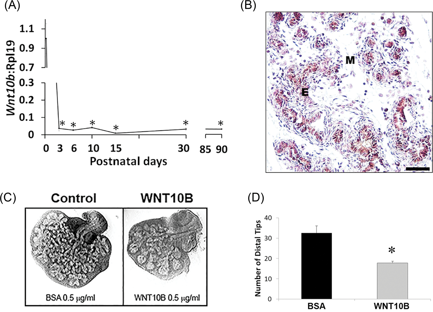
FIGURE 1.

A, Ontogeny of Wnt10b mRNA expression in the rat VP lobes as quantified by real-time reverse transcription PCR. Wnt10b expression was high at birth with a precipitous decline by pnd3 and maintained at nadir levels through adulthood. Each time point represents the mean ± SEM for 3 to 5 replicates. *P < .001 vs pnd1. B, immunohistochemistry of pnd6 VP revealed WNT10B protein localization to epithelial (E) cells. (M) denotes mesenchyme. Scale bar = 50 μM. C, Paired neonatal VP lobes from a single rat were cultured in BSA (control) or WNT10B protein (0.5 μg/mL) for 4 days. WNT10B markedly reduced branching morphogenesis of the epithelium. D, VP distal tip number after 4 days of culture in BSA or WNT10B. Bars represent the mean ± SEM, N = 4, P < .05 vs BSA. BSA, bovine serum albumin; E, epithelial; M, mesenchyme; pnd, postnatal day; VP, ventral prostate
