Abstract
Studying microbial diversity and the effects of external factors on the microbiome could expand our understanding of environmental alterations. Silt and sand are mineral particles that form soil texture and even though most of the soils on earth contain a fraction of them and some other soils form almost by them, their effects on the microbiome remained to elucidate. In this study, the bacterial biodiversity of sand and silt clay soils was investigated. Furthermore, their effects on plant growth have been determined. Our data showed that biodiversity and biomass of microbiome are higher in silt-based soil. It is interesting that the pseudomonas genera only exist in silt-based soil while it is in the absence of sand-based soil. In contrast, B. thuringiensis could be found in sand-based soil while it is not found in silt texture. Our data also demonstrated that there are no significant changes in stress response between the two groups however, differential physiological changes in plants inoculated with silt and sand based bacterial isolates have been observed. This data could indicate that smaller size particles could contain more bacteria with higher biodiversity due to providing more surfaces for bacteria to grow.
Subject terms: Biotechnology, Ecology, Evolution, Microbiology, Plant sciences
Introduction
An essential component of the Earth system is the soil sphere1. The soil microbiome is considered to be the main component of soil2,3. Soil microbiome could influence soil development, organic matter decomposition, geochemical cycles, and energy conversion4. In addition, it could significantly promote plant growth5. The biomass of the microbiome is reducing, and its biodiversity has decreased as a result of the abuse of the environment and resources, while soil bacterial diversity is a critical factor in ecosystem assessment and maintenance of ecological balance6. More research was concentrated on the study and preservation of soil microbial variety, the analysis of diversity traits, and the influence of variables on diversity7,8. Accordingly, these variables can be divided into human interventions and natural variables9,10. While human influences include pesticides, fertilizers, and tillage techniques, natural factors include the type of agricultural vegetation, soil type, temperature, and moisture11–13. Several reports suggest a relationship between soil properties and bacterial populations, although the results of individual studies vary as to the nature, extent, and direction of this relationship. According to certain experimental findings, the texture of the soil is the primary force behind microbial community organization14,15. Greater physical niche space and spatial isolation induced by the structural diversity of the soil environment should promote bacterial diversity16,17. There is evidence that soil bacteria prefer this specific texture18–20. Correlations between the ratio of surface area and bacterial communities have been also observed in marine sediments, suggesting that the surface area to volume ratio of mineral material may influence microbial community formation and activity21,22.
Regardless of the complexity of microbiome changes, there is technical complexity to determine the microbiome itself. Currently, sequencing of 16S rRNA and 18S rRNA genes provides the most accurate identification of bacteria23. Recently, matrix-assisted laser desorption ionization-time of flight mass spectrometry (MALDI-TOF MS) has come into focus as a potential method for the detection and diagnosis of microorganisms. Microorganisms are detected using either whole cells or cell extracts during the MALDI-TOF MS procedure. The method is sensitive, rapid, and affordable in terms of labor and other associated costs in comparison with other techniques. Microbiologists have reported that MALDI-TOF MS is used for a variety of purposes, including identification of bacteria and their strain type, epidemiological studies, and other purposes24–26.
Here, to identify the impact of salt and silt texture on soil, we collected samples from these two specific textures and examined bacterial diversity using MALDI-TOF MS. In addition, we determined the tolerance of bacterial isolates to abiotic stressors (salinity, alkalinity, and thermal stresses). We also investigated the effects of isolates on the growth of Maize, Canola, and Wheat and observed the growth variations of plants inoculated with the same isolates. Our data showed that silt-based soil textures are homing more bacterial types. This texture also harbors pseudomonas genera which is the absence in sand-based soil. Moreover, sand-based soil has a lower number of bacteria and contains B. thuringiensis. Our data could not identify any difference between stress responses of these two groups which might indicate no evolutionary pressure between these groups. Regardless, plant response to bacteria demonstrated that bacteria isolated from sand- and silt-based soils could change the development of plants in favor of their environmental niches. Based on this data, it could be concluded that lower size particle is correlated with higher biomass and biodiversity. This could be a result of a higher surface provided by low size particles which could provide more niches for the growth of bacteria.
Results
Sampling locations of soil samples and characterization of physiochemical treats
Geographical, physical, and over 10 years of synoptic data for two locations along a latitudinal gradient were provided in Table 1. The soil samples were taken from a single longitude across two different latitudes, these points were elected on a strip from semi-arid to arid regions of the north to south, and the exact longitudes with similar pH, rainfall, and temperature. Respectively, the pH spectral was 7.6 to 8.6. The temperature of locations was from 18.1 to 22.2 and the rainfall average has not differed more than 22 mm3. Furthermore, soil samples' texture was determined as 8, 15 and 79% of sand, silt, and clay for location A and 85, 5, and 10% of sand, silt, and clay for location B (Table 1).
Table 1.
Selected geographic and eco-physiological characters of soil samples along with two coastal deserts A and B from 2009 to 2019.
| Location description | A | B |
|---|---|---|
| Geographic characters | ||
| Height (feet) | 142 | 162 |
| Latitude | 37 16.47 | 27 3 40.87 |
| Longitude | 55 1 39.55 | 55 3 59.73 |
| Physical characters | ||
| Percentage of sample weight after sieving | 26.34 | 62.66 |
| pH | 8.69 | 7.12 |
| Texture (particle composition) | ||
| (%) Sand | 8.00 | 85.00 |
| (%) Silt | 79.00 | 5.00 |
| (%) Clay | 13.00 | 10.00 |
| Synoptic characters (10 years av.) | ||
| Temperature (°C) | ||
| Lowest value | 7.80 | 17.70 |
| Max value | 29.10 | 29.40 |
| Ann | 18.10 | 22.20 |
| Total rainfall (mm3) | ||
| Lowest value | 25.30 | 0.00 |
| Max value | 92.20 | 52.50 |
| Ann | 150.20 | 133.25 |
| Wind speed (m/s) | ||
| Lowest value | 2.25 | 3.11 |
| Max value | 2.30 | 3.80 |
| Ann | 2.70 | 3.50 |
Isolation, characterization, and screening of the growth parameters of soil bacteria on selective media and MB medium
After collecting samples, bacterial content of these samples has been isolated via several medium. As shown in Table 2, the selective microbial media have unique and different effects on the growth and morphology of soil isolates. At a glance, it was observed that most isolates have different growth parameters and colony sizes when they are cultured in the same microbial media, however, the growth rate of isolates has shown interesting results in all microbial media. Surprisingly the number of bacteria in location A (silt) was much greater than the bacteria isolated from location B (sand). Also, Table 2, introduced a complete list of bacteria isolated from soil of locations A and B and the influences of selective microbial media and MB medium on their growth parameters. Of the 27 soil bacteria, 17 isolates belonged to location A and 10 to location B that were isolated 1 on DPM, 4 on GYM, 4 on LB, 9 on MHA, 4 on NA, 3 on NA+, and 2 on VRB. Investigation of selective microbial media showed that isolates 3, 66, 1, 31, 32, and 67 had the most growth rate (CFU/ml) after 10 h of culture, respectively. Almost all the isolates’ colonies were cream in color but there were some colonies with colorless, white, cream, yellow, grey, orange, and dark orange. Most colonies were circular and had a smooth or flat surface, with only 30 isolates showing a raised surface (Table 2). While all colonies had an irregular shape with a flat surface and were white on the MB medium. As shown in Table 2, selective microbial media were more effective than MB medium in terms of bacterial growth and morphological characterizations of isolates. At a glance, it was observed that most isolates have different growth parameters and colony sizes when they are cultured in the same microbial media, however, the growth rate of isolates has shown interesting results in all microbial media. Isolates 6, 5, 2, 1, and 132 indicated the highest growth (CFU/ml) on the MB medium, and by comparing the growth of the isolates in both MB and selective media isolates 6, 5, and 65 showed the growth rate equal or higher than other ones (MB medium/ Selective media CFU ratio).
Table 2.
Effect of two coastal deserts on microbial growth parameters and morphological characterization of soil bacteria isolated from locations A and B.
| Location | Isolate | Selective microbial media | Morphological characterization | MB medium | MB medium/selective media CFU ratio | |||||
|---|---|---|---|---|---|---|---|---|---|---|
| Medium name | CFU/ml (× 105) after 10 h | Color | Colony size score | Colony shape | CFU/ml (× 105) after 10 h | Colony size score | ||||
| Top view | Side view | |||||||||
| A | 1 | MHA | 12.00 | Cream | 3 | Circular | Flat | 5.00 | 2 | 0.83 |
| 2 | MHA | 9.80 | Orange | 2 | Circular | Flat | 5.50 | 1 | 0.86 | |
| 3 | MHA | 14.00 | Dark Orange | 2 | Circular | Flat | 4.00 | 1 | 0.95 | |
| 4 | MHA | 8.50 | Cream | 7 | Irregular | Flat | 3.50 | 6 | 0.7 | |
| 5 | MHA | 9.50 | Cream | 2 | Circular | Flat | 7.50 | 1 | 1.25 | |
| 6 | MHA | 4.00 | Colorless | 2 | Circular | Flat | 8.00 | 1 | 1.33 | |
| 65 | NA | 8.00 | Colorless | 3 | Circular | Flat | 4.00 | 2 | 1.00 | |
| 66 | NA | 13.00 | White | 3 | Circular | Flat | 3.80 | 2 | 0.95 | |
| 67 | NA | 11.00 | Cream | 10 | Irregular | Flat | 3.50 | 6 | 0.92 | |
| 75 | NA+ | 6.00 | Cream | 3 | Irregular | Flat | 3.00 | 2 | 0.77 | |
| 92 | LB | 5.00 | Cream | 3 | Circular | Flat | 4.00 | 2 | 0.71 | |
| 93 | LB | 4.50 | Orange | 2 | Circular | Flat | 2.50 | 1 | 0.19 | |
| 112 | VRB | 2.80 | White | 1 | Circular | Flat | 2.30 | 1 | 0.37 | |
| 122 | DPM | 1.00 | Grey | 1 | Circular | Flat | 2.30 | 1 | 0.44 | |
| 131 | GYM | 8.50 | Cream | 10 | Irregular | Flat | 4.80 | 9 | 0.53 | |
| 132 | GYM | 7.00 | Cream | 3 | Circular | Flat | 5.00 | 2 | 0.63 | |
| 133 | GYM | 9.00 | Cream | 5 | Irregular | Flat | 2.00 | 4 | 0.48 | |
| B | 30 | MHA | 9.50 | Cream | 2 | Circular | Raised | 3.00 | 1 | 0.5 |
| 31 | MHA | 12.00 | Cream | 6 | Irregular | Flat | 2.00 | 5 | 0.22 | |
| 32 | MHA | 11.00 | Yellow | 2 | Circular | Flat | 2.10 | 1 | 0.22 | |
| 72 | NA | 4.80 | Cream | 10 | Irregular | Flat | 3.00 | 7 | 0.75 | |
| 87 | NA+ | 4.00 | White | 10 | Irregular | Flat | 2.50 | 9 | 0.39 | |
| 88 | NA+ | 4.50 | Cream | 3 | Circular | Flat | 2.80 | 2 | 0.28 | |
| 102 | LB | 4.00 | Cream | 7 | Circular | Flat | 2.00 | 6 | 0.42 | |
| 103 | LB | 3.80 | Cream | 4 | Circular | Flat | 2.00 | 3 | 0.4 | |
| 119 | VRB | 3.00 | White | 1 | Circular | Flat | 3.00 | 1 | 0.36 | |
| 145 | GYM | 7.50 | Cream | 10 | Irregular | Flat | 3.00 | 8 | 0.43 | |
MALDI TOF–MS and biochemical-based identification and investigation of the impacts of abiotic stresses on bacterial growth
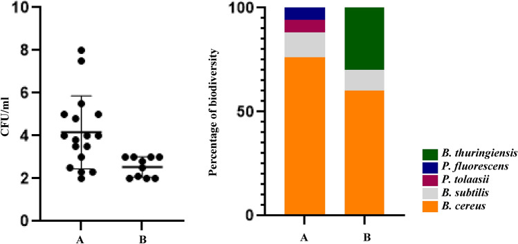
Table 3, showed MALDI-TOF MS results of the 27 soil bacteria isolated from locations A and B. The obtained MALDI-TOF MS profiles were then compared to the reference spectra of the BioTyper database and their similarity was expressed by a BioTyper Log (score). In total, two different genera of Pseudomonas and Bacillus have been identified accordingly, two species belong to the Pseudomonas genus: P. fluorescens and P. tolaasii also three species belong to Bacillus genus: B. cereus, B. thuringiensis, and B. subtilis. The 27 soil colonies isolated from locations A and B along the transect gradient were identified by MALDI TOF MS (2 isolates (7.5%) with Log (score) ≥ 2.3; 25 isolates (92.5%) with Log (score) ≤ 2.3 and ≥ 2.0 (Table 3). Identification data showed isolates were 1 P. fluorescens, 1 P. tolaasii, 17 B. cereus, 4 B. thuringiensis, and 4 B. subtilis. Both locations have the same diversity of Bacillus genus and 3 Bacillus species were isolated from these regions moreover two species of Pseudomonas genus were isolated from location A so it could be said location A had more bacterial diversity. Interestingly, silt-based soil (location A) has higher diversity as well as microbiome biomass (Fig. 1A,B). We also test the stress response of these isolates to see the difference in tolerance in sand versus silt-based soil. Our data showed that there are no significant differences between the sand (location B) and silt soils (location A) (Table 3). The growth of all isolates under abiotic stresses was significantly reduced. Only two isolates No. 1 in cold stress and No. 145 in heat stress were able to grow close to normal condition.
Table 3.
Investigation of bacterial diversity along with two coastal deserts by MALDI TOF and biochemical complementary tests and impacts of cold, dryness, and salinity stresses on soil bacteria isolated from locations A and B, 15 h after inoculation.
| Location | Isolate | Bacterial name | MALDI-TOF MS Score | Biochemical tests | Normal condition CFU/ml (× 105) | Salt stress CFU/ml (× 105) | Drought stress CFU/ml (× 105) | Cold stress CFU/ml (× 105) | pH stress CFU/ml (× 105) | Heat stress CFU/ml (× 105) | |||
|---|---|---|---|---|---|---|---|---|---|---|---|---|---|
| Gram staining | Catalase | KOH | Oxidase | ||||||||||
| A | 1 | B. cereus | 2.15 | − | + | + | + | 30.00 | 17.34 | 13.53 | 22.57 | 6.01 | 5.84 |
| 2 | B. cereus | 2.08 | − | + | − | + | 32.00 | 4.44 | 1.63 | 3.87 | 6.32 | 5.54 | |
| 3 | B. cereus | 2.25 | + | + | − | + | 21.00 | 2.48 | 1.46 | 2.89 | 7.33 | 5.15 | |
| 4 | B. cereus | 2.45 | + | + | + | + | 25.00 | 3.93 | 1.91 | 2.46 | 9.26 | 6.84 | |
| 5 | P. fluorescens | 2.03 | + | + | − | − | 30.00 | 4.25 | 1.78 | 4.81 | 14.96 | 21.67 | |
| 6 | B. cereus | 2.13 | − | + | − | + | 30.00 | 4.39 | 2.44 | 4.33 | 12.14 | 9.93 | |
| 65 | B. cereus | 2.23 | + | + | − | + | 20.00 | 4.23 | 1.27 | 3.34 | 4.8 | 5.18 | |
| 66 | B. cereus | 2.04 | − | + | − | + | 20.00 | 4.28 | 1.56 | 3.57 | 8.74 | 11.6 | |
| 67 | B. cereus | 2.26 | − | + | − | + | 19.00 | 3.58 | 1.27 | 3.01 | 5.23 | 4.35 | |
| 75 | B. cereus | 2.18 | − | + | − | + | 19.50 | 5.57 | 1.15 | 3.01 | 6.95 | 7.71 | |
| 92 | B. cereus | 2.07 | + | + | − | − | 28.00 | 6.05 | 2.25 | 4.81 | 12.86 | 11.55 | |
| 93 | B. cereus | 2.11 | + | + | − | + | 65.00 | 9.17 | 5.07 | 8.38 | 39.02 | 34.77 | |
| 112 | P. tolaasii | 2.03 | + | + | + | + | 31.00 | 5.95 | 2.44 | 4.92 | 7.26 | 6.04 | |
| 122 | B. thuringiensis | 2.07 | + | + | − | + | 26.00 | 5.35 | 1.01 | 2.52 | 15.65 | 11.8 | |
| 131 | B. subtilis | 2.14 | + | + | − | + | 45.00 | 5.11 | 2.25 | 2.78 | 15.58 | 24.61 | |
| 132 | B. cereus | 2.26 | + | + | − | + | 39.50 | 4.97 | 1.98 | 2.17 | 19.45 | 21.71 | |
| 133 | B. subtilis | 2.11 | + | + | − | − | 21.00 | 3.11 | 1.19 | 1.73 | 13.47 | 18.33 | |
| B | 30 | B. cereus | 2.06 | - | + | − | + | 30.00 | 5.93 | 1.51 | 2.65 | 13.11 | 7.77 |
| 31 | B. cereus | 2.00 | + | + | − | + | 45.00 | 8.77 | 2.53 | 3.99 | 15.04 | 24.54 | |
| 32 | B. cereus | 2.17 | + | + | − | + | 48.00 | 9.21 | 2.5 | 6.52 | 10.12 | 14.24 | |
| 72 | B. cereus | 2.38 | + | + | − | + | 20.00 | 4.4 | 1.58 | 3.38 | 6.89 | 11.74 | |
| 87 | B. cereus | 2.09 | + | − | − | − | 32.00 | 4.76 | 2.23 | 3.92 | 17.1 | 10.1 | |
| 88 | B. subtilis | 2.00 | + | + | + | − | 50.00 | 7.11 | 3.21 | 5.46 | 31.27 | 21.5 | |
| 102 | B. thuringiensis | 2.06 | + | + | − | + | 24.00 | 3.29 | 1.29 | 1.84 | 11.71 | 8.23 | |
| 103 | B. thuringiensis | 2.22 | + | + | − | + | 25.00 | 4.86 | 1.99 | 3.98 | 7.27 | 6.54 | |
| 119 | B. thuringiensis | 2.05 | + | + | + | + | 42.00 | 8.22 | 2.33 | 5.01 | 21.02 | 15.81 | |
| 145 | B. subtilis | 2.01 | + | + | − | − | 35.00 | 6.44 | 3.55 | 4.03 | 25.86 | 34.34 | |
Isolates that were undoubtedly identified to genus or species level by a valid MALDI-TOF MS score of ≥ 2.0 are shown. a and b were indicated sampling sites where the bacteria were isolated. Basal media was Muller Hinton for normal and stress conditions.
Figure 1.
Changes in biodiversity and biomass of microbiome in response to silt and sand-based soils (A). Colony counting of microbiome isolated from sampling locations (B). Diversity of microbiome isolates collected from locations A and B.
Effects of isolates on plant growth parameters
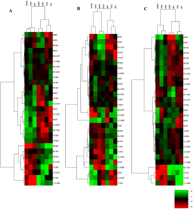
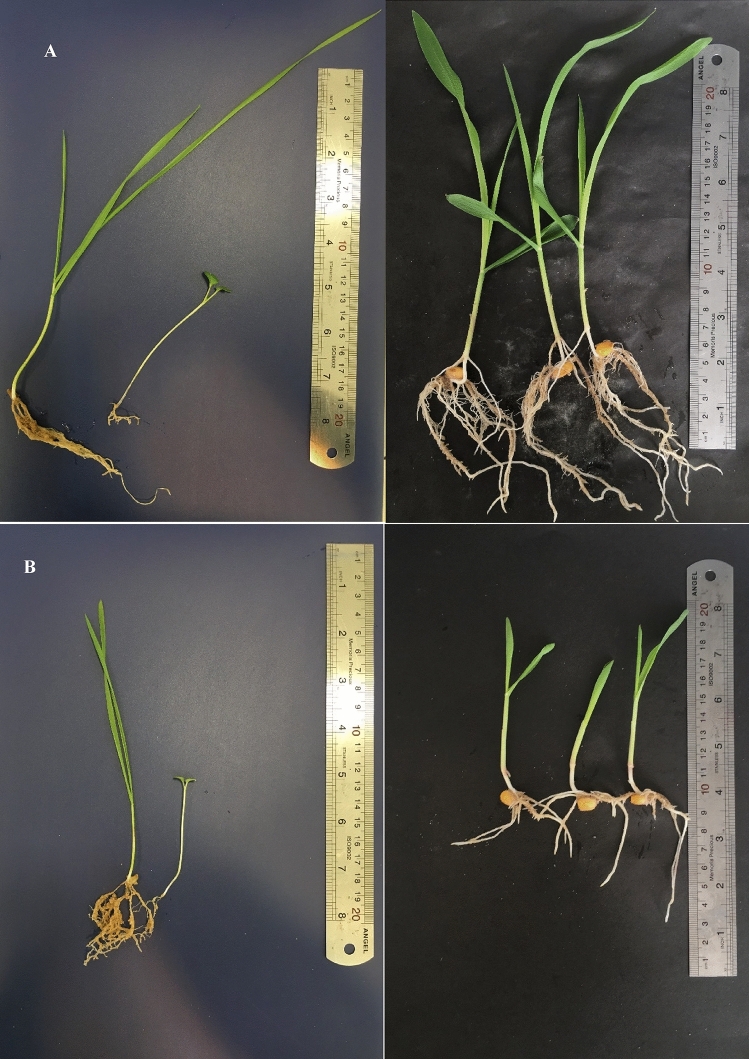
In general, all of the plant types show significant changes in response to inoculation of isolates in comparison to control condition. Plant growth parameters of wheat were strongly influenced by soil types. Results of soil bacteria effect on wheat after 21 days indicated that isolate 92 had significant impacts on shoot dry weight and shoot/root but isolate 145 caused the max reduction of shoot dry weight and root density. Furthermore isolate 93 was the most effective isolate on root weight and it was one of the most significant isolates on shoot density after isolate 103. Interestingly, isolate 133 decreased shoot/root but boosted shoot length also isolate 31 with a significant effect on root length while the reduced length of shoot, and it should be noted that isolate 5 had the best impact on root length (Table 4). By analyzing the average value of each parameter based on the isolation location of bacteria, it was found Location B was more effective on root dry weight and shoot density, while location A influenced shoot length, shoot dry weight, and root density (Table 4, Figs. 2A, 3A,B).
Table 4.
Influence of soil isolates on growth parameters after 21 days of assessment on wheat plants.
| Location | Isolate | Bacterial name | Wheat | ||||||
|---|---|---|---|---|---|---|---|---|---|
| Dry biomass | Length (cm) | ||||||||
| mg | mg/l × 100 | ||||||||
| Shoot | Root | Shoot/root | Shoot density | Root density | Shoot | Root | |||
| A | 1 | B. cereus | 1.96 jk | 34.73 hijklm | 0.22 jkl | 0.69 jklmno | 5.70 ghi | 8.75 fgh | 5.00 bcde |
| 2 | B. cereus | 4.02 fghij | 34.91 hijklm | 0.37 hijkl | 1 fghijk | 11.56 fghi | 10.75 abcd | 3.50 fghi | |
| 3 | B. cereus | 5.36 defg | 30.00 jklm | 0.48 ghi | 0.55 klmno | 17.86 defg | 11.50 ab | 5.50 abc | |
| 4 | B. cereus | 1.83 jk | 11.00 op | 0.18 kl | 0.37 no | 16.67 defgh | 10.00 bcdef | 3.00 hijk | |
| 5 | P. fluorescens | 1.67 k | 88.33 c | 0.19 kl | 1.31 efghi | 1.89 i | 9.00 efgh | 6.75 a | |
| 6 | B. cereus | 4.67 fgh | 80.67 c | 0.5 ghi | 2.02 bcd | 5.68 ghi | 9.50 cdefg | 4.00 defgh | |
| 65 | B. cereus | 7.33 cd | 26.50 lmn | 0.8 ef | 0.98 fghijkl | 27.62 de | 9.25 defg | 2.75 hijk | |
| 66 | B. cereus | 3.57 ghijk | 16.43 no | 0.36 hijkl | 0.43 mno | 21.97 def | 10.00 bcdef | 4.00 defgh | |
| 67 | B. cereus | 7.50 cd | 53.33 de | 0.77 ef | 1.48 ef | 14.06 efghi | 9.75 cdefg | 3.75 efghi | |
| 75 | B. cereus | 5.83 cdef | 5.83 op | 0.60 fgh | 0.19 o | 100.00 b | 9.75 cdefg | 3.00 hijk | |
| 92 | B. cereus | 23.33 a | 45.00 efgh | 2.28 a | 1.50 def | 51.92 c | 10.25 bcdef | 3.00 hijk | |
| 93 | B. cereus | 7.00 cde | 116.00 a | 0.67 efg | 2.24 b | 6.10 ghi | 10.50 abcde | 5.25 bcd | |
| 112 | P. tolaasii | 4.50 fgh | 29.17 klm | 0.41 hijk | 0.85 ijklmn | 15.49 efghi | 11.00 abc | 3.50 fghi | |
| 122 | B. thuringiensis | 3.33 ghijk | 27.50 lm | 0.38 hijkl | 1.17 efghij | 12.22 fghi | 9.00 efgh | 2.50 ijk | |
| 131 | B. subtilis | 14.67 b | 49.83 efg | 1.28 bc | 1.42 efg | 29.79 d | 11.50 ab | 3.50 fghi | |
| 132 | B. cereus | 5.00 efg | 38.57 hijk | 0.49 ghi | 1.5 def | 13.05 fghi | 10.25 bcdef | 2.75 hijk | |
| 133 | B. subtilis | 1.83 jk | 41.33 fghi | 0.15 l | 2.07 bc | 4.45 ghi | 12.00 a | 2.00 jk | |
| B | 30 | B. cereus | 4.64 fgh | 36.31 hijkl | 0.49 ghi | 1.4 efgh | 12.77 fghi | 9.50 cdefg | 2.75 hijk |
| 31 | B. cereus | 8.00 c | 32.00 ijklm | 1.07 cd | 0.47 lmno | 25.00 def | 7.50 h | 6.75 a | |
| 32 | B. cereus | 2.25 ijk | 50.00 efg | 0.23 jkl | 1.67 cde | 4.50 ghi | 10.00 bcdef | 3.00 hijk | |
| 72 | B. cereus | 3.50 ghijk | 84.75 c | 0.36 hijkl | 2.12 bc | 4.08 ghi | 9.50 cdefg | 4.00 defgh | |
| 87 | B. cereus | 4.17 fghi | 25.00 mn | 0.45 ghij | 0.9 ghijklm | 16.67 defgh | 9.25 defg | 2.75 hijk | |
| 88 | B. subtilis | 2.50 hijk | 38.33 hijk | 0.28 ijkl | 1.19 efghij | 6.52 ghi | 9.00 efgh | 3.25 ghij | |
| 102 | B. thuringiensis | 13.93 b | 62.74 d | 1.33 b | 1.05 fghijk | 22.23 def | 10.5 abcde | 6.00 ab | |
| 103 | B. thuringiensis | 3.93 fghij | 104.40 b | 0.39 hijkl | 5.22 a | 3.71 hi | 10.25 bcdef | 2.00 jk | |
| 119 | B. thuringiensis | 1.55 k | 51.07 ef | 0.16 l | 1.09 fghij | 3.03 hi | 9.75 cdefg | 4.75 bcdef | |
| 145 | B. subtilis | 1.55 k | 82.26 c | 0.16 l | 2.12 bc | 1.89 i | 9.75 cdefg | 4.00 defgh | |
| 150 | Control + | 5.00 efg | 40.00 ghij | 0.45 ghij | 0.89 hijklmn | 12.88 fghi | 11.00 abc | 4.50 cdefg | |
| 151 | Control - | 7.00 cde | 4.00 p | 0.85 de | 0.23 o | 175.00 a | 8.25 gh | 1.75 k | |
LSD p = 0.01 value for the parameters on wheat, respectively: 0.003243491, 0.05366804, 0.05586241, 0.1312794, 3.136652, 2.177158, 4.041771; A and B were indicated sampling locations.
Figure 2.
(A–C) Represented hierarchical cluster analysis of the effect of isolates on wheat, canola, and maize plants, respectively. Drown by CLUSTER and Treeview softwares. Hierarchical clustering was done based on Euclidian distance and the complete linkage method. Colors were indicated the type of isolates impacts on plants. Accordingly, red, green, and black colors showed positive, negative, and no-effect isolates, respectively. The horizontal axis indicates plant growth parameters: shoot density (ShD), root density (RD), root dry weight (RDW), root length (RL), shoot dry weight (ShDW), shoot length (ShL) and shoot/root weight (ShR). The vertical axis shows the assayed bacterial isolates.
Figure 3.
Influence of bacterial isolates on the overall growth of wheat, canola, and maize (A) in comparison to uninoculated control condition (B).
Plant growth parameters of Canola were also strongly influenced by the size of the particle. Results of the effect of soil bacteria on canola after 21 days indicate that 103 had significant impacts on shoot dry weight and shoot/root however isolate 72 caused reducing these parameters. However, no isolate was more appropriate for root dry weight, shoot density, and root density than the control. Isolate 131 showed the max shoot length but isolate 32 caused the lowest length of the shoot. Also, isolate 31 indicated the best root length (Table 5). Hierarchical cluster analysis of effects of isolates on canola showed that location B was more effective on root dry weight, shoot/root, and shoot density, while location A had a good influence on shoot length, root length, root density and shoot dry weight (Table 5, Figs. 2B, 3A,B).
Table 5.
Influence of soil isolates on growth parameters after 21 days of assessment on canola plants.
| Location | Isolate | Bacterial name | Canola | ||||||
|---|---|---|---|---|---|---|---|---|---|
| Dry biomass | Length (cm) | ||||||||
| mg | mg/l × 100 | ||||||||
| Shoot | Root | Shoot/root | Shoot density | Root density | Shoot | Root | |||
| A | 1 | B. cereus | 22.68 cdef | 315.98 def | 0.94 bc | 2.5 efgh | 7.20 b | 24.25 def | 12.75 cde |
| 2 | B. cereus | 9.29 l | 212.74 hijklm | 0.34 lm | 2.18 ghij | 4.36 b | 27.25 bc | 9.75 hijkl | |
| 3 | B. cereus | 17.02 hijk | 125.36 nopq | 0.65 fghij | 1.06 klm | 13.58 b | 26.25 cd | 12.00 defg | |
| 4 | B. cereus | 16.43 ijk | 226.43 ghijk | 0.63 hij | 2.21 fghij | 7.26 b | 26.25 cd | 10.25 fghijk | |
| 5 | P. fluorescens | 15.71 jk | 357.14 cd | 0.79 cdefgh | 2.35 fghi | 4.40 b | 20.00 i | 15.25 ab | |
| 6 | B. cereus | 23.93 cde | 96.07 opq | 0.87 cde | 0.87 m | 25.02 b | 27.50 abc | 11.00 efghij | |
| 65 | B. cereus | 21.34 defg | 232.14 ghijk | 0.81 cdefg | 1.83 hij | 9.22 b | 26.25 cd | 12.75 cde | |
| 66 | B. cereus | 25.00 bc | 468.57 b | 0.85 cde | 3.68 c | 5.34 b | 29.50 ab | 12.75 cde | |
| 67 | B. cereus | 15.36 jk | 339.40 de | 0.75 defghi | 2.90 def | 4.58 b | 20.50 hi | 11.75 defgh | |
| 75 | B. cereus | 24.64 bcd | 231.43 ghijk | 0.93 bcd | 2.01 hij | 10.70 b | 26.50 cd | 11.50 defghi | |
| 92 | B. cereus | 15.71 jk | 149.29 mnop | 0.63 ghij | 1.03 klm | 10.53 b | 25.00 cdef | 14.50 abc | |
| 93 | B. cereus | 15.83 jk | 272.50 fgh | 0.58 ijk | 2.37 efghi | 5.82 b | 27.50 abc | 11.50 defghi | |
| 112 | P. tolaasii | 14.73 k | 88.93 pq | 0.58 ijk | 0.89 lm | 16.55 b | 25.75 cde | 10.00 ghijk | |
| 122 | B. thuringiensis | 28.00 b | 156.00 lmno | 1.07 b | 1.58 jkl | 17.95 b | 26.25 cd | 10.00 ghijk | |
| 131 | B. subtilis | 19.46 fghi | 216.34 hijkl | 0.65 fghij | 1.89 hij | 8.98 b | 30.00 a | 11.50 defghi | |
| 132 | B. cereus | 15.00 k | 179.17 jklmn | 0.70 efghi | 2.01 hij | 8.37 b | 21.50 ghi | 9.00 jkl | |
| 133 | B. subtilis | 21.67 cdefg | 321.07 def | 0.82 cdef | 2.79 defg | 6.75 b | 26.50 cd | 11.50 defghi | |
| B | 30 | B. cereus | 18.57 ghij | 203.57 ijklm | 0.73 efghi | 1.90 hij | 9.12 b | 25.50 cdef | 10.75 efghij |
| 31 | B. cereus | 18.57 ghij | 167.14 klmn | 0.81 cdefgh | 1.04 klm | 11.11 b | 23.00 fgh | 16.25 a | |
| 32 | B. cereus | 15.67 jk | 290.83 efg | 0.80 cdefgh | 3.05 cde | 5.38 b | 19.50 i | 9.50 ijkl | |
| 72 | B. cereus | 4.29 m | 260.00 fghi | 0.18 m | 2.89 def | 1.65 b | 24.00 defg | 9.00 jkl | |
| 87 | B. cereus | 19.40 fghi | 414.40 bc | 0.94 bc | 3.38 cd | 4.67 b | 20.50 hi | 12.25 def | |
| 88 | B. subtilis | 18.57 ghij | 272.26 fgh | 0.70 efghi | 2.02 hij | 6.82 b | 26.50 cd | 13.50 bcd | |
| 102 | B. thuringiensis | 9.29 l | 235.83 ghij | 0.40 kl | 2.10 ghij | 3.93 b | 23.50 efg | 11.25 efghi | |
| 103 | B. thuringiensis | 41.67 a | 225.24 ghijk | 1.52 a | 1.76 ij | 18.56 b | 27.50 abc | 12.75 cde | |
| 119 | B. thuringiensis | 20.71 efg | 430.54 b | 0.77 cdefgh | 4.78 b | 4.82 b | 27.00 bc | 9.00 jkl | |
| 145 | B. subtilis | 20.00 fgh | 197.50 ijklm | 0.80 cdefgh | 1.65 jk | 10.13 b | 25.00 cdef | 12.00 defg | |
| 150 | Control + | 10.12 l | 668.45 a | 0.50 jkl | 7.86 a | 1.50 b | 20.50 hi | 8.50 kl | |
| 151 | Control - | 10.18 l | 78.75 q | 0.49 jkl | 1.05 klm | 54.29 a | 20.50 hi | 7.75 l | |
LSD p = 0.01 value for the parameters on canola, respectively: 0.001935741, 0.009622568, 0.01059692, 0.08514634, 1.476113, 1.482904, 2.118454; A and B were indicated sampling locations.
Maize growth patterns show substantial changes due to isolates inoculation. Results of the effect of soil bacteria on Maize after 14 days indicate that 132 and 72 had significant impacts on shoot dry weight, respectively. Furthermore, isolate 132 also had the best effects on shoot/root and shoot density. Isolate 4 and 65 had the most effective isolates on root dry weight and root length parameters, respectively, although isolate 2 had a bad impact on root dry weight and root length. Isolate 92 decreased shoot length nevertheless Isolate 87, in addition to having significant effects on root length, also had the best effect on shoot length (Table 6). By examining the hierarchical clustering based on the isolation and their effect on growth parameters, it was found that location B was more effective on shoot dry weight and shoot density, while location A affected the rest of the parameters (Table 6, Figs. 2C, 3A,B).
Table 6.
Influence of soil isolates on growth parameters after 21 days of assessment on maize.
| Location | Isolate | Bacterial name | Maize | ||||||
|---|---|---|---|---|---|---|---|---|---|
| Dry biomass | Length (cm) | ||||||||
| mg | mg/l × 100 | ||||||||
| Shoot | Root | Shoot/root | Shoot density | Root density | Shoot | Root | |||
| A | 1 | B. cereus | 31.78 b | 658.28 bcdefg | 53.29 b | 0.27 b | 8.22 bc | 13.00 fghi | 8.00 hijkl |
| 2 | B. cereus | 13.48 b | 167.25 g | 127.12 b | 0.19 b | 2.76 c | 7.25 jk | 5.75 lm | |
| 3 | B. cereus | 16.77 b | 207.01 fg | 101.15 b | 0.11 b | 2.65 c | 16.00 abcdef | 8.25 ghijkl | |
| 4 | B. cereus | 24.55 b | 1210.85 a | 23.84 b | 0.24 b | 17.4 a | 10.00 ij | 7.25 jkl | |
| 5 | P. fluorescens | 26.68 b | 310.03 efg | 82.90 b | 0.15 b | 3.02 c | 17.25 abcde | 10.00 bcdefghij | |
| 6 | B. cereus | 43.93 b | 358.90 defg | 129.37 b | 0.24 b | 2.88 c | 18.00 abcde | 12.25 abcd | |
| 65 | B. cereus | 51.93 b | 1016.53 ab | 51.24 b | 0.29 b | 7.72 bc | 18.25 abcd | 13.50 a | |
| 66 | B. cereus | 31.63 b | 671.98 bcdefg | 46.70 b | 0.21 b | 5.78 bc | 15.00 cdefg | 11.75 abcdef | |
| 67 | B. cereus | 34.13 b | 658.66 bcdefg | 52.52 b | 0.20 b | 9.68 b | 16.50 abcdef | 7.00 jklm | |
| 75 | B. cereus | 33.30 b | 656.63 bcdefg | 55.72 b | 0.19 b | 7.61 bc | 17.50 abcde | 8.50 fghijkl | |
| 92 | B. cereus | 13.75 b | 182.58 g | 90.49 b | 0.19 b | 2.46 c | 7.25 jk | 7.00 jklm | |
| 93 | B. cereus | 35.55 b | 837.68 abcde | 47.69 b | 0.21 b | 6.71 bc | 17.75 abcde | 13.00 ab | |
| 112 | P. tolaasii | 41.75 b | 445.83 cdefg | 98.47 b | 0.21 b | 3.7 bc | 19.50 ab | 12.00 abcde | |
| 122 | B. thuringiensis | 37.08 b | 741.13 abcdef | 50.34 b | 0.19 b | 6.39 bc | 19.50 ab | 11.50 abcdefg | |
| 131 | B. subtilis | 35.38 b | 929.08 abc | 38.50 b | 0.18 b | 7.88 bc | 19.25 ab | 11.75 abcdef | |
| 132 | B. cereus | 201.63 a | 455.85 cdefg | 8202.83 a | 1.32 a | 4.59 bc | 17.50 abcde | 9.00 defghijkl | |
| 133 | B. subtilis | 45.18 b | 273.18 fg | 170.98 b | 0.26 b | 2.44 c | 17.50 abcde | 11.25 abcdefgh | |
| B | 30 | B. cereus | 18.69 b | 312.59 efg | 61.03 b | 0.17 b | 4.9 bc | 10.75 hij | 6.25 klm |
| 31 | B. cereus | 77.72 b | 246.00 fg | 436.21 b | 0.57 ab | 3.55 bc | 14.25 defgh | 6.75 jklm | |
| 32 | B. cereus | 18.28 b | 241.80 fg | 80.46 b | 0.16 b | 2.89 c | 11.00 ghij | 8.00 hijkl | |
| 72 | B. cereus | 127.43 ab | 468.13 cdefg | 251.99 b | 0.78 ab | 7.06 bc | 14.50 cdefgh | 7.00 jklm | |
| 87 | B. cereus | 51.88 b | 592.08 bcdefg | 88.04 b | 0.26 b | 4.64 bc | 20.00 a | 12.75 abc | |
| 88 | B. subtilis | 29.00 b | 417.50 cdefg | 69.71 b | 0.19 b | 5.49 bc | 15.75 bcdef | 7.75 ijkl | |
| 102 | B. thuringiensis | 49.48 b | 890.32 abcd | 55.65 b | 0.27 b | 8.09 bc | 18.25 abcd | 11.00 abcdefghi | |
| 103 | B. thuringiensis | 29.59 b | 301.69 fg | 115.93 b | 0.18 b | 4.31 bc | 16.25 abcdef | 7.00 jklm | |
| 119 | B. thuringiensis | 39.12 b | 615.57 bcdefg | 76.49 b | 0.21 b | 6.08 bc | 18.50 abc | 9.50 cdefghijk | |
| 145 | B. subtilis | 35.88 b | 486.64 bcdefg | 73.22 b | 0.21 b | 5.31 bc | 17.00 abcdef | 8.75 efghijkl | |
| 150 | Control + | 31.50 b | 451.28 cdefg | 83.86 b | 0.23 b | 3.88 bc | 14.00 efghi | 11.75 abcdef | |
| 151 | Control - | 21.93 b | 304.23 efg | 184.34 b | 0.30 b | 4.76 bc | 4.75 k | 3.75 m | |
LSD p = 0.01 value for the parameters on maize, respectively: 0.1151398, 0.5349834, 4.409074, 0.007713552, 0.06318265, 4.12234, 3.276811; A and B were indicated sampling locations.
Concerning plant–microbe interactions, it should be noted that the effect of bacteria on plant growth is limited to the plant species or plant-specific. Hierarchical cluster analysis of plant–microbe interactions of three plants maize, canola, and wheat demonstrated all thee plants growth parameters for each plant were evaluated by several isolates and surveyed isolates created unique developmental effects in different plants (Fig. 2).
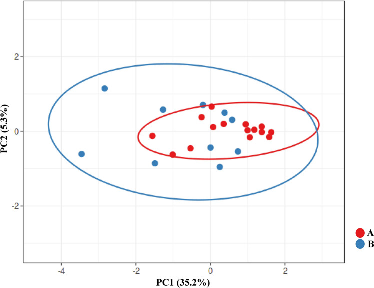
PCA analysis of Plant growth parameters of Wheat, Canola, and Maize inoculated by bacteria isolated from sand (location B) and silt (location A) soils demonstrated that silt-based soil in location A has lower variation among themselves which might indicate lower evolutionary pressure (Fig. 4).
Figure 4.
PCA analysis of all growth parameters of wheat, maize, and canola treated by isolates collected from locations A (silt) and B (sand).
Discussion
Evidence from several indicates that there is a connection between soil texture and microbial populations, although each study's findings on the nature, degree, and direction of this connection vary30. According to specific experimental findings, texture is the primary force behind the organization of microbial communities. For instance, an experiment revealed that altering the particle size distribution had a bigger effect on the organization of the microbial community than did compaction or changing the pH15. Previous research has discovered an association between soil textural heterogeneity as characterized by a single fractal model and microbial biomass31. Due partly to the considerable impact of pH on microbial diversity in natural environments, most field investigations have discovered a significant but lessened impact of texture upon microbial communities. As there is no consistent correlation between soil particle size heterogeneity and texture size classes across landscapes, these seemingly contradictory results might still be explained by a positive link between bacterial diversity and soil particle size heterogeneity31–36.
It has been suggested that a higher particle size could result in higher bacterial biomass30. Although this assumption might be right, the higher surface could give more niche for bacteria to adhere to. In this perspective lowering the size of particle in soils might result in higher biomass and diversity which might be an explanation for why silt-based soil have higher biomass and slightly higher biodiversity. Also, changes in soil texture result in biodiversity and biomass of soil which in turn affect the ecosystem and plants either. As it has been proved, microbes can evolve on short time scales, therefore shifting plant–microbe interactions quickly, boosting plant growth, and altering how we scale ecosystem processes up to longer periods37,38. Regarding the effects of bacteria on agriculture, it is previously known that some microbes have amazing impacts on plant performance, and changing their biodiversity could have a substantial impact on the environment39. For example, Bacillus cereus strains s as plant growth-promoting rhizobacteria have been used as biopesticides or biocontrol agents against various plant diseases40–42 and biofertilizers43. As previously reported Bacillus is an aerobic, rod-shaped, endospore-forming bacteria, and is a major community of the microbial flora in coastal ecosystems44. Moreover, several bacterial genera (pseudomonads and bacilli) have been founded as phosphate solubilizing bacteria their performance under in situ conditions is not reliable and therefore needs to be improved by using either genetically modified strains or co-inoculation techniques45. Plant growth and health-supporting bacteria of the Bacillus group due to their ability to form heat- and desiccation-resistant spores which can even provide a biological solution to the disease suppression of phytopathogenic fungi46,47. In this essence, slight changes in bacterial diversity and their changing forces could have a huge impact on plants and we analyze the effect of isolates on Maize, Canola, and Wheat.
Here we tried to have only one variable which is the texture of the soil. We tried to collect samples from locations with the same physiochemical identity. Our data show that the biomass of silt-base soil is higher than sandy soil. Interestingly, pseudomonas genera were absent in all of the samples from sand-based soil while B. thuringiensis have a substantial percentage of sand-based soil. The stress response of both sampling types shows no difference which might indicate none of the environments and soil textures did not induce chronic stress on the microbiome.
Last but not least our data suggest each silt or sand-based soil isolation influences plants differently and complexly which might have a better outcome in favor of the plants in their locational conditions. The result of the plant–microbe interaction test was expected as silt and sand-based soil show differences in biodiversity and biomass. Based on the heterogeneity of data on this matter more and more research and data should be available to have a better understanding of this complicated subject.
Material and methods
Soil sampling and determination of soil physical properties and synoptic data
Soil samples were taken from two coastal deserts in the north and south of Iran. Details of their geographic distribution and eco-physiological characterization were shown in Table 1. A total of 2 kg of soil samples were collected from 2 distinct sampling locations ranging in depth from 0 to 30 cm, and the samples were dried for 3 days at room temperature and in the dark before sifting. The soil samples were sieved using a 2 mm sieve to remove stones and other inert material before being stored in zip-top bags. Table 1 lists the soil samples' physical characteristics, including soil texture (sand 2–0.02 mm; silt 0.02–0.002 mm; clay 0.002 mm), pH, and the proportions of clay, silt, and sand. Synoptic data from the past 10 years (2009–2019), including the average annual temperature, maximum temperature, minimum temperature, average rainfall, average annual wind speed, and maximum wind speed, were obtained from the I.R.OF Iran Meteor (http://www.irimo.ir/far/index.php).
Bacterial isolation and effect of manure-based medium on their growth
According to Chen et al. 2005, the soil-borne bacteria were isolated using direct-spreading method. For this essence soil samples were treated through a series of dilutions. The mixture of 1 g of soil sample was vortexed for 1 min after being suspended in 2 ml of sterile physiological saline (0.9% w/v NaCl). The mixture was then diluted serially (typically 10–1 to 10–7), and level 100 μl of the diluted soil samples were scattered on the surface of solidified plates using glass spreaders. The samples were then incubated for 1 to 3 days at 30 °C in an inverted posture without light. For bacterial isolation, we used eleven culture media including Nutrient Agar (NA), Nutrient Agar plus MnSO4 (NA + MnSO4), LB, Moller Hinton Agar (MHA), Acidithiobacillus (APH) medium, Violet Red Bile Lactose (VRB) agar medium, GYM Streptomyces medium, DPM medium, Azospirillum medium, Azotobacter medium and Manure based medium (MB).
To prepare MB medium, dry animal manure and distilled water (1:6 w/v) were combined to create MB medium, which was then let to sit at room temperature for 16 h. The resulting mixture was then centrifuged at 5000 rcf for 30 min after being filtered twice. The next stage involved adding Hoagland salts (10% w/v) to the final extract, adjusting the medium's pH to 5.8 ± 0.02, and autoclaving it for 20 min at 121 °C and 1.5 kPa. Before sterilization, bacteriological agar (1.5 w/v) was employed as a gelling agent to solidify the medium.
After bacterial isolation on NA, NA+ MnSO4, LB, MHA, APH, VRB, GYM, DPM, and Azospibrillum media, the growth of all isolates was evaluated on an MB medium. To investigate isolates biomass in the same condition, we elected MB medium. First, the bacteria were grown in the liquid form of NA, NA+ MnSO4, LB, MHA, APH, VRB, GYM, DPM, and Azospirillum and Azotobacter media at 30 °C for 48 h, then 103 cells of each isolate were transferred to 48 wells plates containing MB medium, and plates were incubated at 30 °C for 10 h. Then, the growth of bacteria was read at an optical density (OD) of 630 nm 10 h after inoculation, the experiment was performed with three replicates. In the following step, CFU/ml equivalent to each OD was obtained by inoculating the uniform amount of liquid culture of the isolates on the solid form of MB medium at 30 °C for 16 h.
Phenotypic characterization and biochemical identification of bacterial isolates
The morphological analysis of the cell shape, colony (i.e., shape, color, and size), and biochemical tests were used to identify the bacterial isolates. Biochemical characterization was carried out By using gram staining, KOH27, oxidase, and catalase tests. For this essence, following Bartholomew's method28, gram staining of bacteria was studied 48 h after inoculation on MHA, and the non-staining KOH method was used to confirm the results. Using 0.5 ml of a 10% hydrogen peroxide solution, a catalase test was conducted, and the generation of gas bubbles was monitored. Using biochemical oxidase discs, the oxidative activity of 27 isolates was investigated.
Effect of abiotic stresses on bacterial isolates
To determine the effect of abiotic stresses on isolates alkaline (MH medium with pH 10), salinity (MH medium supplemented with the final concentration of 100 mM NaCl), osmotic [MH medium supplemented with 25% polyethylene glycol (PEG) Mn6000], and thermal stresses (MH medium incubated at 15 °C for cold stress and 60 °C for heat stress) were screened. For all experiments, the incubation period was 15 h, and plates were kept in a dark condition.
MALDI-TOF MS identification of isolates
Soil bacterial isolates were subcultured twice on MHA and incubated at 30 °C for 24 h before MALDI-TOF MS measurement. Then ∼0.1 µg of cell material was directly transferred from a bacterial colony or smear of colonies to a MALDI target spot. After drying at laboratory temperature, sample spots were overlaid with 1 μl of matrix solution (10 mg/mL a-cyano-4-hydroxycinnamic acid in 50% acetonitrile and 2.5% trifluoroacetic acid) and each measurement was carried out in triplicate (technical replicates). MS analysis was performed on an Autoflex MALDI-TOF mass spectrometer (Bruker Daltonics, Germany) using Flex Control 3.4 software (Bruker Daltonics, Germany). Calibration was carried out with the use of the Bacterial Test Standard (Bruker Daltonics, Germany). Soil isolates with a valid MALDI-TOF MS score of 2 were undoubtedly assigned to the genus/species level. For bacterial classification and identification, BioTyper 3.1 software (Bruker Daltonics, Germany) equipped with MBT 6903 MPS Library (released in April 2016), the MALDI Biotyper Preprocessing Standard Method, and the MALDI Biotyper MSP Identification Standard Method adjusted by the manufacturer (Bruker Daltonics, Germany) were used. Only the highest score value of all mass spectra belonging to individual cultures (biological and technical replicates) was recorded25. The score between 2.3 and 3.00 shows highly probable species-level identification and between 2.0 and 2.29 represents genus-level identification and probable species level of identification. A score between 1.7 and 1.99 indicates probable genus-level identification29.
Effects of bacterial isolates on plants growth
The Seed and Plant Improvement Institute of Karaj (Karaj, Iran; http://www.spii.ir/homepage.aspx?site=DouranPortal&tabid=1&lang=faIR) provided the maize, canola, and wheat seeds (Zea mays. Var Kosha; Brassica napus Var Nima; Triticum aestivum Var Kalate). In greenhouse trials, 2 × 10³ cells/seed of soil-borne isolates cultured in a manure-based medium were inoculated to maize, canola, and wheat plants. During the studies, sand that had been acid washed and autoclaved was used for planting. For three weeks, seedlings were kept under a 16/8 h day/night photoperiod with a 25 °C temperature. Three replications of a complete randomized block design were used for the colonization experiment's treatments. Under the bacterial treatments, measurements were made of the plant growth parameters including shoot dry biomass (mg), root dry biomass (mg), shoot length (cm), root length (cm), shoot density (mg/cm), root density (mg/cm), and shoot/root weight (mg). Samples were dried at 60 °C for three days to measure dry biomass.
Statistical analysis
Statistical analysis was done by R software (version 4.1.3). One-way analysis of variance (ANOVA) was used to determine the significance of the experiment, and Fisher's protected Least Significant Difference (LSD) test with a P-value of 0.01 was performed to separate the means. Furthermore, PCA analysis has been carried out based on the Clustvis package and the SVD imputation approach.
Ethics approval and consent to participate
All authors agree to the ethics and consent to participate in this article and declare that this submission follows the policies of Scientific Reports. Accordingly, the material is the author's original work, which has not been previously published elsewhere. The paper is not being considered for publication elsewhere. All authors have been personally and actively involved in substantial work leading to the paper and will take public responsibility for its content.
Ethics for research involving plants
All authors confirmed that experimental research and field studies on plants, including receiving the seeds from the Seed and Plant Improvement Institute of Karaj, complied with relevant institutional, national, and international guidelines and legislation. Furthermore, methods were conducted according to the relevant guidelines and regulations.
Acknowledgements
The authors acknowledge the technical support staff of the Department of Cellular and Molecular Biology, Faculty of Life Sciences and Biotechnology, Shahid Beheshti University, and Department of Microbiology and Immunology, Clinical and Diagnostic Immunology, KU Leuven University.
Author contributions
M.Z. and H.A. convinced of the presented idea, M.Z. carried out the experiment and wrote the manuscript. H.A. and M.S. supervised the project. All authors discussed the results and contributed to the final manuscript.
Funding
This research received no specific grant, funding, equipment, or supplies from any funding agency in the public, commercial, or not-for-profit sectors.
Data availability
The datasets used and/or analyzed during the current study are available from the corresponding author upon reasonable request.
Competing interests
The authors declare no competing interests.
Footnotes
Publisher's note
Springer Nature remains neutral with regard to jurisdictional claims in published maps and institutional affiliations.
References
- 1.Amundson R. Factors of soil formation in the 21st century. Geoderma. 2021;391:114960. doi: 10.1016/j.geoderma.2021.114960. [DOI] [Google Scholar]
- 2.Nannipieri P, Ascher J, Ceccherini M, Landi L, Pietramellara G, Renella G. Microbial diversity and soil functions. Eur. J. Soil Sci. 2003;54(4):655–670. doi: 10.1046/j.1351-0754.2003.0556.x. [DOI] [Google Scholar]
- 3.Wang G, Or D. A hydration-based biophysical index for the onset of soil microbial coexistence. Sci. Rep. 2012;2(1):1–5. doi: 10.1038/srep00881. [DOI] [PMC free article] [PubMed] [Google Scholar]
- 4.Ling LL, Schneider T, Peoples AJ, Spoering AL, Engels I, Conlon BP, Mueller A, Schäberle TF, Hughes DE, Epstein S, Jones M. A new antibiotic kills pathogens without detectable resistance. Nature. 2015;517(7535):455–459. doi: 10.1038/nature14098. [DOI] [PMC free article] [PubMed] [Google Scholar]
- 5.Zhang LN, Wang DC, Hu Q, Dai XQ, Xie YS, Li Q, Liu HM, Guo JH. Consortium of plant growth-promoting rhizobacteria strains suppresses sweet pepper disease by altering the rhizosphere microbiota. Front. Microbiol. 2019;10:1668. doi: 10.3389/fmicb.2019.01668. [DOI] [PMC free article] [PubMed] [Google Scholar]
- 6.Bardgett RD, Van Der Putten WH. Belowground biodiversity and ecosystem functioning. Nature. 2014;515(7528):505–511. doi: 10.1038/nature13855. [DOI] [PubMed] [Google Scholar]
- 7.Torsvik V, Øvreås L. Microbial diversity and function in soil: From genes to ecosystems. Curr. Opin. Microbiol. 2002;5(3):240–245. doi: 10.1016/S1369-5274(02)00324-7. [DOI] [PubMed] [Google Scholar]
- 8.Trivedi P, Rochester IJ, Trivedi C, Van Nostrand JD, Zhou J, Karunaratne S, Anderson IC, Singh BK. Soil aggregate size mediates the impacts of cropping regimes on soil carbon and microbial communities. Soil Biol. Biochem. 2015;91:169–181. doi: 10.1016/j.soilbio.2015.08.034. [DOI] [Google Scholar]
- 9.Mishra A, Singh L, Singh D. Unboxing the black box—One step forward to understand the soil microbiome: A systematic review. Microb. Ecol. 2022 doi: 10.1007/s00248-022-01962-5. [DOI] [PMC free article] [PubMed] [Google Scholar]
- 10.Berg G, Smalla K. Plant species and soil type cooperatively shape the structure and function of microbial communities in the rhizosphere. FEMS Microbiol. Ecol. 2009;68(1):1–13. doi: 10.1111/j.1574-6941.2009.00654.x. [DOI] [PubMed] [Google Scholar]
- 11.Ochoa-Hueso R. Global change and the soil microbiome: A human-health perspective. Front. Ecol. Evol. 2017;5:71. doi: 10.3389/fevo.2017.00071. [DOI] [Google Scholar]
- 12.Tischer C, Kirjavainen P, Matterne U, Tempes J, Willeke K, Keil T, Apfelbacher C, Täubel M. Interplay between natural environment, human microbiota and immune system: A scoping review of interventions and future perspectives towards allergy prevention. Sci. Total Environ. 2022 doi: 10.1016/j.scitotenv.2022.153422. [DOI] [PubMed] [Google Scholar]
- 13.Khan KS, Ali MM, Naveed M, Rehmani MIA, Shafique MW, Ali HM, Abdelsalam NR, Ghareeb RY, Feng G. Co-application of organic amendments and inorganic P increase maize growth and soil carbon, phosphorus availability in calcareous soil. Front. Environ. Sci. 2022 doi: 10.3389/fenvs.2022.949371. [DOI] [Google Scholar]
- 14.Sessitsch A, Weilharter A, Gerzabek MH, Kirchmann H, Kandeler E. Microbial population structures in soil particle size fractions of a long-term fertilizer field experiment. Appl. Environ. Microbiol. 2001;67(9):4215–4224. doi: 10.1128/AEM.67.9.4215-4224.2001. [DOI] [PMC free article] [PubMed] [Google Scholar]
- 15.Sleutel S, Bouckaert L, Buchan D, Van Loo D, Cornelis WM, Sanga HG. Manipulation of the soil pore and microbial community structure in soil mesocosm incubation studies. Soil Biol. Biochem. 2012;45:40–48. doi: 10.1016/j.soilbio.2011.09.016. [DOI] [Google Scholar]
- 16.Wilpiszeski RL, Aufrecht JA, Retterer ST, Sullivan MB, Graham DE, Pierce EM, Zablocki OD, Palumbo AV, Elias DA. Soil aggregate microbial communities: Towards understanding microbiome interactions at biologically relevant scales. Appl. Environ. Microbiol. 2019;85(14):e00324–e419. doi: 10.1128/AEM.00324-19. [DOI] [PMC free article] [PubMed] [Google Scholar]
- 17.Nishiyama M, Sugita R, Otsuka S, Senoo K. Community structure of bacteria on different types of mineral particles in a sandy soil. Soil Sci. Plant Nutr. 2012;58(5):562–567. doi: 10.1080/00380768.2012.729226. [DOI] [Google Scholar]
- 18.Gardner T, Acosta-Martinez V, Calderón FJ, Zobeck TM, Baddock M, Van Pelt RS, Senwo Z, Dowd S, Cox S. Pyrosequencing reveals bacteria carried in different wind-eroded sediments. J. Environ. Qual. 2012;41(3):744–753. doi: 10.2134/jeq2011.0347. [DOI] [PubMed] [Google Scholar]
- 19.Hemkemeyer M, Dohrmann AB, Christensen BT, Tebbe CC. Bacterial preferences for specific soil particle size fractions revealed by community analyses. Front. Microbiol. 2018;9:149. doi: 10.3389/fmicb.2018.00149. [DOI] [PMC free article] [PubMed] [Google Scholar]
- 20.Poll C, Thiede A, Wermbter N, Sessitsch A, Kandeler E. Micro-scale distribution of microorganisms and microbial enzyme activities in a soil with long-term organic amendment. Eur. J. Soil Sci. 2003;54(4):715–724. doi: 10.1046/j.1365-2389.2003.00569.x. [DOI] [Google Scholar]
- 21.Wang J, Xiong Z, Kuzyakov Y. Biochar stability in soil: Meta-analysis of decomposition and priming effects. Gcb Bioenergy. 2016;8(3):512–523. doi: 10.1111/gcbb.12266. [DOI] [Google Scholar]
- 22.Sokol NW, Slessarev E, Marschmann GL, Nicolas A, Blazewicz SJ, Brodie EL, Firestone MK, Foley MM, Hestrin R, Hungate BA, Koch BJ. Life and death in the soil microbiome: How ecological processes influence biogeochemistry. Nat. Rev. Microbiol. 2022 doi: 10.1038/s41579-022-00695-z. [DOI] [PubMed] [Google Scholar]
- 23.Clarridge III JE. Impact of 16S rRNA gene sequence analysis for identification of bacteria on clinical microbiology and infectious diseases. Clin. Microbiol. Rev. 2004;17(4):840–862. doi: 10.1128/CMR.17.4.840-862.2004. [DOI] [PMC free article] [PubMed] [Google Scholar]
- 24.Dec M, Puchalski A, Urban-Chmiel R, Wernicki A. 16S-ARDRA and MALDI-TOF mass spectrometry as tools for identification of Lactobacillus bacteria isolated from poultry. BMC Microbiol. 2016;16(1):1–16. doi: 10.1186/s12866-016-0732-5. [DOI] [PMC free article] [PubMed] [Google Scholar]
- 25.Strejcek M, Smrhova T, Junkova P, Uhlik O. Whole-cell MALDI-TOF MS versus 16S rRNA gene analysis for identification and dereplication of recurrent bacterial isolates. Front. Microbiol. 2018;9:1294. doi: 10.3389/fmicb.2018.01294. [DOI] [PMC free article] [PubMed] [Google Scholar]
- 26.Pandey A, Jain R, Sharma A, Dhakar K, Kaira GS, Rahi P, Dhyani A, Pandey N, Adhikari P, Shouche YS. 16S rRNA gene sequencing and MALDI-TOF mass spectrometry based comparative assessment and bioprospection of psychrotolerant bacteria isolated from high altitudes under mountain ecosystem. SN Appl. Sci. 2019;1(3):1–12. doi: 10.1007/s42452-019-0273-2. [DOI] [Google Scholar]
- 27.Suslow, T.V., Schroth, M.N., & Isaka, M. Application of a rapid method for Gram differentiation of plant pathogenic and saprophytic bacteria without staining. Phytopathology (USA) (1982).
- 28.Bartholomew JW. Variables influencing results, and the precise definition of steps in Gram staining as a means of standardizing the results obtained. Stain Technol. 1962;37(3):139–155. doi: 10.3109/10520296209117723. [DOI] [PubMed] [Google Scholar]
- 29.Ashfaq MY, Al-Ghouti MA, Qiblawey H, Rodrigues DF, Hu Y, Zouari N. Isolation, identification and biodiversity of antiscalant degrading seawater bacteria using MALDI-TOF-MS and multivariate analysis. Sci. Total Environ. 2019;656:910–920. doi: 10.1016/j.scitotenv.2018.11.477. [DOI] [PubMed] [Google Scholar]
- 30.Seaton FM, George PB, Lebron I, Jones DL, Creer S, Robinson DA. Soil textural heterogeneity impacts bacterial but not fungal diversity. Soil Biol. Biochem. 2020;144:107766. doi: 10.1016/j.soilbio.2020.107766. [DOI] [Google Scholar]
- 31.Hu Y, Xiang D, Veresoglou SD, Chen F, Chen Y, Hao Z, Zhang X, Chen B. Soil organic carbon and soil structure are driving microbial abundance and community composition across the arid and semi-arid grasslands in northern China. Soil Biol. Biochem. 2014;77:51–57. doi: 10.1016/j.soilbio.2014.06.014. [DOI] [Google Scholar]
- 32.Constancias F, Terrat S, Saby NP, Horrigue W, Villerd J, Guillemin JP, Biju-Duval L, Nowak V, Dequiedt S, Ranjard L, ChemidlinPrévost-Bouré N. Mapping and determinism of soil microbial community distribution across an agricultural landscape. MicrobiologyOpen. 2015;4(3):505–517. doi: 10.1002/mbo3.255. [DOI] [PMC free article] [PubMed] [Google Scholar]
- 33.Naveed M, Herath L, Moldrup P, Arthur E, Nicolaisen M, Norgaard T, Ferré TP, de Jonge LW. Spatial variability of microbial richness and diversity and relationships with soil organic carbon, texture and structure across an agricultural field. Appl. Soil. Ecol. 2016;103:44–55. doi: 10.1016/j.apsoil.2016.03.004. [DOI] [Google Scholar]
- 34.Tecon R, Or D. Biophysical processes supporting the diversity of microbial life in soil. FEMS Microbiol. Rev. 2017;41(5):599–623. doi: 10.1093/femsre/fux039. [DOI] [PMC free article] [PubMed] [Google Scholar]
- 35.Yao X, Zhang N, Zeng H, Wang W. Effects of soil depth and plant–soil interaction on microbial community in temperate grasslands of northern China. Sci. Total Environ. 2018;630:96–102. doi: 10.1016/j.scitotenv.2018.02.155. [DOI] [PubMed] [Google Scholar]
- 36.Pasternak Z, Al-Ashhab A, Gatica J, Gafny R, Avraham S, Minz D, Gillor O, Jurkevitch E. Spatial and temporal biogeography of soil microbial communities in arid and semiarid regions. PLoS ONE. 2013;8(7):e69705. doi: 10.1371/journal.pone.0069705. [DOI] [PMC free article] [PubMed] [Google Scholar]
- 37.Chave J. The problem of pattern and scale in ecology: What have we learned in 20 years? Ecol. Lett. 2013;16:4–16. doi: 10.1111/ele.12048. [DOI] [PubMed] [Google Scholar]
- 38.Classen AT, Sundqvist MK, Henning JA, Newman GS, Moore JA, Cregger MA, Moorhead LC, Patterson CM. Direct and indirect effects of climate change on soil microbial and soil microbial-plant interactions: What lies ahead? Ecosphere. 2015;6(8):1–21. doi: 10.1890/ES15-00217.1. [DOI] [Google Scholar]
- 39.Jacoby R, Peukert M, Succurro A, Koprivova A, Kopriva S. The role of soil microorganisms in plant mineral nutrition—Current knowledge and future directions. Front. Plant Sci. 2017;8:1617. doi: 10.3389/fpls.2017.01617. [DOI] [PMC free article] [PubMed] [Google Scholar]
- 40.Prasad R, Deo RC, Li Y, Maraseni T. Weekly soil moisture forecasting with multivariate sequential, ensemble empirical mode decomposition and Boruta-random forest hybridizer algorithm approach. CATENA. 2019;177:149–166. doi: 10.1016/j.catena.2019.02.012. [DOI] [Google Scholar]
- 41.Zhang X, Xin X, Zhu A, Yang W, Zhang J, Ding S, Mu L, Shao L. Linking macroaggregation to soil microbial community and organic carbon accumulation under different tillage and residue managements. Soil Tillage Res. 2018;178:99–107. doi: 10.1016/j.still.2017.12.020. [DOI] [Google Scholar]
- 42.Silva HSA, da Silva Romeiro R, Macagnan D, de Almeida Halfeld-Vieira B, Pereira MCB, Mounteer A. Rhizobacterial induction of systemic resistance in tomato plants: Non-specific protection and increase in enzyme activities. Biol. Control. 2004;29(2):288–295. doi: 10.1016/S1049-9644(03)00163-4. [DOI] [Google Scholar]
- 43.Shah R, Amaresan N, Patel P, Jinal HN, Krishnamurthy R. Isolation and characterization of Bacillus spp. endowed with multifarious plant growth-promoting traits and their potential effect on tomato (Lycopersicon esculentum) seedlings. Arab. J. Sci. Eng. 2020;45(6):4579–4587. doi: 10.1007/s13369-020-04543-1. [DOI] [Google Scholar]
- 44.Yousuf J, Thajudeen J, Rahiman M, Krishnankutty SP, Alikunj A, Abdulla MH. Nitrogen fixing potential of various heterotrophic Bacillus strains from a tropical estuary and adjacent coastal regions. J. Basic Microbiol. 2017;57(11):922–932. doi: 10.1002/jobm.201700072. [DOI] [PubMed] [Google Scholar]
- 45.Sharma SB, Sayyed RZ, Trivedi MH, Gobi TA. Phosphate solubilizing microbes: Sustainable approach for managing phosphorus deficiency in agricultural soils. Springerplus. 2013;2(1):1–14. doi: 10.1186/2193-1801-2-587. [DOI] [PMC free article] [PubMed] [Google Scholar]
- 46.Kumar P, Dubey RC, Maheshwari DK. Bacillus strains isolated from rhizosphere showed plant growth promoting and antagonistic activity against phytopathogens. Microbiol. Res. 2012;167(8):493–499. doi: 10.1016/j.micres.2012.05.002. [DOI] [PubMed] [Google Scholar]
- 47.Radhakrishnan R, Hashem A, Abd Allah EF. Bacillus: A biological tool for crop improvement through bio-molecular changes in adverse environments. Front. Physiol. 2017;8:667. doi: 10.3389/fphys.2017.00667. [DOI] [PMC free article] [PubMed] [Google Scholar]
Associated Data
This section collects any data citations, data availability statements, or supplementary materials included in this article.
Data Availability Statement
The datasets used and/or analyzed during the current study are available from the corresponding author upon reasonable request.


