

Abstract
Many implant materials have been used in various dental applications depending on their efficacy and availability. A dental implant must possess the required characteristics, such as biocompatibility, corrosion & wear resistance, adequate mechanical properties, osseointegration, etc., to ensure its safe and optimum use. This review analyzes various aspects of titanium (Ti) and Ti alloys, including properties, manufacturing processes, surface modifications, applications as dental implants, and limitations. In addition, it also presents a perception of recent advances in Ti-based implant materials and the futuristic development of innovative dental implants.
Keywords: Dental implant, Titanium alloy, Surface modification, Corrosion resistance, Osseointegration, Biocompatibility, Antibacterial activity
Graphical abstract
Dental implant; Titanium alloy; Surface modification; Corrosion resistance; Osseointegration, Biocompatibility; Antibacterial activity.
1. Introduction
Titanium (Ti) and Ti alloys have increased extensively since the early 1980s. It has become the most accepted metallic biomaterial for its distinct properties and numerous biomedical uses (Özcan et al., 2012; Vizureanu et al., 2020; Takeuchi et al., 2020). Most of the time, metallic biomaterials are utilized for their high load-bearing capacity and fatigue strength to sustain the regular movements’ loads applied to them (Gegner et al., 2014). Titanium has been presented as one of the most encouraging designing biomaterials for its low modulus of elasticity, low specific weight, extraordinary resistance to corrosion, outstanding strength-to-weight ratio, good tribological properties, and exceptional biocompatibility (Hatamleh et al., 2018; Mutombo, 2018). Titanium alloys have the highest biocompatibility for biomedical applications than any metallic contents. However, because of the trend of osteogenesis, they are graded as bioinert materials compared to bioceramics like zirconia, alumina, hydroxyapatite, and combinations (Niinomi et al., 2008; Hoque et al., 2013, 2014; Ragurajan et al., 2018; Golieskardi et al., 2019). Current dentistry aims to reinstate the patient to usual purpose, health, aesthetics, and speech irrespective of the stomatognathic system's injury, atrophy, or disease. As a result, prosthetics in dentistry are one of the most excellent options for persons who usually are in appropriate oral health but have lost their teeth because of periodontal illness, an injury, or some other reasons (Oshida et al., 2010; Golieskardi et al., 2020). Many implants of many designs are now made from pure titanium and its alloys.
Until now, most metallic implants have been manufactured using traditional methods like hot rolling, investment casting, forging, and machining. However, numerous advanced manufacturing approaches are also utilized as all the implant alloys cannot be efficiently handled into the ultimate form in a similar method (Trevisan et al., 2017). Compared to traditional dental casting, titanium prostheses can be better made-up utilizing CAD/CAM (computer-aided design and computer-aided manufacturing) (Ohkubo et al., 2008). Nowadays, an innovative technique, 3D printing/Additive Manufacturing (AM), is customized to manufacture dental implants rapidly utilizing computer-aided design (Mohd and Abid, 2019). 3D printing/AM has demonstrated microscale resolution for the fabrication of implants through unclear efficiency of this process, but a potential approach for manufacturing dental implants (Thaisa and Andréa, 2019).
The metal ion release causes corrosion-related biological problems, such as toxicity, carcinogenicity, and hypersensitivity. The discharge of metal elements from the implant material to different body organs and peri-implant tissues was caused by biocorrosion, tribocorrosion, and their combination, which is a natural occurrence in the oral setting (Barão et al., 2021). While biofilms or high fluoride concentrations are existent, this effect is amplified. The presence of metallic particles activates T-lymphocytes, neutrophils, and macrophages, increasing the production of cytokines and metallic proteases. Furthermore, vanadium, aluminum, and Ti–6Al–4V particles are toxic and mutagenic, causing Alzheimer's disease, osteomalacia, and neurological issues (Kirmanidou et al., 2016). Ti and Ti alloys have noteworthy applications in orthopedics and dentistry. Hence, many implants are being introduced into the market daily. This review aims to determine why and how this material has progressed significantly, especially CAD/CAM. It is essential to study the interaction of Ti with the biological environment to decide what characteristics make this material and its alloys attractive as an orthodontic treatment material.
3D printing (3DP) is an emerging technology for dental implants, overcoming numerous dental difficulties, including diastema, crown damage, and tooth loss, because it plays a vital role in preventive/restorative dentistry. 3DP can attain close control of (i) multiple compositions, (ii) microstructure, (iii) mechanical attributes, and (iv) biological methods of attached tissues and organs with the implants. Indeed, it focuses on an exceptional attribution in dentistry for implant and restoration applications because of the significance of 3DP via CAD/CAM for manufacturing and implantation. It is plausible that Ti material with desired features for curing dental distortions increases the speed with lesser effort (Gagg et al., 2013; Unnikrushnan et al., 2021).
This study aims to describe the different uses of titanium and its alloys in Dentistry, along with its historical development, manufacturing procedures, and surface modification techniques. Various mechanical and physiological properties of Ti alloys are abridged in this review. It also discusses state-of-the-art and future perspectives about its utilization which will provide an overview for future manufacturers, researchers, and academicians.
2. Overview of commercially pure titanium (CP–Ti)
In 1791, Williams Gregor, a British mineralogist, discovered Titanium in Black Magnetic Sand' in Cornwall and termed it 'Menachite.' In 1795 it was rediscovered by a German mineralogist and scientific expert, Martin H. Klaproth, who named it Titanium. He deduced from the metal's properties that it was similar to the metal discovered by Gregor. An evacuee from Luxembourg, Dr. Wilhelm Kroll, is known as "The Father of the Titanium Industry." He introduced the Kroll, a useful Titanium production process. The US Bureau of Mines used the Kroll procedure to yield Titanium (Gosavi et al., 2013). The mechanical characteristics of CP-Ti (commercially pure titanium) are enumerated in Table 1 (Masa and Braunitzer, 2017).
Table 2.
Constituents of pure titanium (CP–Ti) grades 1–4 (Jorge et al., 2013).
| Components (%) | Fe max. | C max. | O max. | N max | H max. | ∑ | Ti (%) | |
|---|---|---|---|---|---|---|---|---|
| ASTM grades | 1 | 0.2 | 0.1 | 0.18 | 0.03 | 0.015 | 0.525 | 99.47 |
| 2 | 0.3 | 0.1 | 0.25 | 0.03 | 0.015 | 0.695 | 99.3 | |
| 3 | 0.3 | 0.1 | 0.35 | 0.05 | 0.015 | 0.855 | 99.13 | |
| 4 | 0.5 | 0.1 | 0.4 | 0.05 | 0.015 | 1.095 | 98.93 | |
Ti - titanium, H - hydrogen, N - nitrogen, O - oxygen, C - carbon, Fe - iron.
Table 1.
Mechanical properties of CP-Ti (Masa and Braunitzer, 2017).
| Properties | Tensile strength (MPa) | Yield strength (0.2% offset; MPa) | Elongation (%) | Reduction of area (%) |
|---|---|---|---|---|
| CP-Ti Grade 4 | 550 | 485 | 15 | 25 |
| CP-Ti Grade 3 | 450 | 380 | 18 | 30 |
| CP-Ti Grade 2 | 345 | 275 | 20 | 30 |
| CP-Ti Grade 1 | 240 | 170 | 24 | 30 |
According to the pureness and the processing elements like Fe, C, O, N, and H, pure titanium is available in four grades, as presented in Table 2 (John Nicholson, 2020).
3. Properties of Ti and Ti alloys
The essential quality of Ti as biocompatible material is that it does not cause an unfavorable reaction when used in services. It's also necessary to have superior mechanical qualities, osseointegration, excellent wear, and corrosion resistance.
3.1. Biocompatibility
Implants are considered exceptionally non-toxic and unlikely to originate inflammation or allergic reactions (Williams et al., 2008). According to the tissue reaction, implants are classified as “bio tolerant,” which shows distant osteogenesis (formation of bone with ancillary interaction to the material), “bio-inert,” which shows contact osteogenesis, and “bioactive,” which shows bonding osteogenesis. While implants are revealed to human tissues and fluids, the factors that affect biocompatibility are (i) thrombosis, which is linked to blood coagulation and platelet adhesion to the surface of implants, and (ii) fibrous tissue encapsulation of implants placed in soft tissues (Viteri et al., 2013). Figure 1 depicts some of the biological effects that may cause by Ti implants.
Figure 1.

Biological effect of an implant.
3.2. Mechanical properties
Hardness, rigidity, stretching, and Young's modulus is the most important mechanical features that aid in material selection. A crack generated by a mechanical malfunction on an implant is biomechanical unsuitability (Viteri et al., 2013; Luo et al., 2020; Zhang et al., 2022). Dental appliances must withstand forces during mastication; dental alloys must meet minimum mechanical properties requirements. Mechanical properties (Yadav et al., 2022) of some of the significant Ti alloys employed in dental and surgical treatment are shown in Figure 2 (Sarraf et al., 2022).
Figure 2.

Mechanical characteristics of numerous titanium alloys for dental implants (Sarraf et al., 2022).
3.2.1. Strength
Because today's exercises consistently put significant mechanical pressure on bones and joints, the design of an embed should be as reliable as possible. Titanium's quality and unbending nature contrast with noble combinations commonly used in dentistry (Jiang et al., 2014). Titanium composites can be made by combining them with other metals, such as iron, vanadium, or aluminum, to change their mechanical characteristics (Morinaga et al., 2018).
3.2.2. Flexibility
The embedded materials must be adaptable to maintain a strategic distance from the pressure-protecting bones; due to this trademark, implant adaptability is critical. Titanium is applied for low modulus use when beta alloyed (along with niobium and zirconium), and its thermal development coefficient is comparable to that of humanoid bone, reducing the risk of embed disappointment. A higher modulus is required when alpha-beta alloyed (as with Ti) is applied for uses (for example, bone plates) (Elias et al., 2019). Titanium implants are thought to have extraordinary adaptability than cobalt-chromium cast fastens, allowing them to connect with more resounding undermines or be used in situations where smaller implants are required, like on premolar teeth ( Sun et al., 2022).
3.2.3. Density
Cp Ti's thickness (4.5 g/cm3) is a fraction of the thickness estimated for many other base metals. Titanium is lighter (around 56 percent thick) than treated steel but has twice the yielding quality and nearly doubles the extreme elasticity. This stimulates the highest weight proportion of any metal suitable for clinical use (Gosavi et al., 2013).
3.2.4. Non-magnetic
Cp-Ti, like all-titanium compounds, is unappealing. The charge levels of ferromagnetic and non-ferromagnetic materials are different. In addition, Ti is not distinguished by the metal locator, interfering with other metals (Emsley et al., 2011). Patients with Ti implants can be securely investigated with MRI as Ti is non-ferromagnetic.
3.2.5. Low thermal coefficient of expansion
The low, warm coefficient of development of Ti, as opposed to the more significant part of other metals, makes it suitable for earthenware or glass materials. It also effectively helps Ti adheres to metal-glass or metal-artistic seals (Gosavi et al., 2013).
3.2.6. Low modulus of elasticity
The modulus of the flexibility of Ti is many times greater than that of smaller bone. The immenseness of the plan in the best possible circulation of mechanical pressure move is the property of Ti. The flexibility modulus of the combination is generally higher than that of Ti, which is about 5.6 times that of minimal bone (Philip et al., 2017).
3.3. Shape memory
In the shape memory alloy of Ti and Nickel, in 1968, Johnson and Alicandri introduced NiTi as an implant material (Castleman et al., 1976). NiTinol, as a super-elastic or pseudo-elastic alloy, for biomedical applications, is made for orthopedic and orthodontic implants (DI et al., 1996; Sharma et al., 2008). Again, Nitinol is used in endodontics to shape and clean root canals throughout root canal procedures. However, embedded materials are the main driver of clean titanium and Ti–6Al–4V composites. Lately, a form of Ti composite made of non-harmful components such as Sn, Mo, Zr, Ta, Nb, or with a lower module of flexibility and higher quality, has grown (Kurod et al., 1998).
3.4. Chemical properties
In the oral cavity, significant variations in pH and temperature occur regularly. Dampness, corrosive conditions, and antacid or other compound operators may all degrade the stuff. Different materials such as water, oxygen, chloride, and sulfur are available that cause mechanical dissatisfaction in dental compounds due to ingestion, significantly affecting the material's durability and consistency (Iijima et al., 2010). Water-synthesized Ti has the greatest biocompatibility of all metals. No matter how you look at it, the research on the consequence of toxins and surface medicines of Ti implants on bony joining was groundbreaking (Bhola et al., 2010). Because of its warmth sensitivity, Ti has a typically negative reversible ability). Ti and Ti combinations can change effectively, prompting unconstrained re-passivation regardless of whether the obstruction layer gets disturbed (Koijumi et al., 2019; Alkhadashi et al., 2020).
Regarding dental implant methods (implant fixings and abutments) and prostheses (dental metal-ceramic crowns and changeable denture frameworks), Ti and its alloys have long been the preferred materials. After coming into contact with the surrounding environment, Ti has extremely high resistance to corrosion in physiological solutions. The Ti also has outstanding biocompatibility due to the development of defensive titanium oxide (TiO2) film on the metal surface when interacting with the external surroundings (Souza et al., 2015).
3.5. Wear behavior
Wear is common at the junction of counterfeit joints. Between the embed and the bone meddle, the lower layer of the bone undergoes a change cycle that causes osteolysis. Titanium's superior corrosion confrontation, overall biocompatibility, and low Young's modulus comparable to traditional hardened steels and cobalt-based composites make it an excellent choice for biomedical uses (Ding et al., 2018; Revathi et al., 2017). Extraction of restorative Ti surfaces can occur during the chewing process due to frictional surface contact with opposing surfaces, hard particles initiated from nutritional sources, food bolus, toothbrushes, and paste (Lambrechts et al., 2006).
Munching friction on Ti can erode the TiO2 coating, resulting in material loss and probable failure of dental implants and prostheses. Metallic ions are generated due to wear and corrosion processes, and particles instigating from Ti have been identified in the adjacent tissues and linked to inflammatory processes. As seen in Figure 3, various tribocorrosion processes can occur during contact between ductile metallic Ti and a strong inert counter-body (alumina). (Souza et al., 2015).
Figure 3.

Tribocorrosion processes of Ti implants (Souza et al., 2015).
3.6. Osseointegration
At all stages of bone recovery around oral Ti inserts, irritation, recovery, and redesigning occur with a conceivable cover. Both mending types show a minor departure from the cell and sub-atomic level in the absence of proximity of Ti implants. The Ti implant surface affects the underlying arrangements of protein adsorption, platelet grip, hemostasis, aggravation, and osteogenic cell reaction (Tan et al., 2006; Elias et al., 2010). One of the primary initial phases in the cell reaction to a biomaterial is cell connection to the implant's surface. Attachment receptors, such as integrin, mediate the cell-cell connection through a protein-rich layer. When an implant is precisely positioned inside the bone, several natural, physical, synthetic, warm, and other factors influence whether osseointegration occurs.
The timetable for osseointegration of dental implants is depicted in Figure 4. Within four weeks, the development of new bone is noticeable on the implant's surface, which connects with bone that has already developed on the host bone (distant osteogenesis). At some point between 8 and 12 weeks, the peri-implant boundary is entirely substituted by mature lamellar bone in direct connection with the implant surface, bringing osseointegration to an end (Wang et al., 2015).
Figure 4.

Osseointegration Timeline of dental implants (Wang et al., 2015).
Due to its outstanding biocompatibility, high quality, and break sturdiness, Ti, and its compounds have been widely applied for dental and orthopedic implants under load-bearing circumstances (Li et al., 2020). Despite direct clinging to the bone, they don't produce a synthetic bond with bone tissue (Figure 5(a-b)). However, despite its excellent biocompatibility, the hydroxyapatite covering has a few drawbacks, including delamination of the covering layer from the substrate, challenges in regulating the structure of the covering layer, and debasement of the covering layer itself, which can result in garbage becoming a wellspring of third body degradation (Liu et al., 2005). By using appropriate embed surface medicines, electrochemical testimony, plunging, and physical fume statement methods, steady bone tissue can achieve a stable and robust issue that remains to be worked out in association with the bone-embed interface (Kim et al., 1996). Ti–6Al–4V implants, with medical success rates of up to 99% at ten years, have been used to fabricate dental implants since around 1981 are biocompatible with bone and the gingival tissues and are experiencing osseointegration (John Nicholson, 2020).
Figure 5.

(a) TEM images of osseointegration of dental implants b) SEM image of the bone-implant interface after 3-week (James et al., 2017).
3.7. Surface characteristics and wettability
Appropriate surface roughness promotes interfacial adhesion and decreases the chance of ionic leak and peri-implantitis. 1–2 mm of roughness may be used to establish the stability of these two characteristics. External topographies have been widely studied to determine their influence on Osseointegration and the functional integrity of dental implants. The impact of surface quality on gene regulation and neighboring skeletal surface reactions is critical in implant quality. As a result, enhancing the implant surface roughness with Nano roughness is required to give numerous bonding locations for cell amalgamation, resulting in effectiveness and facilitating high-speed Osseointegration (Wennerberg et al., 2009; Albrektsson et al., 2016; Salerno et al., 2015; Kandavalli et al., 2021).
Surfaces with a wetting tension >30 mN/m are hydrophobic, while those below 30 mN/m are hydrophilic (Gittens et al., 2014). A hydrophilic surface with a water contact angle of 40–70 is excellent for bio-response among human body fluids and cells (Wang et al., 2020). The dental implant volume affects the implant's biogenesis when the contact angle is changed. It also increases contact with biological substances and cells (Gittens et al., 2014). Dental implants with extremely hydrophilic and irregular surface interfaces are still the best candidates for osseointegration (Sawase et al., 2008). Hydrophilic surfaces are preferred over hydrophobic surfaces due to their affinity for cells and biological substances. Chemically treated Ti surfaces with increased hydrophilicity and surface-free energy can abridge hydrocarbon contamination (Rupp et al., 2006; Elias et al., 2008).
3.8. Antibacterial property
Infections induced by microbes that are introduced by the implant are key contributors to the failure of implants. Since it begins immediately after bacterial adherence, the creation of biofilm is the primary cause of infections. Once a biofilm has formed on an implant, it is tough to eradicate any germs that may have colonized there. Therefore, the antibacterial qualities of the implant play a significant role in ensuring the implant's success over a long period. Coatings made of silver (Ag), coatings implanted with fluoride (F) or chloride (Cl), coatings made of titanium nitride (TiN), coatings made of hydroxyapatite, coatings made of Hyaluronic acid, and coatings made of titanium dioxide (TiO2) all exhibited potent antibacterial activity (Esteves et al., 2022; Parfenova et al., 2022).
4. Manufacturing of Ti and Ti alloy dental implants
4.1. Casting
Titanium has a greater melting point than traditional dental metals, and high-temperature melting processes, like arc melting, are required. Furthermore, as titanium oxidizes easily at high temperatures, ambient gases must remain inert during melting and casting. Even the mold's oxide component should have been resistant to molten titanium. Furthermore, new techniques were required to provide acceptable casting pressure due to titanium's low density. As a result, entirely new dental casting equipment with Ti was required. The production of a partial denture employing Ti casting in the United States was described in 1978 (Waterstrat et al.,1978). An arc melting pressure casting equipment with an argon environment was invented in Japan, and to generate the required casting pressure, vacuum pressure or centrifugation was developed ((Hamanaka et al., 1989; Ida et al., 1982). The reports on the castability of Ti and Ti-alloys are summarized in Table 3.
Table 3.
Research of the Ti cast-ability (Koizumi et al., 2019).
| Casting Machine | Investment material | Casting metal | Ref |
|---|---|---|---|
| Centrifugal casting machine | MgO | Ti–6Al–7Nb, Ti–15Mo–5Zr–3Al, CP-Ti |
(Koizumi et al., 2018) |
| MgO, Al2O3 + MgO |
CP-Ti | (Reza et al., 2010) | |
| MgO | Ni–Cr, Au alloy, CP-Ti | (Watanabe et al., 2000) | |
| Argon gas pressure casting machine | SiO2 | Co–Cr, Ti–6Al–7Nb, CP-Ti |
(Wang et al., 1999) |
| SiO2, ZrSiO4 | CP-Ti | (Takahashi et al., 1993) | |
| MgO, SiO2 | Ti–6Al–4V, CP Ti, etc. | (Ida et al., 1982) |
Titanium is very responsive at high temperatures and easily responds to oxygen. As a result, it reacts with oxides like silica (SiO2), the major constituent of most investment casting materials. The Ti investment casting material must maintain its reliability at high melting temperatures, be chemically constant, not oxidize Ti, and exhibit casting shrinkage-compensating qualities. Steady oxides such as zirconia (ZrO2), magnesia (MgO), and alumina (Al2O3) were researched as refractory ingredients as a replacement for SiO2 to produce investment materials that fit such characteristics (Kudoh et al., 1996). When it came to combining the best casting machinery with the best casting venture materials, the radial projecting apparatus combined with MgO-based speculation materials free of phosphate produced excellent Ti castability (Ida et al., 1982; Reza et al., 2010).
A Ti casting framework (Figure 6 (a-c)) can also be utilized for more durable dental prostheses. Mechanical characteristics of Ti–6Al–7Nb composite have been reported to be of sufficient quality and lengthening: Tensile quality was rough twice that of Ti and only a few percent lower than that of the Ti–6Al–4V compound. Simultaneously, it stretched nearly 40% more than the Ti–6Al–4V combination (Pieralini et al., 2010). The Ti–6Al–7Nb amalgam consumption resistance was investigated using long-distance inundation and anodic polarisation techniques, and it was discovered that this composite would be preferable to the Ti–6Al–4V compound, which was comparable to Ti in terms of erosion resistance (Rodrigues et al., 2012). With fewer casting imperfections than a one-chamber vacuum projecting framework designed for Ti, the liquid Ti–6Al–7Nb mixture progressed almost identically to pure Ti and better than cobalt-chromium amalgam (Srimaneepong et al., 2005; Wang et al., 1999).
Figure 6.

(a) Ti framework; (b) Clinical fit over the implant abutments; (c) Installed prosthesis (Silveira et al., 2012).
4.2. Additive Manufacturing/3D printing of Ti dental implants
In metal powder-based added substance manufacturing methods, various parts are delivered by softening metal powder layer by layer under the influence of an electron or a laser (Fang et al., 2017). The 3-dimensional (3D) CAD (computer-aided design) model is the starting point for Additive Manufacturing innovations, which can be created in a CAD framework or acquired through research, such as 3D scanners or PC tomography. Figure 7 shows a schematic demonstrating the design and manufacturing procedure of a patient-distinctive mandibular prosthetic device for maxillofacial restoration.
Figure 7.

Design and production of a patient-distinctive mandibular prosthetic implant (Sarraf et al., 2022).
Additive Manufacturing techniques produce tissue, organs, prosthetics, implants, and anatomical models. Powder-based cycles, such as laser powder bed combination (LPBF), also known as selective laser melting (SLM) and Electron Beam Melting (EBM) and coordinated vitality testimony (Directed Energy Deposition (DED), for example, Laser metal deposition (LMD), are progressively being used in various biomedical sectors (Trevisan et al., 2017). Some of the dental prostheses manufactured by 3DP/AM are shown in Table 4. Also, Figures 8 and 9 illustrate Ti frameworks for the edentulous jaw.
Table 4.
Ti and Ti alloys dental prosthesis depending on various 3DP/AM fabrication systems (Revilla et al., 2020; Huang et al., 2022).
| 3DP/AM Techniques | Applications | Significant Findings | References |
|---|---|---|---|
| Selective Laser Melting (SLM) | Dental Implant | Superficial roughness (Ra) (SLM): 10.65 ± 2.3 μm Water contact angle: 86.97 ± 2.65° |
Shaoki et al., 2016 |
| Surgical guides and implants in bimaxillary orthognathic surgery | High Accuracy left-right (0.3 mm), up-down (0.33 mm), and anterior-posterior positioning (0.7mm) | (Heufelder et al., 2017) | |
| Electron Beam Melting (EBM) | Root Analog | Micro porosities characteristics of the specimens stimulated bone in growth and improved bone bonding. |
(Ramakrishnaiah et al., 2017) |
| Cutting guides and fixation plates in bimaxillary orthognathic surgery | Post-operative nerve parenthesis was recovered within 2–3 months Achieve good final occlusion. |
(Li et al., 2017) | |
| Direct Metal Laser Sintering (DMLS) | Immediate dental implant in the anterior maxilla | Efficient aesthetic integration (1-year follow-up) | (Figliuzzi et al., 2019) |
| Immediate dental implant | 100% survival rate (1-year follow-up) | (Mangano et al., 2014) | |
| Direct Laser Metal Fusion (DLMF) | Root Analog | The specimen was clinically steady with no contamination, | Figliuzzi et al., 2012) |
| Dental implant | 94.3% success rate and 94.5% survival rate (3-year follow-up) | (Tunchel et al., 2016) |
Figure 8.

Laser-welded Ti framework (Örtorp et al., 1999).
Figure 9.

Ti-metal framework for implant-supported prostheses (Revilla et al., 2020).
Uncoated standards embed fatigue resistance increases by 1/3 compared to coated implants made with 3D printing (Gruner et al., 2001). Co-sintering forerunner particles, powder plasma splashing over a high-thickness center, Ti strands sintering, and stable state foaming by extension of argon-filled pores are some of the techniques that have been proposed for obtaining porous Ti structures (Li et al., 2006). Late 3D printing/added substance fabricating (3DP/AM) innovations can now comprehend Ti platforms, giving them unlimited control over the outside shape math and the interconnected pore system (Traini et al., 2008).
Diverse in vitro studies have inspected the cell reaction to the outside of AM/3DP implants after watching the development of the human fibrin cluster and the actions of human mesenchymal foundational microorganisms and human osteoblasts (Mangano et al., 2009, 2010, 2011). Furthermore, numerous animal studies (Alkhadashi et al., 2020) and humans (Jiang et al., 2014) have documented the bone reaction to 3DP/AM Ti implants. In addition, more than some animals and humans have archived histological morphometrics about the bone reaction after AM/3DP Ti implants (Witek et al., 2012; Stubinger et al., 2013; Mangano et al., 2013; Shibli et al., 2013).
3DP/AM, the most attractive methods for the production of implants: (i) swiftness, (ii) precisely controlled the requirement of the patient, (iii) exact shape and dimension, (iv) quality of different living tissues, (v), especially on techniques and procedures, and (vi) effectual variables for porous scaffolds, density, taking pore size, etc., into concern. It is postulated that 3DP is becoming a popular method for Ti, Ti6Al4V, shape memory alloys, and cobalt-chromium alloys. AM implants would comply with custom-built designs, rapid production, biocompatibility, suitable strength, elastic modulus, surface conditions, etc. (Attarilar et al., 2021).
Ti–6Al–4V implants have excellent biological and mechanical properties for applications because failure rates over substantial periods are meager and survive for over ten years. Therefore, it is plausible that CAD/CAM-based 3D printing could make these implants more effective for the critical development of osseointegration of releasing bone cells (20–50 nm) which initiates the steps that result in bone formation (John Nicholson, 2020).
The yield strength, tensile strength, and hardness values were compared under the Ti alloy Ti-64 for 3D printing (SLM) and metallurgical process (static tests). Mechanical properties are higher for additive manufacturing than conventional methods (rolled bars). Table 5 lists some of the significant properties that 3D printing can achieve after advancements:
Table 5.
Comparison of Mechanical properties obtained for Ti-64 received by 3D printing and metallurgical methods (Karolewska and Liga, 2019; Quintana and Tong, 2017; Rehounek et al., 2018; Kim et al., 2020).
| Fabrication Methods | Yield Strength (Sy0,2, MPa) | Tensile Strength (Su, MPa) | Hardness (H, GPa) | Tensile Strain (%) | Flexural Strength (MPa) | Flexural Modulus (GPa) |
|---|---|---|---|---|---|---|
| 3D printing (SLM) | 1225.7 | 1360 | 5.19 | 12.33 | 2200 | 40 |
| Metallurgical method | 1107 | 1126 | 4.58 | - | - | - |
| Cast Ti | - | - | - | 5.88 | - | - |
A comparison of surface characteristics of Additive Manufactured (AM) samples and other surface-modified samples (AM + sand-blasted and AM + polished). SEM image confirmed that the Additive Manufactured sample contained spherical particles (unsintered powder) on the surface, but surface treatment (AM + sand-blasted and AM + polished) removed the unsintered spherical particles. The surface roughness (Ra) from the measurements is shown in Table 6 below (Kim et al., 2020).
Table 6.
Surface roughness of additive manufactured Ti alloy.
| Specimens | Surface roughness (μm) |
|---|---|
| AM specimen | 8.3 ± 0.2 |
| AM + sand-blasted | 2.3 ± 0.3 |
| AM + polished | 0.6 ± 0.1 |
5. Surface modification of Ti implants
Topology and surface geography is critical for achieving dental implants' short- and long-term benefits (Guéhennec et al., 2007; Lee et al., 2014). Surface treatment improves wettability and thus (Nakae et al., 2005) enhances bone connection with the surface of dental implants. Adequate cell adhesion, proliferation, and accelerated osseointegration are required for an implant to succeed (Sollazzo et al., 2008).
5.1. Surface roughening
The surface roughness of dental inserts influences the rate of osseointegration and biomechanical fixation, according to a study by Brett et al. who discovered that the quality articulation of osteoblast cells augmented with an escalation in surface roughness (Brett et al., 2004). These qualities were dubbed harshness. Surface roughness is separated into three levels based on the size of highlights: large-scale, micro, and nano-sized topologies (Guéhennec et al., 2007). Geographical highlights in millimeters to many microns are acquired at the Macro level. In this case, high harshness promotes early mending and obsession and long-term mechanical security; however, it increases the risk of periimplantitis and ionic spillage (Becker et al., 2000). The micro topographic profile of inserts for surface alteration ranges from 1-10 m, but the ideal surface should theoretically be bonded with hemispheric destruction around 1.5 m top bottom and 4 m in diameter (Hansson et al., 1999). Geology in the nanometer ranges is essential for protein adsorption, osteoblastic cell attachment, and osseointegration speed (Brett et al., 2004).
Roughening dental implant surfaces can be done by various methods (Shayganpour et al., 2015; Muthaiah et al., 2022). Roughness sizes vary depending on the surface treatment. The topography and characteristics of implant surfaces had a role in osseointegration and the long-term survivability of dental implants. Numerous surface treatment approaches of diverse dental implant schemes are summarized in Table 7, and Figure 10 (a-d) compares the SEM images of some of the treated Ti surfaces (Rimal U et al., 2018).
Table 7.
Numerous surface modification approaches for dental implant systems (Rimal U et al., 2018).
| Surface Treatment |
Materials and method |
Special Feature |
Reference |
|---|---|---|---|
| Ablative Procedure | |||
| Acid Etching: Etching with strong acids which produces micro pits of 0.5–2 μm | HCL, HNO3, HF, H2SO4 | By forming Ti hydride, Escalate the hydrogen concentration | (Degidi et al., 2003; Iwaya et al., 2008) |
| Anodizing: titanium oxide layer is roughened by the electrochemical cycle | Strong acids (HF, H3PO, HNO3, H2SO4) are utilized for galvanostatic-anodization of Ti surface at high current density (200A/m2) or potential (100 V). | Oxide thickness: between 600 and 1000 nm. | (Sul et al., 2001) (Marenzi et al., 2019) |
| Grit Blasting: Pressurized Particles are projected onto the implant surface by Nozzle | Hydroxyapatite (HA), Aluminum dioxide, and Titanium dioxide | Further acid etching is required. | (Shemtov-Yona et al., 2014) |
| Laser Treatment: Can be used to change the surface of an implant for osseointegration or to treat periimplantitis. | High-power laser strikes on the defensive layer that covers the metallic surface. | The embed shows a honeycomb design with tiny pores | (Berardi et al., 2008) |
| Additive Procedures | |||
| Plasma-spraying: Used to lay down a thick layer of coating materials. | Thermal spraying of Ti and Hydroxyapatite | A film thickness of a few millimeters to a few micrometers is formed | (Ong et al., 2004) |
| Electrophoretic Deposition of Hydroxyapatite: HA is an osteoconductive material that can frame a reliable connection between the bone and the embed. | It consists of two steps: the dispersion of charged particles in a liquid solvent under the influence of a functional electric field (electrophoresis), and the congealing of particles to create an adherent layer on the electrode (deposition) | The coating obtained by this technique is finally sintered at 800 °C. | (Meng et al., 2008) |
| Sputter Deposition: A robust methodology in taking out material-property discontinuities to build grip quality is by evaluating the material synthesis close to the interfaces or through the coatings. | Radiofrequency magnetron sputtering methods | Significantly increase adhesion strength | (Ding, 2003) |
| Sol-Gel Coating: Ti implants are immersed into the gel and quickly hermos-cycled at 600 °C | The precursor is typically produced in ethanol at the sol stage before being thermally cycled into a gel with a specified viscosity. | Several coatings and thermos cycling is required to attain the preferred thickness of the coat. | (Jaafar et al., 2020) |
| Biomimetic Precipitation: This procedure considers the nucleation and development of bone-like precious stones on a pretreated substrate | Calcium phosphate under physiological conditions (37 °C and pH = 7.4). | This strategy can be altered for the fuse of medications or development features onto the embed surface along these lines making the inserts osteoinductive and osteoconductive. | (Kreller et al., 2021) |
Figure 10.

Surface morphology of Treated Ti surface (a) sandblasted and etched with warm HCl (Kim et al., 2008); (b) Plasma sprayed (Jemat et al., 2015); (c) Anodized (Elias, 2011); (d) Sol-gel coating (Jaafar et al., 2020).
5.2. Chemical, electrochemical, and thermal modifications
A few trial results on substance, electrochemical, warm, and blends for modifying the Ti surface to improve surface compound, mechanical, and natural responses. Endo-treated NiTi in 30% NHO3, which was then heated at 400 °C for 0.75 h before the NHO3 treatment, followed by boiling in water for 6–14 h (Endo et al., 1995). The treated NiTi surfaces were tested for disintegration resistance in cow-like serum. It was discovered that (i) Because of stable rutile oxide (TiO2) development, those stems that were thermally treated had significantly lower metal particle discharge, and (ii) Glutaraldehyde was used to covalently immobilize human plasma fibronectin (a glue protein) onto an alkylaminosilane derivate of NiTi substrate and (iii) According to the XPS spectra, gamma-aminopropyltriethoxysilane (-APS) was clung to the surface via metallosiloxane securities (Ti–O–Si) framed using a buildup response between the silanol end of -APS and the outside of the hydroxyl gathering, with an exceptionally cross-connected siloxane arrange shaped after warming the silanized surface to 100 °C. Given these findings, it was hypothesized that human plasma fibronectin was immobilized in the body, promoting fibroblast spreading; suggesting that this concoction change could be a compelling method for controlling metal/cell associations (Endo et al., 1995; Krozer et al., 1999). CPTi was first impacted with 354–500 m enormous cornmeal in a study done by Rupp et al. (Krozer et al., 1999), followed by (1) HNO3/HF/HCl scratching, (2) H2SO4l/HC drawing, (3) HF/H2SO4/HCl/oxalic corrosive + killed, and (4) HF/H2SO4/HCl/oxalic corrosive (Macdonald et al., 2004) The microstructure, compound arrangement, and wettability of thermally and artificially adjusted Ti–6Al–4V circles were investigated, and the results were linked to the level of adsorption between radiolabeled fibronectin and the Ti–6Al–4V combination surface, as well as the subsequent bond of osteoblast-like cells. After water and hydrogen plasma drenching particle implantation (PI3) (Xie et al., 2005, Rohanizadeh et al., 2004), the surface bioactivity of titanium was investigated.
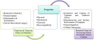
6. Applications of Ti and Ti alloys in dentistry
Recently, titanium has sparked much attention in dentistry. Since its outstanding resistance to corrosion, biocompatibility, and favorable mechanical and physical qualities, titanium (Ti) uses in dentistry (Figure 11) have increased considerably in recent years. Some of the commonly used Titanium alloys and their properties are shown in Table 8.
Figure 11.

Titanium and Ti Alloys in dentistry.
Table 8.
Properties of Ti alloys (Osman et al., 2015).
| Material | Modulus (GPa) | Ultimate Tensile Strength (MPa) | Yield Strength (MPa) | Elongation (%) | Density (g/cc) | Type of Alloy |
|---|---|---|---|---|---|---|
| Ti–6Al–4V- ELI | 113 | 860 | 795 | 10 | 4.4 | α + β |
| Ti–6Al–4V | 113 | 930 | 860 | 10 | 4.4 | α + β |
| Ti–6Al–7Nb | 114 | 900–1050 | 880–950 | 8–15 | 4.4 | α + β |
| Ti–5Al-2.5Fe | 112 | 1020 | 895 | 15 | 4.4 | α + β |
| Ti–15Zr–4Nb–2Ta-0.2Pd | 94–99 | 715–919 | 693–806 | 18–28 | 4.4 | α + β |
| Ti–29Nb–13Ta-4.6Zr | 80 | 911 | 864 | 13.2 | 4.4 | β |
6.1. Titanium in dental implants
Dental implants are frequently utilized to support prostheses and supplant missing teeth. Figure 12 shows an exceptional fitting between a Ti-based abutment (de Holanda Cavalcanti Pereira et al., 2022) and an implant that has adequate mechanical strength and masticatory force distribution in an implant-supported prosthesis (Souza et al., 2015). As shown in Figure 13, endosseous implants may be used in the invaded jaw and splint with a screw-retained full-arch renovation. Modern implants are frequently designed with a screw thread by which the components are fixed with the mandible or maxilla bone (Baumgarten, 2018).
Figure 12.

Titanium-based abutment-implant joint (Souza et al., 2015).
Figure 13.

Endosseous titanium implant for hybrid restoration of Maxillary and mandibular (Baumgarten, 2018).
6.2. Titanium in restorative dentistry
Dental restoration (Figure 14) replaces or repairs damaged teeth lost due to decay or injury. Restorations on individual teeth are often filled with filler materials to cure decay. More wide-ranging refurbishments, such as dental bridges, crowns, and implant-aided restorations, are also done with patient-specific restoration techniques ((Baumgarten, 2018; Močnik and Kosec, 2021).
Figure 14.

Titanium in restorative dentistry a) Filling of Dental decay, b) Ti crown covered with porcelain c) Spatula (Philip et al., 2017).
Due to its unattractive appearance, Ti and its alloys are rarely employed for treating decay. However, because of their excellent durability and affordable price, dental restoration with titanium crowns is an option many individuals opt for. An alloy of nickel, chrome, and titanium with 4%–6% titanium makes up the inner structure of a titanium crown, which has an outer shell composed of Ceramco3 porcelain. The mouth's saliva does not affect the crown, nor does the oral environment cause it to oxidize. This is the greatest option, especially for those with severe metal allergies (Haag, 2011).
Moreover, Dental composite filling instruments (spatula) are often made with Ti alloy or coated with Ti alloy. For composite resins and glass ionomers, TiN-coated stainless steel instruments produced no decrease in adhesion compared to non-coated, clean, polished stainless steel instruments (Philip et al., 2017).
6.3. Orthodontic applications
Orthodontic braces are affixed to the teeth, and orthodontic wiring is threaded through the bracket and ligated to hold it in place. Recently, orthodontics (Figure 15) has extensively used temporary anchorage devices (TADs). Most of these devices are embedded in the patients' bone/soft tissues, so they should be biologically compatible. Most of the TADs are made of titanium alloys. They have been used as anchorage devices to move teeth in directions that generally are difficult to move without additional conventional anchorage methods. Although Stainless steel has been used as cheaper TADs than titanium-based TADs, Titanium still provides superior properties to Stainless steel TADs (Rokaya et al., 2021).
Figure 15.

Orthodontic applications.
6.4. Endodontic applications
Endodontic therapy entails removing contaminated tissue from a tooth’s root canal system, contouring and disinfecting the canals, and refilling the canals with the appropriate ingredients. In endodontics, using rotary files (Figure 16) made of nickel-titanium (NiTi) has revolutionized root canal treatment (Dobrzański et al., 2022).
Figure 16.

Endodontic application of Rotary file (Philip et al., 2017).
6.5. Maxillofacial and oral surgery applications
Oral and maxillofacial surgical treatment includes temporomandibular joint, bone grafting, orthognathic, dent alveolar, and correcting the face's fractures and soft tissue deformities. Titanium and its alloys significantly contribute to each of these fields. Many of these treatments necessitate an implanted maneuver to either exchange or stabilize a lost portion or sections, allowing for smooth healing. Because Ti appears clinically inactive, producing little or no tissue inflammation, it has primarily replaced stainless steel and Vitallium in maxillofacial and oral surgery.
Figure 17 (a) shows a Ti–6Al–4V arch bar fastened into both the maxilla and mandible used to alleviate the jaws after the restoration of a mandibular rupture (IIzuka et al., 2006). Maintaining space under the soft tissue for the extra volume of the placed graft is crucial when bone grafting is essential to enlarge the size of the bone. Figure 17 (b) shows that Ti mesh is frequently employed to keep the distance between a particulate xenograft and a deficient maxilla (De Angelis et al., 2015).
Figure 17.

(a) Repair of mandibular fracture. (b) A particulate xenograft is used to enhance a defective maxilla (Baumgarten, 2018).
7. Limitations of Ti and Ti alloys as dental implants
Implants made by Ti have an extensive track of expectable medical outcomes, with a total accomplishment rate of 98.8% for a long time (Lindquist et al., 1996). This can be attributed to CpTi and Ti–6Al–4V's great biocompatibility, significant bone, mild tissue reaction, and satisfactory quality and consumption obstruction. Recently, there has been progress in searching for replacements for Ti–6Al–4V and CpTi to counteract a few disadvantages of Ti dental implants. Those drawbacks can be grouped into stylish tests, medical issues, and progress in bio-artistic events. Test of taste: Patients can benefit from improved modern reproduction because fired materials are white and imitate natural teeth better than dark Ti. When the delicate mucosa is of thin biotype or retreats after some time, using white artistic implants would block Ti implants' dim shine (Den et al., 2008; Cosyn et al., 2012). In writing, the expansion of keratinized tissue around inserts is accounted for using autografting with subepithelial connective tissue or xenogeneic collagen framework of porcine root (Schneider et al., 2011; Esposito et al., 2012). However, there is no concrete evidence that joining thin tissue thickens it. Medicinal issues: Ti is not currently considered an entirely bioinert material; rather, it may be an allergen, as evidenced by a few studies (Evrard et al., 2010; Pigatto et al., 2009). Raised concerned with Ti alloys have been discovered in the region of oral inserts (Koutayas et al., 2009), pee (Jacobs et al., 1998), serum, and local lymph hubs (Onodera et al., 1993), all of which are potentially harmful to the human body. In any case, the clinical relevance of the above discoveries is unclear, as various studies have previously confirmed Titanium's dependability for long-term usage in the oral circumstance (Wenz et al., 2008). These phenomena, combined with the aesthetic matter, have led some dental patients who are mentally fearful of demanding treatment with metal-free dental implants.
8. Recent advances in Ti-based implant materials
Several examples in titanium-based biomaterials ' imaginative work demonstrate the ability to make mixes made of no allergenic and nontoxic segments with inspiring mechanical characteristics (low modulus and high caliber) and workability. Other non-toxic ingredients, like Zr, Pd, Nb, Ta, Mo, and Fe, are being developed to replace vanadium and aluminum in future developments (Niinomi et al., 2003). These are composites with a lower modulus of flexibility (55–85 GPa), similar to bone (17–28 GPa). This lower adaptability modulus is appealing because it allows for more precise weight movement at the bone-installation interface. Additionally, these composites are designed to achieve higher caliber and quality than ᾳ + β mixes. Another compound (Roxolid®, Straumann, and Basel, Switzerland) for collecting slight separation across embeds has recently been introduced to dentistry. The foundation is the blend's two-part itemization of 83–87% Ti and 13–17% zirconium. With a 953 MPa inflexibility rating and 40% higher weariness strength than Ti–6Al–4V and CpTi, this composite has been determined to have superior mechanical properties (Grandin et al., 2015). When zirconia is added to Ti, osseointegration improves, and the combined material is more biocompatible than pure Ti (Bernhard et al., 2009). Ti-12.5Zr-2.5Nb-2.5Ta is another good Ti mixture for careful installation (TZNT). When compared to standard Ti mixes, this composite had an exclusive benefit in that its modulus of flexibility was closer to that of human bone (100 Gpa–120 Gpa) and had a nearly identical affirmation strain (0.65%) as human bones (0.67%) (Ikarashi et al., 2005). The addition of Zr, Nb, and Ta to the compound, for example, has shown no adverse tissue reactions or toxicity and has a high resistance to disintegration (Li et al., 2003, 2004).
9. Futuristic development of innovative dental implants
The Ti–6Al–4V compound is a better substitute for CPTi because it has better mechanical and consumption resistance and a lower flexibility modulus (Long et al., 1998). However, numerous attempts have been made to enhance its characteristics, particularly those associated with versatile modulus, consumption obstruction, and biocompatibility. Furthermore, there have been reports that aluminum and vanadium particles can cause neurological issues like Alzheimer's disease and unfriendly tissue responses over a long period (Brunette et al., 2001). Hence, there is a pressing need to create new Ti amalgams, primarily by adding non-cytotoxic components such as niobium, molybdenum, tantalum, and zirconium. Parallel composite Zr–Ti is the most promising. Both in vitro (Eisenbarth et al., 2004) and in vivo (Saldana et al., 2007) evaluations have shown that zirconium as an endosseous embed has excellent biocompatibility. Regardless of the certainty of metal particle discharge throughout biocorrosion, the harmfulness of Zr particles is recognized to be negligible due to the deficiency of biomolecule blend (Hanawa et al., 2004). Zr is synthetically similar to Ti; when added to a firm arrangement with Ti, it is considered an unbiased component because it has an indistinguishable allotropic change and a comparable stage progress temperature. Prior research has shown that combining zirconium with robust arrangements can reduce the martensitic change temperature of Ti (Ho et al., 2008). Titanium's liquefying temperature can also be reduced by expanding zirconium, lowering projecting and swaging costs (Ho et al., 2009). Because of the vast temperature dissimilarity between the liquid composite and a much more excellent venture, lowering Titanium's liquefying temperature could reduce its interaction with oxygen and decrease the possibility of defective filling and resulting porosity improvement (Eboungabeka et al., 2021). Correa et al. (2014) recently looked into the biocompatibility, mechanical characteristics, and microstructure of Ti–Zr alloys containing 15, 10, and 5% zirconium. They discovered that Ti–Zr alloys are primarily composed of the α-phase (with hcp arrangement) and have significantly higher miniature hardness values than CPTi. The versatility modulus of the composites was sensitive to zirconium fixations while remaining within the range of regular Ti amalgams (Correa et al., 2014), and the amalgams had no cytotoxic possessions on osteoblastic cells under the conditions examined.
Ti–Zr–Nb–Ta (TZNT) is another promising dental implant combination (Nag et al., 2009). Quaternary Ti alloys are considered superior to business ternary (Samuel et al., 2010) and twofold composites in erosion resistance (Karthega et al., 2007). Metals such as Zr, Ta, and Nb have also been excellent biocompatibility and osteoconductivity (Payer et al., 2010; Rosalbino et al., 2011), with no known adverse effects on humans. The Ti–Nb–Zr–Ta composites were found to have similar cell multiplication but greater cell separation than the Ti–6Al–4V composites (Samuel et al., 2010). In comparison to Ti, Ti–20Nb–10Zr–5Ta (TNZT) has improved inactivity, hardness, and extreme rigidity (883 MPa), as well as a lower versatile modulus (59 GPa) (Milosev et al., 2013; Popa et al., 2012). Furthermore, this alloy appeared to have no adverse situation on cell endurance, apoptosis acceptance, development interruption, or antacid phosphatase movement compared to Ti, indicating that Ti–20Nb–10Zr–5Ta in dental applications is feasible (Milosev et al., 2014).
10. Futuristic surface modification of Ti-based dental implants
Recent surveys have proposed a few procedures (Bauer et al., 2013) to improve the speed and quality of osseointegration of Ti implants. These future patterns concern (1) Surface roughness can be adjusted at the nanoscale to improve protein adsorption and cell grip (Zhang et al., 2013), (2) biomimetic calcium phosphate coatings for improving osteoconduction (Shadanbaz et al., 2012), (3) combining natural medications to speed up the bone remedial progression in the peri-embed area (Choo et al., 2013; Zhang et al.,2014) and (4) natural segments with nanoparticles (Qiao et al., 2015; Memarzadeh et al., 2015), Chlorhexidine and polysaccharides are two examples, with chitosan receiving special attention (Wang et al., 2004, 2006; Scharnweber et al., 2007). A technique of current interest is Nano topographical alterations on the embed surface to persuade inherent osteoinductive motioning of the surface follower cells. Existing evidence for Nano topography suggests that basic osseointegration steps can be regulated by nanoscale changes in the embed surface (Puckett et al., 2008; Park et al., 2009; McNamara et al., 2010). The embed's surface collaboration with particles and proteins is altered due to these changes (i.e., adsorption, arrangement, bioactivity, etc.). These connections can greatly impact subatomic and cell exercises and the osseointegration cycle. Endosseous inserts are altered Nano topographically using various strategies and approaches (Subramanian et al., 2012). Physical techniques for compaction of nanoparticles to produce surfaces with nanoscale grain limits (Webster and Ejiofor, 2004), Chemical Treatment (Cooper et al., 2006), creative sandblasting/acid etching (Ellingsen et al., 2006), optical lithography (Domanski et al., 2012), galvanostatic anodization (Sul et al., 2005), and crystal deposition (Lee et al., 2006, 2012). Because these techniques are arbitrary cycles, it's challenging to regulate the consistency and dispersion of nanostructures on the embedded surfaces. Scientists have devised a new covering strategy fueled by the regular biomineralization cycle. Precipitation of calcium phosphate apatite precious stones onto the Ti surface from reenacted body liquids (simulated body fluid (SBF)) framed a covering at room temperature in this biomimetic strategy. A few techniques have been developed to speed up the statement of coatings from watery arrangements (Shadanbaz et al., 2012; Bose et al., 2012). In preclinical relative models, the osseointegration of Ti inserts coated with biomimetic calcium phosphate has been studied (Habibovic et al., 2002, 2005). In any case, the osseointegration of biomimetic Ti dental inserts has yet to be compared to other surface medicines in preclinical models. Bone-invigorating agents, such as growth factors, could be applied outside of Ti dental inserts to improve the bone-recovery measure locally. Recent research has revealed that bioactive specialists may improve the embedded surface's osteogenic properties (Schwartz-Filho et al., 2012; Bougas et al., 2014). Bioactive inserts have been considered to create a biochemical bond between the bone tissue and the Ti-embedded surface rather than just a physical one (Sul et al., 2010). A bioactive embed surface can promote various sub-atomic collaborations, potentially forming a substantial connection between the bone and the embed surface (Sul et al., 2013). Fibronectin, fibroblast growth factor (FGF), type I collagen, Bone morphogenetic proteins (BMPs), and arginine-glycine-aspartic acid (RDG peptide) are promising bioactive sub-atomic up-and-comers with high osteogenic potential, according to a few studies (Jimbo et al., 2007).
A blend of RGD peptides (aspartate glycine and arginine) was used to create a lattice using acrylate stays and the Nano mechanical dock of collagen I filaments (Schliephake et al., 2008), causing increased bone-embed contact and bone thickness during the early stages of peri-embed bone development effectively after one month. With the ability to promote cell bonding via integrin transmembrane receptors important for cell-extracellular grid collaborations, arginyl glycyl aspartic corrosive tripeptide has been thoroughly researched in vitro and preclinical creature models (Hanawa et al., 2010). However, the cost of producing these bioactive particles and specialized and administrative issues have prompted researchers to investigate alternative bioactive atoms, like the bone-preparing hormone nutrient D (Kelly et al., 2009).
Amorphous material, diamond-like carbon (DLC), is used to coat dental implants by deposition of an intermediate layer for experimental study with high inherent biocompatibility with bone. This process is known as chemical vapor deposition (CVD) onto heated CPTi abutment screws and includes electrodeposition. DLC produces enhanced biocompatibility and improved corrosion resistance, with in vitro experimental indication of achievement. However, it requires in vivo despite this promise before clinical practice (patients) (Huacho et al., 2017). Hovsepian et al. (2022) reported the structural response and load-bearing capacity of TiN/NbN superlattice coatings deposited on medical-grade CoCrMo alloy substrate. The coatings were applied by a mixed High Power Impulse Magnetron Sputtering-Unbalanced Magnetron Sputtering (HIPIMS-UBM) process. The coated CoCrMo alloy was studied under localized load to investigate the crack formation mechanism. The high toughness of the superlattice structured TiN/NbN coatings was obtained, which, combined with their high adhesion on the substrate, made the coated medical grade CoCrMo alloy a potential candidate for medical implant applications.
Implant placement in dentistry and orthopedics focuses on improving the osseointegration and biocompatibility properties of Ti and Ti-based composites by the versatile surface modification layer-by-layer technique. In future applications in Ti-based composite implants, the morphology and chemical structure could be characterized by (i) ultraviolet-visible, (ii) atomic force microscopy, (iii) Fourier-transform infrared spectroscopies, (iv) scanning electron microscopy, and (v) micro-Raman spectroscopy. Films of poly (sodium 4-styrene sulfonate) (PSS) and Ti dioxide (TiO2) nanoparticles multilayers might enhance the screw topography and chemical surface properties with beneficial effects for osteoblast activity to improve dental implants (Igor et al., 2021).
11. Conclusions
In dentistry, Ti and Ti alloys are considered to be the materials of excellence due to their optimum physical, mechanical and biochemical properties. Because of the generally increased costs, handling of Ti and Ti alloys using advanced additive manufacturing (AM) was given insufficient attention by physicians until a few years ago. However, advanced AM methods like LMD, EBM, SLM etc. are getting progressively well-known in dental applications due to their ability to produce implants with customized designs and surface morphologies that allow easy cell multiplication required for bone ingrowth. Various advances have aided the osseointegration in physical and chemical surface modification approaches. However, still there is a great need for long-term clinical research to compare the performance of different coatings, and evaluate the success rate of innovative implant coatings. In conclusion, Ti and Ti alloys are anticipated to remain the mainstream implant materials in dentistry with the advancement of continual research.
Declarations
Author contribution statement
All authors listed have significantly contributed to the development and the writing of this article.
Funding statement
This research did not receive any specific grant from funding agencies in the public, commercial, or not-for-profit sectors.
Data availability statement
No data was used for the research described in the article.
Declaration of interests statement
The authors declare no conflict of interest.
Additional information
No additional information is available for this paper.
Acknowledgements
The authors would like to thank their respective organizations for supporting this work.
Contributor Information
Md Enamul Hoque, Email: enamul1973@gmail.com.
Huaizhong Xu, Email: xhz2008@kit.ac.jp.
References
- Albrektsson T., Canullo L., Cochran D., De Bruyn H. Peri-implantitis": a complication of a foreign body or a man-made "disease". Facts and fiction. Clin. Implant Dent. Relat. Res. 2016 Aug;18(4):840–849. doi: 10.1111/cid.12427. Epub 2016 May 30. PMID: 27238274. [DOI] [PubMed] [Google Scholar]
- Alkhadashi A., Güven M., Erol F., Yıldırım G. The effect of different combinations of surface treatments and bonding agents on the shear bond strength between titanium alloy and lithium disilicate glass-ceramic. Int. J. Periodontics Restor. Dent. 2020;40(2):271–276. doi: 10.11607/prd.3893. [DOI] [PubMed] [Google Scholar]
- Attarilar Shokouh, Ebrahimi Mahmoud, Djavanroodi Faramarz, Fu Yuanfei, Wang Liqiang, Yang Junlin. 3D printing technologies in metallic implants: a thematic review on the techniques and procedures. Intl. J. Bioprinting. 2021;7(1) doi: 10.18063/ijb.v7i1.306. [DOI] [PMC free article] [PubMed] [Google Scholar]
- Barão V.A.R., Ramachandran R.A., Matos A.O., Badhe R.V., Grandini C.R., Sukotjo C., Ozevin D., Mathew M. Prediction of tribocorrosion processes in titanium-based dental implants using acoustic emission technique: initial outcome. Mater. Sci. Eng. C. 2021;123 doi: 10.1016/j.msec.2021.112000. [DOI] [PubMed] [Google Scholar]
- Bauer S., Schmuki P., von der Mark K., Park J. Engineering biocompatible implant surfaces Part I: materials and surfaces. Prog. Mater. Sci. 2013;58:261–326. [Google Scholar]
- Baumgarten H.S. Titanium in Medical and Dental Applications. Elsevier; 2018. Why titanium in dental applications? pp. 495–504. [Google Scholar]
- Becker W., Becker B.E., Ricci A., Bahat O., Rosenberg E., Rose L.F.…Israelson H. A prospective multicenter clinical trial comparing one- and two-stage titanium screw-shaped fixtures with one-stage plasma-sprayed solid-screw fixtures. Clin. Implant Dent. Relat. Res. 2000;2(3):159–165. doi: 10.1111/j.1708-8208.2000.tb00007.x. [DOI] [PubMed] [Google Scholar]
- Berardi D., Colagiovanni M., Scoccia A., Raffaelli L., Manicone P.F., Perfetti G. Evaluation of a new laser surface implant: scanning electron microscopy/energy dispersive X-ray and X-ray photoelectron spectroscopy analyses. J. Biol. Regul. Homeost. Agents. 2008;22(3):161–167. [PubMed] [Google Scholar]
- Bernhard N., Berner S., de Wild M., Wieland M. The binary Ti-Zr Alloy - a newly developed Ti alloy for the use in dental implants. Forum Implantol. 2009;5:30–39. [Google Scholar]
- Bhola R.B., Olson D.L. Electrochemical evaluation of wrought titanium –15 molybdenum alloy for dental implant applications in phosphate buffer saline. Port. Electrochim. Acta. 2010;28(2) S. M., B. M. [Google Scholar]
- Bose S., Tarafder S. Calcium phosphate ceramic systems in growth factor and drug delivery for bone tissue engineering: a review. Acta Biomater. 2012;8:1401–1421. doi: 10.1016/j.actbio.2011.11.017. [DOI] [PMC free article] [PubMed] [Google Scholar]
- Bougas K., Jimbo R., Vandeweghe S., Tovar N., Baldassarri M., Alenezi A., Janal M., Coelho P.G., Wennerberg A. In vivo evaluation of a novel implant coating agent: laminin-1. Clin. Implant Dent. Relat. Res. 2014;16(5):728–735. doi: 10.1111/cid.12037. [DOI] [PubMed] [Google Scholar]
- Brett P., Harle J., Salih V., Mihoc R., Olsen I., Jones F., Tonetti M. Roughness response genes in osteoblasts. Bone. 2004;35(1):124–133. doi: 10.1016/j.bone.2004.03.009. [DOI] [PubMed] [Google Scholar]
- Brunette D.M., Tengvall P., Textor M., Thomsen P. Springer; Berlin: 2001. Titanium in Medicine: Material Science, Surface Science, Engineering, Biological Responses and Medical Applications. [Google Scholar]
- Choo T., Marino V., Bartold P.M. Effect of PDGF-BB and betatricalcium phosphate (b-TCP) on bone formation around dental implants: a pilot study in sheep. Clin. Oral Implants Res. 2013;24:158–166. doi: 10.1111/j.1600-0501.2011.02345.x. [DOI] [PubMed] [Google Scholar]
- Cooper L.F., Zhou Y., Takebe J., Guo J., Abron A., Holmen A., Ellingsen J.E. Fluoride modification effects on osteoblast behavior and bone formation at TiO2 grit-blasted c.p. Titanium Endosseous Implan. Biomat. 2006;27:926–936. doi: 10.1016/j.biomaterials.2005.07.009. [DOI] [PubMed] [Google Scholar]
- Correa D.R.N., Vicente F.B., Donato T.A.G., Arana-Chavez W.E., Buzalaf M.A.R., Grandini C.R. The effect of the solute on the structure, selected mechanical properties, and biocompatibility of Ti–Zr system alloys for dental applications. Mater. Sci. Eng. C. 2014;34:354–359. doi: 10.1016/j.msec.2013.09.032. [DOI] [PubMed] [Google Scholar]
- Cosyn J., Hooghe N., De Bruyn H. A systematic review on the frequency of advanced recession following single immediate implant treatment. J. Clin. Periodontol. 2012;39(6):582–589. doi: 10.1111/j.1600-051X.2012.01888.x. [DOI] [PubMed] [Google Scholar]
- De Angelis N., De Lorenzi M., Benedicenti S. Surgical combined approach for alveolar ridge augmentation with titanium mesh and rhPDGF-BB: a 3-year clinical series. Int.J. Periodontics Restorative Dent. 2015;35(2):230–237. doi: 10.11607/prd.2234. [DOI] [PubMed] [Google Scholar]
- De Holanda Cavalcanti Pereira A.K., de Oliveira Limirio J.P.J., Cavalcanti do Egito Vasconcelos B., Pellizzer E.P., Dantas de Moraes S.L. Mechanical behavior of titanium and zirconia abutments at the implant-abutment interface: a systematic review. J. Prosthet. Dent. 2022 doi: 10.1016/j.prosdent.2022.01.006. [DOI] [PubMed] [Google Scholar]
- Degidi M., Petrone G., Iezzi G., Piattelli A. Bone contact around acid-etched implants: a histological and histomorphometrical evaluation of two human-retrieved implants. J. Oral Implantol. 2003;29(1):13–18. doi: 10.1563/1548-1336(2003)029<0013:BCAAIA>2.3.CO;2. [DOI] [PubMed] [Google Scholar]
- Den Hartog L., Huddleston Slater J.J.R., Vissink A., Meijer H.J.A., Raghoebar G.M. Treatment outcome of immediate, early and conventional single-tooth implants in the aesthetic zone: a systematic review to survival, bone level, soft-tissue, aestheticsand patient satisfaction. J. Clin. Periodontol. 2008;35(12):1073–1086. doi: 10.1111/j.1600-051X.2008.01330.x. [DOI] [PubMed] [Google Scholar]
- Ding S.-J. Properties and immersion behavior of magnetron-sputtered multi-layered hydroxyapatite/titanium composite coatings. Biomaterials. 2003;24(23):4233–4238. doi: 10.1016/s0142-9612(03)00315-6. [DOI] [PubMed] [Google Scholar]
- Ding Z., Wang L., Zhang C., Zhang L. Development and Application of Biomedical Titanium Alloys; 2018. Surface Modification of Biomedical Titanium Alloys; pp. 149–173. [Google Scholar]
- Dobrzański L.A., Dobrzański L.B., Dobrzańska-Danikiewicz A.D., Dobrzańska J. Nitinol type Alloys general characteristics and applications in endodontics. Processes. 2022;10(1):101. [Google Scholar]
- Domanski M., Luttge R., Lamers E., Walboomers X.F., Winnubst L., Jansen J.A., Gardeniers J.G.E. Submicron- Eboungabeka AD, marcelle ER, trigo E, nsongo T (2021); reactivity of titanium dental implants in salivary environment in the presence of foods consumed in the republic of the Congo. Adv. Mater. Phys. Chem. 2012;11:93–99. [Google Scholar]
- Eboungabeka AD, Marcelle ER, Trigo E, Nsongo T (2021); Reactivity of titanium dental implants in salivary environment in the presence of foods consumed in the Republic of the Congo. Advances in Mat. Phy. Chem., 11, 93–99.
- Eisenbarth E., Velten D., Muller M., Thull R., Breme J. Biocompatibility of beta-stabilizing elements of Titanium alloys. Biomaterials. 2004;25:5705–5713. doi: 10.1016/j.biomaterials.2004.01.021. [DOI] [PubMed] [Google Scholar]
- Elias C.N. Implant Dentistry - A Rapidly Evolving Practice. IntechOpen; 2011. Factors affecting the success of dental implants. [Google Scholar]
- Elias C.N., Meirelles L. Improving osseointegration of dental implants. Expet Rev. Med. Dev. 2010;7(2):241–256. doi: 10.1586/erd.09.74. [DOI] [PubMed] [Google Scholar]
- Elias C.N., Oshida Y., Lima J.H.C., Muller C.A. Relationship between surface properties (roughness, wettability and morphology) of titanium and dental implant removal torque. J. Mech. Behav. Biomed. Mater. 2008;1(3):234–242. doi: 10.1016/j.jmbbm.2007.12.002. [DOI] [PubMed] [Google Scholar]
- Elias C.N., Fernandes D.J., Souza F.M., Monteiro E.D., Biasi R.S. Mechanical and clinical properties of titanium and titanium-based alloys (Ti G2, Ti G4 cold worked nanostructured and Ti G5) for biomedical applications. J. Mater. Res. Technol. 2019;8(1):1060–1069. [Google Scholar]
- Ellingsen J.E., Thomsen P., Lyngstadaas S.P. Advances in dental implant materials and tissue regeneration. Periodontol. 2006;41(2000):136–156. doi: 10.1111/j.1600-0757.2006.00175.x. [DOI] [PubMed] [Google Scholar]
- Emsley J. Oxford University Press; Oxford: 2011. Natures Building Blocks: an A-Z Guide to the Elements. [Google Scholar]
- Endo K. Chemical modification of metallic implant surfaces with biofunctional proteins. Dent. Mater. J. 1995;14(2) doi: 10.4012/dmj.14.199. [DOI] [PubMed] [Google Scholar]
- Esposito M., Maghaireh H., Grusovin M.G., Ziounas I., Worthington H.V. Interventions for replacing missing teeth: management of soft tissues for dental implants. Cochrane Database Syst. Rev. 2012 doi: 10.1002/14651858.CD006697.pub2. [DOI] [PMC free article] [PubMed] [Google Scholar]
- Esteves G.M., Esteves J., Resende M., Mendes L., Azevedo A.S. Antimicrobial and antibiofilm coating of dental implants—past and new perspectives. Antibiotics. 2022;11(2):235. doi: 10.3390/antibiotics11020235. [DOI] [PMC free article] [PubMed] [Google Scholar]
- Evrard L.W.D., Parent D. Allergies to dental metals. Titanium: a new allergen. Rev. Med. Brux. 2010;31(1):44–49. [PubMed] [Google Scholar]
- Fang Z.Z., Paramore J.D., PS, Ravi K.S., Chandran Y.Z. Powder metallurgy of titanium – past, present, and future. Int. Mater. Rev. 2017;63(7):407–459. [Google Scholar]
- Figliuzzi M., Mangano F., Mangano C. A novel root analogue dental implant using CT scan and CAD/CAM:selective laser melting technology. Int. J. Oral Maxillofac. Surg. 2012;41:858–862. doi: 10.1016/j.ijom.2012.01.014. [DOI] [PubMed] [Google Scholar]
- Figliuzzi M., Giudice A., Rengo C., Fortunato L. A direct metal laser sintering (DMLS) root analogue implant placed in the anterior maxilla. Case Report. Annali Italiani di Chirurgia. 2019;8 [PubMed] [Google Scholar]
- Gagg G., Ghassemieh E., Wiria F.E. Effects of sintering temperature on morphology and mechanical characteristics of 3D printed porous titanium used as dental implant. Mater. Sci. Eng. C. 2013;33(7):3858–3864. doi: 10.1016/j.msec.2013.05.021. [DOI] [PubMed] [Google Scholar]
- Gan Huang, Libo Wu, Jie Hu, Xiongming Zhou, Fei He, Li Wan, Shu-Ting Pan. "Main applications and recent research progresses of additive manufacturing in dentistry". BioMed Res. Int., 2022 doi: 10.1155/2022/5530188. vol. 2022, Article ID 5530188, 26 pages, 2022. [DOI] [PMC free article] [PubMed] [Google Scholar]
- Gegner J. INTECH; Rijeka, Croatia: 2014. Tribology - Fundamentals and Advancements. [Google Scholar]
- Gittens R.A., Scheideler L., Rupp F., Hyzy S.L., Geis-gerstorfer J., Schwartz Z., et al. A review on the wettability of dental implant surfaces II: biologicaland clinical aspects. Acta Biomater. 2014;10:2907–2918. doi: 10.1016/j.actbio.2014.03.032. [DOI] [PMC free article] [PubMed] [Google Scholar]
- Golieskardi M., Satgunam M., Ragurajan D., Hoque M.E., Ng A.M.W., Shanmuganantha L. Advanced 3Y-TZP bioceramic doped with Al2O3 and CeO2 potentially for biomedical implant applications. Mater. Technol. 2019;34(8):480–489. [Google Scholar]
- Golieskardi M., Satgunam M., Ragurajan D., Hoque M.E., Ng A.M.W. Microstructural, tribological, and degradation properties of Al2O3- and CeO2-doped 3 mol.% yttria-stabilized zirconia bioceramic for biomedical applications. J. Mater. Eng. Perform. 2020;29:2890–2897. [Google Scholar]
- Gosavi S., Gosavi S., Alla R. Vol. 6. 2013. Titanium: A Miracle Metal in Dentistry. [Google Scholar]
- Grandin H.M., Berner S., Dart M. A review of titanium zirconium (TiZr) alloys for use in endosseous dental implants. Materials. 2015;5:1348–1360. [Google Scholar]
- Gruner H. In: Titanium in Medicine: Material Science, Surface Science, Engineering, Biological Responses and Medical Applications. Brunette D.M., Tengvall P., Textor M., Thomsen P., editors. Springer; Berlin, Germany: 2001. Thermal spray coatings on titanium; pp. 375–416. Engineering Materials. [Google Scholar]
- Guéhennec L.L., Soueidan A., Layrolle P., Amouriq Y. Surface treatments of titanium dental implants for rapid osseointegration. Dent. Mater. 2007;23(7):844–854. doi: 10.1016/j.dental.2006.06.025. [DOI] [PubMed] [Google Scholar]
- Haag P. Malmö University, Faculty of Odontology; 2011. Porcelain Veneering of Titanium: Clinical and Technical Aspects (Doctoral Dissertation. [PubMed] [Google Scholar]
- Habibovic P., Barrere F., van Blitterswijk C.A., de Groot K., Layrolle P. Biomimetic hydroxyapatite coating on metal implants. J. Am. Ceram. Soc. 2002;85:517–522. [Google Scholar]
- Habibovic P., Li J., van der Valk C.M., Meijer G., Layrolle P., van Blitterswijk C.A., de Groot K. Biological performance of uncoated and octacalcium phosphate-coated Ti6Al4V. Biomaterials. 2005;26:23–36. doi: 10.1016/j.biomaterials.2004.02.026. [DOI] [PubMed] [Google Scholar]
- Hamanaka H., Doi H., Yoneyama T., Okuno O. Dental casting of titanium and Ni-Ti alloys by a new casting machine. J. Dent. Res. 1989;68(11):1529–1533. doi: 10.1177/00220345890680111201. [DOI] [PubMed] [Google Scholar]
- Hanawa T. Metal ion release from metal implants. Mater. Sci. Eng. C. 2004;24:745–752. [Google Scholar]
- Hanawa T. Biofunctionalization of Titanium for dental implant. Jpn. Dent Sci Rev. 2010;46:93–101. [Google Scholar]
- Hansson S., Norton M. The relation between surface roughness and interfacial shear strength for bone-anchored implants. A mathematical model. J. Biomech. 1999;32(8):829–836. doi: 10.1016/s0021-9290(99)00058-5. [DOI] [PubMed] [Google Scholar]
- Hatamleh M.M., Wu X., Alnazzawi A., Watson J., Watts D. Surface characteristics and biocompatibility of cranioplasty titanium implants following different surface treatments. Dent. Mater. 2018;34(4):676–683. doi: 10.1016/j.dental.2018.01.016. [DOI] [PubMed] [Google Scholar]
- Heufelder M., Wilde F., Pietzka S., et al. Clinical accuracy of waferless maxillary positioning using customized surgical guides and patient specific osteosynthesis in bimaxillary orthognathic surgery. J. Cranio-Maxillo-Fac. Surg. 2017;45(9):1578–1585. doi: 10.1016/j.jcms.2017.06.027. [DOI] [PubMed] [Google Scholar]
- Ho W.F., Chen W.K., Wu S.C., Hsu H.C. Structure, mechanical properties, and grindability of dental Ti–Zr alloys. J. Mater. Sci. Mater. Med. 2008;19:3179–3186. doi: 10.1007/s10856-008-3454-x. [DOI] [PubMed] [Google Scholar]
- Ho W.F., Chen W.K., Pan C.H., Wu S.C., Hsu H.C. Structure, mechanical properties and grindability of dental Ti–10Zr–X alloys. Mater. Sci. Eng. C. 2009;29:36–43. doi: 10.1007/s10856-008-3454-x. [DOI] [PubMed] [Google Scholar]
- Hoque M.E., Shehryar M., Islam K.M.N. Processing and characterization of cockle shell calcium carbonate (CaCO3) bioceramic for potential application in bone tissue engineering. J. Mater. Sci. Eng. 2013;2(4):132. [Google Scholar]
- Hoque M.E., Sakinah N., Chuan Y.L., Ansari M.N.M. Synthesis and characterization of hydroxyapatite bioceramic. Int. J. Sci. Eng. Technol. 2014;3(5):458. [Google Scholar]
- Hovsepian P.E., Sugumaran A.A., Rainforth M., Qi J., Khan I., Ehiasarian A.P. Microstructure and load bearing capacity of TiN/NbN superlattice coatings deposited on medical grade CoCrMo alloy by HIPIMS. J. Mech. Behav. Biomed. Mater. 2022;132 doi: 10.1016/j.jmbbm.2022.105267. [DOI] [PubMed] [Google Scholar]
- Huacho P.M.M., Nogueira M.N.M., Basso F.G., Junior M.J., Francisconi R.S., Spolidorio D.M.P. Analyses of biofilm on implant abutment surfaces coating with Diamond-Like carbon and biocompatibility. Braz. Dent. J. 2017;28:317–323. doi: 10.1590/0103-6440201601136. [DOI] [PubMed] [Google Scholar]
- Ida K., Togaya T., Tsutsumi S., Takeuchi M. Effect of magnesia investments in the dental casting of pure titanium or titanium alloys. Dent. Mater. J. 1982;1(1):8–21. doi: 10.4012/dmj.1.8. [DOI] [PubMed] [Google Scholar]
- Igor L. Kitagawa, Celina M. Miyazaki, Letícia Pitol-Palin, Roberta Okamoto, Luana M. R. de Vasconcellos, Carlos J. L. Constantino, and Paulo N. Lisboa-Filho (2021), Titanium-based alloy surface modification with TiO2 and poly(sodium 4-styrenesulfonate) multilayers for dental implants. Appl. Bio Mater., 4, 4, 3055–3066. [DOI] [PubMed]
- Iijima M., Yuasa T., Endo K., Muguruma T., Ohno H., Mizoguchi I. Corrosion behavior of ion implanted nickel-titanium orthodontic wire in fluoride mouth rinse solutions. Dent. Mater. J. 2010;29(1):53–58. doi: 10.4012/dmj.2009-069. [DOI] [PubMed] [Google Scholar]
- Iizuka T., Hallermann W., Seto I., Smolka W. A titanium arch bar for maxillimandibular fixation in maxillofacial surgery. J. Oral Maxillofac. Surg. 2006;64:989–992. doi: 10.1016/j.joms.2005.11.050. [DOI] [PubMed] [Google Scholar]
- Ikarashi Y., Toyoda K., Kobayashi E., Doi H., Yoneyama T., Hamanaka H., et al. Improved biocompatibility of Titanium- Zirconium (Ti-Zr) alloy: tissue reaction and sensitization Ti-Zr alloy compared with pure Ti and Zr in rat implantation study. Mater. Trans. 2005;46:2260–2267. [Google Scholar]
- Iwaya Y., Machigashira M., Kanbara K., Miyamoto M., Noguchi K., Izumi Y., Ban S. Surface properties and biocompatibility of acid-etched titanium. Dent. Mater. J. 2008;27(3):415–421. doi: 10.4012/dmj.27.415. [DOI] [PubMed] [Google Scholar]
- Jaafar A., Hecker C., Árki P., Joseph Y. Sol-gel derived hydroxyapatite coatings for titanium implants: a review. Bioengineering. 2020;7(4):127. doi: 10.3390/bioengineering7040127. [DOI] [PMC free article] [PubMed] [Google Scholar]
- Jacobs J.J., Skipor A.K., Patterson L.M., Hallab N.J., Paprosky W.G., Black J., Galante J.O. Metal release in patients who have had a primary total hip arthroplasty.A prospective, controlled, longitudinal study. J. Bone Jt Surg. Am. 1998;80:1447–1458. doi: 10.2106/00004623-199810000-00006. [DOI] [PubMed] [Google Scholar]
- James Tedesco, Bryan E., Lee J., Lin Alex Y.W., Binkley Dakota M., Delaney Kathleen H., Kwiecien Jacek M., Grandfield Kathryn. Osseointegration of a 3D printed stemmed titanium dental implant: a pilot study. Int. J. Dentistry. 2017;2017 doi: 10.1155/2017/5920714. Article ID 5920714, 11 pages. [DOI] [PMC free article] [PubMed] [Google Scholar]
- Jemat A., Ghazali M.J., Razali M., Otsuka Y. Surface modifications and their effects on titanium dental implants. BioMed Res. Int. 2015:1–11. doi: 10.1155/2015/791725. [DOI] [PMC free article] [PubMed] [Google Scholar]
- Jiang C., Huang Z.H. Grain size effect on mechanical properties of titanium alloy. Key Eng. Mater. 2014;626:548–552. [Google Scholar]
- Jimbo R., Sawase T., Shibata Y., Hirata K., Hishikawa Y., Tanaka Y., Bessho K., Ikeda T., Atsuta M. Enhanced osseointegration by the chemotactic activity of plasma fibronectin for cellular fibronectin positive cells. Biomaterials. 2007;28(24):3469–3477. doi: 10.1016/j.biomaterials.2007.04.029. [DOI] [PubMed] [Google Scholar]
- Jorge J.R.P., Barão V.A., Delben J.A., et al. Titanium in dentistry: historical development, state of the art and future perspectives. J. Indian Prosthodont. Soc. 2013;13:71–77. doi: 10.1007/s13191-012-0190-1. [DOI] [PMC free article] [PubMed] [Google Scholar]
- Kandavalli S.R., Wang Q., Ebrahimi M., Gode C., Djavanroodi F., Attarilar S., Liu S. A brief review on the evolution of metallic dental implants: history, design, and application. Front. Mater. 2021;8 [Google Scholar]
- Karolewska K., Liga B. Comparison analysis of titanium alloy Ti6Al4V produced by metallurgical and 3D printing method. AIP Conf. Proc. 2019;2077 [Google Scholar]
- Karthega M., Raman V., Rajendran N. Influence of potential on the electrochemical behaviour of b Titanium alloys in Hank’s solution. Acta Biomater. 2007;3(207):1019–1023. doi: 10.1016/j.actbio.2007.02.009. [DOI] [PubMed] [Google Scholar]
- Kelly J., Lin A., Wang C.J., Park S., Nishimura I. Vitamin D and bone physiology: demonstration of vitamin Def in an implant osseointegration rat model. J. Prosthodont. 2009;18(6):473–478. doi: 10.1111/j.1532-849X.2009.00446.x. [DOI] [PubMed] [Google Scholar]
- Kim H., Miyaji F., Kokubo T., Nakamura T. Preparation of bioactive Ti and its alloys via simple chemical surface treatment. J. Biomed. Mater. Res. 1996;32(3):409–417. doi: 10.1002/(SICI)1097-4636(199611)32:3<409::AID-JBM14>3.0.CO;2-B. [DOI] [PubMed] [Google Scholar]
- Kim H., Choi S.-H., Ryu J.-J., Koh S.-Y., Park J.-H., Lee I.-S. The biocompatibility of SLA-treated titanium implants. Biomed. Mater. 2008;3(2) doi: 10.1088/1748-6041/3/2/025011. [DOI] [PubMed] [Google Scholar]
- Kim J.-H., Kim M.-Y., Knowles J.C., Choi S., Kang H., Parki S.-H., Park S.-M., Kim H.-W., Park J.-T., Lee J.-H., Lee H.-H. Mechanophysical and biological properties of a 3D-printed titanium alloy for dental applications. Dent. Mater. 2020;36:945–958. doi: 10.1016/j.dental.2020.04.027. [DOI] [PubMed] [Google Scholar]
- Kirmanidou Y., Sidira M., Drosou M.-E., Bennani V., Bakopoulou A., Tsouknidas A., et al. New Ti-alloys and surface modifications to improve the mechanical properties and the biological response to orthopedic and dental implants: a review. BioMed Res. Int. 2016:1–21. doi: 10.1155/2016/2908570. [DOI] [PMC free article] [PubMed] [Google Scholar]
- Koizumi H., Ishii T., Okazaki T., Kaketani M., Matsumura H., Yoneyama T. Castability and mechanical properties of Ti-15Mo-5Zr-3Al alloy in dental casting. J. Oral Sci. 2018;60(2):285–292. doi: 10.2334/josnusd.17-0280. [DOI] [PubMed] [Google Scholar]
- Koizumi H., Takeuchi Y., Imai H., Kawai T., Yoneyama T. Application of titanium and titanium alloys to fixed dental prostheses. J. Prosthodontic Res. 2019;63(3):266–270. doi: 10.1016/j.jpor.2019.04.011. [DOI] [PubMed] [Google Scholar]
- Koutayas S.O., Vagkopoulou T., Pelekanos S., Koidis P., Strub J.R. Zirconia in dentistry. Part 2. Evidence-based clinical breakthrough. Eur. J. Esthetic Dent. 2009;4:348–380. [PubMed] [Google Scholar]
- Kreller T., Sahm F., Bader R., Boccaccini A.R., Jonitz-Heincke A., Detsch R. Biomimetic calcium phosphate coatings for bioactivation of titanium implant surfaces: methodological approach and in vitro evaluation of biocompatibility. Materials. 2021;14:3516. doi: 10.3390/ma14133516. [DOI] [PMC free article] [PubMed] [Google Scholar]
- Krozer A., Hall J., Ericsson L. Chemical treatment of machined titanium surfaces. An in vitro study. Clin. Oral Implants Res. 1999;10(3):204–211. doi: 10.1034/j.1600-0501.1999.100303.x. [DOI] [PubMed] [Google Scholar]
- Kurod D., Niinomi M., Morinaga M., Kato Y., Yashiro T. Design and mechanical properties of new â type titanium alloys for implant materials. Material Sci. Eng. 1998;243(1-2):244–249. [Google Scholar]
- Lambrechts P., Debels E., Landuyt V., Peumans M., Meerbeek B.V. How to simulate wear? Overview of existing methods. Dent. Mater. 2006;22:693–701. doi: 10.1016/j.dental.2006.02.004. [DOI] [PubMed] [Google Scholar]
- Lee J.W., Roe M.J., Hoque M.E., Spowage A.C., Scotchford C.A., Grant D.M., Brown P.D. Kuala Lumpur; Malaysia: 2014. Characterisation of silica-doped hydroxyapatite biomedical coatings on titanium for biomedical applications. 9th International Materials Technology Conference and Exhibition (IMTCE 2014) [Google Scholar]
- Lee S.H., Kim H.W., Lee E.J., Li L.H., Kim H.E. Hydroxyapatite–TiO2 hybrid coating on Ti implants. J. Biomater. Appl. 2006;20:195–208. doi: 10.1177/0885328206050518. [DOI] [PubMed] [Google Scholar]
- Lee W.T., Koak J.Y., Lim Y.J., Kim S.K., Kwon H.B., Kim M.J. Stress shielding and fatigue limits of poly-ether-ether-ketone dental implants. J. Biomed. Mater. Res. B. 2012;100:1044–1052. doi: 10.1002/jbm.b.32669. [DOI] [PubMed] [Google Scholar]
- Li J., Zhou L., Li Z.C., Chen D.J. Study of a new titanium alloy for surgical implant application. Rare Met. Mater. Eng. 2003;32:398. [Google Scholar]
- Li S.J., Yanga R., Lia S., Hao Y.L., Cui Y.Y., Niinomi M., et al. Wear characteristics of Ti-Nb-Ta-Zr and Ti-6Al-4V alloys for biomedical applications. Wear. 2004;257:869–876. [Google Scholar]
- Li B., Shen S., Jiang W., et al. A new approach of splint-less orthognathic surgery using a personalized orthognathic surgical guide system: a preliminary study. Int. J. Oral Maxillofac. Surg. 2017;46(10):1298–1305. doi: 10.1016/j.ijom.2017.03.025. [DOI] [PMC free article] [PubMed] [Google Scholar]
- Li J., Jansen J.A., Walboomers X.F., van den Beucken J.J.J.P. Mechanical aspects of dental implants and osseointegration: a narrative review. J. Mech. Behav. Biomed. Mater. 2020;103 doi: 10.1016/j.jmbbm.2019.103574. [DOI] [PubMed] [Google Scholar]
- Lindquist L.W., Carlsson G.E., Jemt T. A prospective 15-year follow-up study of mandibular fixed prostheses supported by osseointegrated implants. Clinical results and marginal bone loss. Clin. Oral Implants Res. 1996;7(4):329–336. doi: 10.1034/j.1600-0501.1996.070405.x. [DOI] [PubMed] [Google Scholar]
- Liu F., Wang F., Shimizu T., Igarashi K., Zhao L. Formation of hydroxyapatite on Ti–6Al–4V alloy by microarc oxidation and hydrothermal treatment. Surf. Coating. Technol. 2005;199(2-3):220–224. doi: 10.1263/jbb.100.100. [DOI] [PubMed] [Google Scholar]
- Long M., Rack H.J. Titanium alloys in total joint replacement—a materials science perspective. Biomaterials. 1998;(19):1621–1639. doi: 10.1016/s0142-9612(97)00146-4. [DOI] [PubMed] [Google Scholar]
- Luo H., Wu Y., Diao X., Shi W., Feng F., Qian F., Umeda J., Kondoh K., Xin H., Shen J. Mechanical properties and biocompatibility of titanium with a high oxygen concentration for dental implants. Mater. Sci. Eng. C. 2020;117 doi: 10.1016/j.msec.2020.111306. [DOI] [PubMed] [Google Scholar]
- Macdonald D., Rapuano B., Deo N., Stranick M., Somasundaran P., Boskey A. Thermal and chemical modification of titanium–aluminum–vanadium implant materials: effects on surface properties, glycoprotein adsorption, and MG63 cell attachment. Biomaterials. 2004;25(16):3135–3146. doi: 10.1016/j.biomaterials.2003.10.029. [DOI] [PubMed] [Google Scholar]
- Mangano C., Raspanti M., Traini T., Piattelli A., Sammons R. Stereo imaging and cytocompatibility of a model dental implant surface formed by direct laser fabrication. J. Biomed. Mater. Res. Part A. 2009;88(3):823–831. doi: 10.1002/jbm.a.32033. [DOI] [PubMed] [Google Scholar]
- Mangano C., De Rosa A., Desiderio V., et al. The osteoblastic differentiation of dental pulp stem cells and bone formation on different titanium surface textures. Biomaterials. 2010;31(13):3543–3551. doi: 10.1016/j.biomaterials.2010.01.056. [DOI] [PubMed] [Google Scholar]
- Mangano C., Piattelli A., Raspanti M., et al. Scanning electron microscopy (SEM) and X-ray dispersive spectrometry evaluation of direct laser metal sintering surface and human bone interface: a case series. Laser Med. Sci. 2011;26(1):133–138. doi: 10.1007/s10103-010-0831-8. [DOI] [PubMed] [Google Scholar]
- Mangano C., Piattelli A., Mangano F., et al. Histological and synchrotron radiation-based computed microtomography study of 2 human-retrieved direct laser metal formed titanium implants. Implant Dent. 2013;22(2):175–181. doi: 10.1097/ID.0b013e318282817d.. [DOI] [PubMed] [Google Scholar]
- Mangano F.G., De Franco M., Caprioglio A., Macchi A., Piattelli A., Mangano C. Immediate, non-submerged, root-analogue direct laser metal sintering (DLMS) implants: a 1-year prospective study on 15 patients. Laser Med. Sci. 2014;29(4):1321–1328. doi: 10.1007/s10103-013-1299-0. [DOI] [PubMed] [Google Scholar]
- Marenzi G., Spagnuolo G., Sammartino J.C., Gasparro R., Rebaudi A., Salerno M. Micro-scale surface patterning of titanium dental implants by anodization in the presence of modifying salts. Materials. 2019;12:1753. doi: 10.3390/ma12111753. [DOI] [PMC free article] [PubMed] [Google Scholar]
- Masa R., Braunitzer G. International Magazine of Oral Implantology; 2017. Hungary Implants Titanium and its Alloys in Dental Implantology; pp. 1868–3207. [Google Scholar]
- McNamara L.E., McMurray R.J., Biggs M.J.P., Kantawong F., Oreffo R.O.C., Dalby M.J. Nanotopographical control of stem cell differentiation. J. Tissue Eng. 2010 doi: 10.4061/2010/120623. [DOI] [PMC free article] [PubMed] [Google Scholar]
- Memarzadeh K., Sharili A.S., Huang J., Rawlinson S.C.F., Allaker R.P. Nanoparticulate zinc oxide as a coating material for orthopedic and dental implants. J. Biomed. Mater. Res., Part A. 2015;103A:981–989. doi: 10.1002/jbm.a.35241. [DOI] [PubMed] [Google Scholar]
- Meng X., Kwon T.-Y., Kim K.-H. Hydroxyapatite coating by electrophoretic deposition at dynamic voltage. Dent. Mater. J. 2008;27(5):666–671. doi: 10.4012/dmj.27.666. [DOI] [PubMed] [Google Scholar]
- Milosȇv I., Zĕrjav G., Calderon Moreno J.M., Popa M. Electrochemical properties, chemical composition and thickness of passive film formed on novel Ti–20Nb–10Zr–5Ta alloy. Electrochim. Acta. 2013;99:176–189. [Google Scholar]
- Milošev I., Hmeljak J., erjavG Z.ˇ, Cör A., CalderonMoreno J.M., Popa M. Quaternary Ti–20Nb–10Zr–5Ta alloy during immersion in simulated physiological solutions: formation of layers, dissolution and biocompatibility. J. Mater. Sci. Mater. Med. 2014;25:1099–1114. doi: 10.1007/s10856-014-5144-1. [DOI] [PubMed] [Google Scholar]
- Močnik P., Kosec T. A critical appraisal of the use and properties of nickel–titanium dental alloys. Materials. 2021;14(24):7859. doi: 10.3390/ma14247859. [DOI] [PMC free article] [PubMed] [Google Scholar]
- Mohd J., Abid H. Current status and applications of additive manufacturing in dentistry: a literature-based review. J. Oral Bio. Craniofacial Res. 2019;9(3):179–185. doi: 10.1016/j.jobcr.2019.04.004. [DOI] [PMC free article] [PubMed] [Google Scholar]
- Morinaga M. The molecular orbital approach and its application to biomedical titanium alloy design. Titanium Med. Dental App. 2018:39–64. [Google Scholar]
- Muthaiah V.M.S., Indrakumar S., Suwas S., Chatterjee K. Surface engineering of additively manufactured titanium alloys for enhanced clinical performance of biomedical implants: a review of recent developments. Bioprinting. 2022;25 [Google Scholar]
- Mutombo K. Research and Development of Ti and Ti alloys: past, present and future. IOP Conf. Ser. Mater. Sci. Eng. 2018;430 [Google Scholar]
- Nag S., Banerjee R., Fraser H.L. Intra-granular alpha precipitation in Ti-Nb-Zr-Ta biomedical alloys. J. Mater. Sci. 2009;44(3):808–815. [Google Scholar]
- Nakae H., Yoshida M., Yokota M. Effects of roughness pitch of surfaces on their wettability. J. Mater. Sci. 2005;40(9-10):2287–2293. [Google Scholar]
- Nicholson John W. Titanium alloys for dental implants: a review. Prosthesis. 2020;2:100–116. [Google Scholar]
- Niinomi M. Recent research and development in titanium alloys for biomedical applications and healthcare goods. Sci. Technol. Adv. Mater. 2003;4:445–454. [Google Scholar]
- Niinomi M. Biologically and mechanically biocompatible titanium alloys. Mater. Trans. 2008;49(10):2170–2178. [Google Scholar]
- Ohkubo C., Hanatani S., Hosoi T. Present status of titanium removable dentures - a review of the literature. J. Oral Rehabil. 2008;35(9):706–714. doi: 10.1111/j.1365-2842.2007.01821.x. [DOI] [PubMed] [Google Scholar]
- Ong J.L., Carnes D.L., Bessho K. Evaluation of titanium plasma-sprayed and plasma-sprayed hydroxyapatite implants in vivo. Biomaterials. 2004;25(19):4601–4606. doi: 10.1016/j.biomaterials.2003.11.053. [DOI] [PubMed] [Google Scholar]
- Onodera K., Ooya K., Kawamura H. Titanium lymph node pigmentation in the reconstruction plate system of a mandibular bone defect. Oral Surg. Oral Med. Oral Pathol. 1993;75:495–497. doi: 10.1016/0030-4220(93)90177-6. [DOI] [PubMed] [Google Scholar]
- Örtorp A., Jemt T., Odont Dr. Clinical experiences of implant-supported prostheses with laser-welded titanium frameworks in the partially edentulous jaw: a 5-year follow-up study. Clin. Implant Dent. Relat. Res. 1999;1(2):84–91. doi: 10.1111/j.1708-8208.1999.tb00096.x. [DOI] [PubMed] [Google Scholar]
- Oshida Y., Tuna E.B., Aktören O., Gençay K. Dental implant systems. Int. J. Mol. Sci. 2010;11(4):1580–1678. doi: 10.3390/ijms11041580. [DOI] [PMC free article] [PubMed] [Google Scholar]
- Osman Reham B., Swain Michael V. A critical review of dental implant materials with an emphasis on titanium versus zirconia. Materials. 2015;8(3):932–958. doi: 10.3390/ma8030932. [DOI] [PMC free article] [PubMed] [Google Scholar]
- Özcan M., Hämmerle C. Titanium as a reconstruction and implant material in dentistry: advantages and pitfalls. Materials. 2012;5(9):1528–1545. [Google Scholar]
- Parfenova L.V., Galimshina Z.R., Gil’fanova G.U., Alibaeva E.I., Danilko K.V., Pashkova T.M., Kartashova O.L., Farrakhov R.G., Mukaeva V.R., Parfenov E.V., Nagumothu R., Valiev R.Z. Hyaluronic acid bisphosphonates as antifouling antimicrobial coatings for PEO-modified titanium implants. Surface. Interfac. 2022;28 [Google Scholar]
- Park J., Bauer S., Schlegel K.A., Neukam F.W., Mark K.V., Schmuki P. TiO2 nanotube surfaces: 15 nm - an optimal length scale of surface topography for cell adhesion and differentiation. Small. 2009;5:666–671. doi: 10.1002/smll.200801476. [DOI] [PubMed] [Google Scholar]
- Payer M., Lorenzoni M., Jakse N., Kirmeier R., Dohr G., Stopper M., Pertl C. Cell growth on different Zirconia and Titanium surface textures: a morpholgic in vitro study. J. Dent. Implants. 2010;4:338–351. (in German) [Google Scholar]
- Philip G.B., MJ E.G., R J. Titanium and its role in dentistry. Int. J. Sci. Res. Pub. 2017;7(5):602–608. [Google Scholar]
- Pieralini A.R., Benjamin C.M., Ribeiro R.F., Scaf G., Adabo G.L. The effect of coating patterns with spinel-based investment on the castability and porosity of titanium cast into three phosphate-bonded investments. J. Prosthodont. 2010;19(7):517–522. doi: 10.1111/j.1532-849X.2010.00629.x. [DOI] [PubMed] [Google Scholar]
- Pigatto P.D., Guzzi G., Brambilla L., Sforza C. Titanium allergy associated with dental implant failure. Clin. Oral Implants Res. 2009;20(8):857. doi: 10.1111/j.1600-0501.2009.01749.x. [DOI] [PubMed] [Google Scholar]
- Popa M., Vasilescu E., Drob P., Raducanu D., Calderon Moreno J.M., Ivanescu S., Vasilescu C., Drob S.I. Microstructure, mechanical, and anticorrosive properties of a new Ti–20Nb– 10Zr–5Ta alloy based on nontoxic and nonallergenic elements. Met. Mater. Int. 2012;18:639–645. [Google Scholar]
- Puckett S., Pareta R., Webster T.J. Nano rough micron patterned titanium for directing osteoblast morphology and adhesion. Int. J. Nanomed. 2008;3:229–241. doi: 10.2147/ijn.s2448. [DOI] [PMC free article] [PubMed] [Google Scholar]
- Qiao S., Cao H., Zhao X., Lo H., Zhuang L., Gu Y., Shi J., Liu X., Lai H. Ag-plasma modification enhances bone apposition around titanium dental implants: an animal study in Labrador dogs. Int. J. Nanomed. 2015;10:653–664. doi: 10.2147/IJN.S73467. [DOI] [PMC free article] [PubMed] [Google Scholar]
- Quintana O.A., Tong W. Effects of oxygen content on tensile and fatigue performance of Ti-6Al-4 V manufactured by selective laser melting. J. Miner. Met. Mater. Soc. 2017;69:2693–2697. [Google Scholar]
- Ragurajan D., Golieskardi M., Satgunam M., Hoque M.E., Ng A.M.W., Ghazali M.J., Ariffin A.K. Advanced 3Y-TZP bioceramic doped with Al2O3 and MnO2 particles potentially for biomedical applications: study on mechanical and degradation properties. J. Mater. Res. Technol. 2018;7(4):432–442. [Google Scholar]
- Ramakrishnaiah R., Al kheraif A.A., Mohammad A., et al. Preliminary fabrication and characterization of electron beam melted Ti-6Al-4V customized dental implant. Saudi J. Biol. Sci. 2017;24:787–796. doi: 10.1016/j.sjbs.2016.05.001. [DOI] [PMC free article] [PubMed] [Google Scholar]
- Rehounek L., Hájková P., Vakrcka P., Jíra A. Acta Polytechnica CTU Proceedings. Vol. 15. 2018. Geometry and mechanical properties of a 3d-printed titanium microstructure; pp. 104–108. [Google Scholar]
- Revathi A., Borrás A.D., Muñoz A.I., Richard C., Manivasagam G. Degradation mechanisms and future challenges of titanium and its alloys for dental implant applications in oral environment. Mater. Sci. Eng. C. 2017;76:1354–1368. doi: 10.1016/j.msec.2017.02.159. [DOI] [PubMed] [Google Scholar]
- Revilla-León M., Sadeghpour M., Özcan M. A review of the applications of additive manufacturing technologies used to fabricate metals in implant dentistry. J. Prosthodont. 2020;29:579–593. doi: 10.1111/jopr.13212. [DOI] [PubMed] [Google Scholar]
- Reza F., Takahashi H., Iwasaki N., Tamaki Y. Effects of investment type and casting system on permeability and castability of CP titanium. J. Prosthet. Dent. 2010;104(2):114–121. doi: 10.1016/S0022-3913(10)60103-5. [DOI] [PubMed] [Google Scholar]
- Rimal U., Joshi S., Shrestha P. Surface modification of titanium dental implants: a review. J. Nepalese Prosthodontic Soc. 2018;1 No. 1/January-June. [Google Scholar]
- Rodrigues R.C., Almeida E.P., Faria A.C., Macedo A.P., Da Gloria Chiarello De Mattos Maria, Ribeiro R.F. Effect of different investments and mold temperatures on titanium mechanical properties. J. Prosthodontic Res. 2012;56(1):58–64. doi: 10.1016/j.jpor.2011.01.002. [DOI] [PubMed] [Google Scholar]
- Rohanizadeh R., Al-Sadeq M., Legeros R.Z. Preparation of different forms of titanium oxide on titanium surface: effects on apatite deposition. J. Biomed. Mater. Res. 2004;71A(2):343–352. doi: 10.1002/jbm.a.30171. [DOI] [PubMed] [Google Scholar]
- Rokaya D., Srimaneepong V., Qin J. In: Metal, Metal Oxides and Metal Sulphides for Biomedical Applications. Rajendran S., Naushad M., Durgalakshmi D., Lichtfouse E., editors. Vol. 58. Springer; Cham: 2021. Modification of titanium alloys for dental applications. (Environmental Chemistry for a Sustainable World). [Google Scholar]
- Rosalbino F., Maccio` D., Giannoni P., Quarto R., Saccone A. Study of the in vitro corrosion behavior and biocompatibility of Zr– 2.5Nb and Zr–1.5Nb–1Ta (at%) crystalline alloys. J. Mater. Sci. Mater. Med. 2011;22:1293–1302. doi: 10.1007/s10856-011-4301-z. [DOI] [PubMed] [Google Scholar]
- Rupp F., Scheideler L., Olshanska N., De Wild M., Wieland M., Geis-Gerstorfer J. Enhancing surface free energy and hydrophilicity throughchemical modification of microstructured titanium implant surfaces. J. Biomed. Mater. Res. A. 2006;76:323–334. doi: 10.1002/jbm.a.30518. [DOI] [PubMed] [Google Scholar]
- Saldanã L., Me´ndez-Vilas A., Jiang L., Multigner M., Gonza`lez- Carrasco J.L., Pe`rez-Prado M.T., Gonza`lez-Martı`n M.L., Munuera L., Vilaboa N. In vitro biocompatibility of an ultrafine grained zirconium. Biomaterials. 2007;28:4343–4354. doi: 10.1016/j.biomaterials.2007.06.015. [DOI] [PubMed] [Google Scholar]
- Salerno M., Itri A., Frezzato M., Rebaudi A. Surface microstructure of dental implants before and after insertion: an in vitro study by means of scanning probe microscopy. Implant Dent. 2015 Jun;24(3):248–255. doi: 10.1097/ID.0000000000000244. [DOI] [PubMed] [Google Scholar]
- Samuel S., Nag S., Nasrazadani S., Ukirde V., El Bouanani M., Mohandas A., Nguyen K., Banarjee R. Corrosion resistance and in vitro response of laser-deposited Ti–Nb–Zr–Ta alloys for orthopedic implant applications. J. Biomed. Mater. Res. 2010;94A:1251–1256. doi: 10.1002/jbm.a.32782. [DOI] [PubMed] [Google Scholar]
- Sarraf M., Rezvani Ghomi E., Alipour S., Ramakrishna S., Liana Sukiman N. A state-of-the-art review of the fabrication and characteristics of titanium and its alloys for biomedical applications. Bio-Design Manuf. 2022;5(2):371–395. doi: 10.1007/s42242-021-00170-3. [DOI] [PMC free article] [PubMed] [Google Scholar]
- Sawase T., Jimbo R., Baba K., Shibata Y., Ikeda T., Atsuta M. Photoinduced hydrophilicity enhances initial cell behavior and early bone apposition. Clin. Oral Implants Res. 2008;19:491–496. doi: 10.1111/j.1600-0501.2007.01509.x. [DOI] [PubMed] [Google Scholar]
- Scharnweber D., Flossel M., Born R., Worch H. Adjusting the chlorhexidine content of calcium phosphate coatings by electrochemically assisted co-deposition from aqueous solutions. J. Mater. Sci. Mater. Med. 2007;18:391–397. doi: 10.1007/s10856-006-0704-7. [DOI] [PubMed] [Google Scholar]
- Schliephake H., Scharnweber D. Chemical and biological functionalization of Titanium for dental implants. J. Mater. Chem. 2008:2404–2414. [Google Scholar]
- Schneider D., Grunder U., Ender A., Hämmerle C.H.F., Jung R.E. Volume gain and stability of peri-implant tissue following bone and soft tissue augmentation: 1-year results from a prospective cohort study. Clin. Oral Implants Res. 2011;22(1):28–37. doi: 10.1111/j.1600-0501.2010.01987.x. [DOI] [PubMed] [Google Scholar]
- Schwartz-Filho H.O., Bougas K., Coelho P.G., Xue Y., Hayashi M., Faeda R.S., Marcantonio R.A., Ono D., Kobayashi F., Mustafa K., Wennerberg A., Jimbo R. The effect of laminin-1-doped nanoroughened implant surfaces: gene expression and morphological evaluation. Int. J. Biomater. 2012 doi: 10.1155/2012/305638. [DOI] [PMC free article] [PubMed] [Google Scholar]
- Shadanbaz S., Dias G.J. Calcium phosphate coatings on magnesium alloys for biomedical applications: a review. Acta Biomater. 2012;8:20–30. doi: 10.1016/j.actbio.2011.10.016. [DOI] [PubMed] [Google Scholar]
- Shaoki A., Xu J.Y., Sun H., et al. Osseointegration of three-dimensional designed titanium implants manufactured by selective laser melting. Biofabrication. 2016;27 doi: 10.1088/1758-5090/8/4/045014. [DOI] [PubMed] [Google Scholar]
- Sharma M., Kumar A.R., Singh N., Adya N., Saluja B. Electrochemical corrosion behavior of dental/implant alloys in artificial saliva. J. Mater. Eng. Perform. 2008;17(5):695–701. [Google Scholar]
- Shayganpour A., Rebaudi A., Cortella P., Diaspro A., Salerno M. Electrochemical coating of dental implants with anodic porous titania for enhanced osteointegration. Beilstein J. Nanotechnol. 2015;6:2183–2192. doi: 10.3762/bjnano.6.224. [DOI] [PMC free article] [PubMed] [Google Scholar]
- Shibli J.A., Mangano C., Mangano F., et al. Bone-to-implant contact around immediately loaded direct laser metal-forming transitional implants in human posterior maxilla. J. Periodontol. 2013;84(6):732–737. doi: 10.1902/jop.2012.120126. [DOI] [PubMed] [Google Scholar]
- Sollazzo V., Pezzetti F., Scarano A., Piattelli A., Bignozzi C., Massari L., Carinci F. Zirconium oxide coating improves implant osseointegration in vivo. Dent. Mater. 2008;24(3):357–361. doi: 10.1016/j.dental.2007.06.003. [DOI] [PubMed] [Google Scholar]
- Souza J.C.M., Henriques M., Teughels W., Ponthiaux P., Celis J.-P., Rocha L.A. Wear and corrosion interactions on titanium in oral environment: literature review. J. Bio. Tribo. Corros. 2015;1:13. [Google Scholar]
- Srimaneepong V., Yoneyama T., Kobayashi E., Doi H., Hanawa T. Mechanical strength and microstructure of laser-welded Ti-6Al-7Nb alloy castings. Dent. Mater. J. 2005;24(4):541–549. doi: 10.4012/dmj.24.541. [DOI] [PubMed] [Google Scholar]
- Stubinger S., Mosch I., Robotti P., et al. Histological and biomechanical analysis of porous additive manufactured implants made by direct metal laser sintering: a pilot study in sheep. J. Biomed. Mater. Res. Part B: App. Biomat. 2013;101(7):1154–1163. doi: 10.1002/jbm.b.32925. [DOI] [PubMed] [Google Scholar]
- Subramanian K., Tran D., Nguyen K.T. In: Emerging Nanotechnologies in Dentistry:materials, Processes, and Applications. Subramani K., Ahmed W., editors. Elsevier; Oxford: 2012. Cellular responses to nanoscale surface modifications of titanium implants for dentistry and bone tissue engineering applications; pp. 115–117. [Google Scholar]
- Sul Y.-T., Johansson C.B., Jeong Y., Albrektsson T. The electrochemical oxide growth behaviour on titanium in acid and alkaline electrolytes. Med. Eng. Phys. 2001;23(5):329–346. doi: 10.1016/s1350-4533(01)00050-9. [DOI] [PubMed] [Google Scholar]
- Sul Y.T., Johansson C., Wennerberg A., Cho L.R., Chang B.S., Albrektsson T. Optimum surface properties of oxidized implants for reinforcement of osseointegration: surface chemistry, oxide thickness, porosity, roughness, and crystal structure. Int. J. Oral Maxillofac. Implants. 2005;20:349–359. [PubMed] [Google Scholar]
- Sul Y.T., Johansson C., Albrektsson T. A novel in vivo method for quantifying the interfacial biochemical bond strength of bone implants. J. R. Soc. Interface. 2010;7(42):81–90. doi: 10.1098/rsif.2009.0060. [DOI] [PMC free article] [PubMed] [Google Scholar]
- Sul Y.T., Kwon D.H., Kang B.S., Oh S.J., Johansson C. Experimental evidence for interfacial biochemical bonding in osseointegrated titanium implants. Clin. Oral Implants Res. 2013;24:8–19. doi: 10.1111/j.1600-0501.2011.02355.x. [DOI] [PubMed] [Google Scholar]
- Sun F., Lv L.-T., Xiang D.-D., Ba D.-C., Lin Z., Song G.-Q. Effect of central screw taper angles on the loosening performance and fatigue characteristics of dental implants. J. Mech. Behav. Biomed. Mater. 2022;129 doi: 10.1016/j.jmbbm.2022.105136. [DOI] [PubMed] [Google Scholar]
- Takahashi J., Zhang J., Okazaki M. Effect of casting methods on castability of pure titanium. Dent. Mater. J. 1993;12(2) doi: 10.4012/dmj.12.245. [DOI] [PubMed] [Google Scholar]
- Takeuchi Y., Tanaka M., Tanaka J., Kamimoto A., Furuchi M., Imai H. Fabrication systems for restorations and fixed dental prostheses made of titanium and titanium alloys. J. Prosthodontic Res. 2020;64(1):1–5. doi: 10.1016/j.jpor.2019.07.008. [DOI] [PubMed] [Google Scholar]
- Tan K.S., Qian L., Rosado R., Flood P.M., Cooper L.F. The role of titanium surface topography on J774A.1 macrophage inflammatory cytokines and nitric oxide production. Biomaterials. 2006;27(30):5170–5177. doi: 10.1016/j.biomaterials.2006.05.002. [DOI] [PubMed] [Google Scholar]
- Thaisa T.O., Andréa C.R. Fabrication of dental implants by the additive manufacturing method: a systematic review. J. Prosthet. Dent. 2019;122(3):270–274. doi: 10.1016/j.prosdent.2019.01.018. [DOI] [PubMed] [Google Scholar]
- Traini T., Mangano C., Sammons R.L., Mangano F., Macchi A., Piattelli A. Direct laser metal sintering as a new approach to fabrication of an isoelastic functionally graded material for manufacture of porous titanium dental implants. Dent. Mater. 2008;24(11):1525–1533. doi: 10.1016/j.dental.2008.03.029. [DOI] [PubMed] [Google Scholar]
- Trevisan F., Calignano F., Aversa A., Marchese G., Lombardi M., Biamino S., et al. Additive manufacturing of titanium alloys in the biomedical field: processes, properties and applications. J. Appl. Biomater. Funct. Mater. 2017;16(2):57–67. doi: 10.5301/jabfm.5000371. [DOI] [PubMed] [Google Scholar]
- Tunchel S., Blay A., Kolerman R., Mijiritsky E., Shibli J.A. 3D printing/additive manufacturing single titanium dental implants: a prospective multicenter study with 3 Years of follow-up. Int. J. Dentistry. 2016:1–9. doi: 10.1155/2016/8590971. [DOI] [PMC free article] [PubMed] [Google Scholar]
- Unnikrushnan BSheela, Usha Preethi G., Joseph Manu M., Savio Melo Jose, Sreelekha T Thankappan, Tripathi Nair Anuj. Woodhead Publishing Series in Biomaterials; 2021. 3D Printing in Dental Implants; pp. 83–104. [Google Scholar]
- Viteri V.S., Fuentes E. Vol. 5. Tribology - Fundamentals and Advancements; 2013. Titanium and Titanium Alloys as Biomaterials; pp. 154–181. [Google Scholar]
- Vizureanu P., Bălțatu M.S. Titanium-Based Alloys for Biomedical Applications. Vol. 74. Materials Research Foundations; Millersvile, USA: 2020. The state of the art on Biomaterials used in the human body; pp. 4–21. [Google Scholar]
- Wang T.J., EK H.D. Castability of Ti-6Al-7Nb alloy for dental casting. J. Med. Dent. Sci. 1999;46(1):9–13. T. Y. [PubMed] [Google Scholar]
- Wang J., de Boer J., de Groot K. Preparation and characterization of electrodeposited calcium phosphate/chitosan coating on Ti6Al4V plates. J. Dent. Res. 2004;83:296–301. doi: 10.1177/154405910408300405. [DOI] [PubMed] [Google Scholar]
- Wang J., van Apeldoorn A., de Groot K. Electrolytic deposition of calcium phosphate/chitosan coating on Titanium alloy: growth kinetics and influence of current density, acetic acid, and chitosan. J. Biomed. Mater. Res. 2006;76:503–511. doi: 10.1002/jbm.a.30542. [DOI] [PubMed] [Google Scholar]
- Wang Y., Zhang Y., Miron R.J. Health, maintenance, and recovery of soft tissues around implants. Clin. Implant Dent. Relat. Res. 2015;18(3):618–634. doi: 10.1111/cid.12343. [DOI] [PubMed] [Google Scholar]
- Wang Q., Zhou P., Liu S., Attarilar S., Ma R.L.-W., Zhong Y., et al. Multi-scale surface treatments of titanium implants for rapid osseointegration:a review. Nanomaterials. 2020;10:1244. doi: 10.3390/nano10061244. [DOI] [PMC free article] [PubMed] [Google Scholar]
- Watanabe I., Woldu M., Watanabe K., Okabe T. Effect of casting method on castability of titanium and dental alloys. J. Mater. Sci. Mater. Med. 2000;11:547–553. doi: 10.1023/a:1008972018025. [DOI] [PubMed] [Google Scholar]
- Waterstrat R.M., Rupp N.W., Franklin O. Production of a cast titanium-base partial denture. J. Dent. Res. 1978;57:254. [Google Scholar]
- Webster TJ, Ejiofor JU. Increased osteoblast adhesion on nanophase metals: Ti, Ti6Al4V, and CoCrMo. Biomaterials. 2004;25:4731–4739. doi: 10.1016/j.biomaterials.2003.12.002. [DOI] [PubMed] [Google Scholar]
- Wennerberg A., Albrektsson T. Effects of titanium surface topography on bone integration: a systematic review. Clin. Oral Implants Res. 2009 Sep;20(Suppl 4):172–184. doi: 10.1111/j.1600-0501.2009.01775.x. [DOI] [PubMed] [Google Scholar]
- Wenz H.J., Bartsch J., Wolfart S., Kern M. Osseointegration and clinical success of Zirconia dental implants: a systematic review. Int. J. Prosthodont. (IJP) 2008;21:27–36. [PubMed] [Google Scholar]
- Williams D.F. On the mechanisms of biocompatibility. Biomaterials. 2008;29(20):2941–2953. doi: 10.1016/j.biomaterials.2008.04.023. [DOI] [PubMed] [Google Scholar]
- Witek L., Marin C., Granato R., et al. Characterization and in vivo evaluation of laser sintered dental endosseous implants in dogs. J. Biomed. Mater. Res. B Appl. Biomater. 2012;100(6):1566–1573. doi: 10.1002/jbm.b.32725. [DOI] [PubMed] [Google Scholar]
- Xie Y., Liu X., Huang A., Ding C., Chu P.K. Improvement of surface bioactivity on titanium by water and hydrogen plasma immersion ion implantation. Biomaterials. 2005;26(31):6129–6135. doi: 10.1016/j.biomaterials.2005.03.032. [DOI] [PubMed] [Google Scholar]
- Yadav R., Meena A., Patnaik A. Biomaterials for dental composite applications: a comprehensive review of physical, chemical, mechanical, thermal, tribological, and biological properties. Polym. Adv. Technol. 2022;33(6):1762–1781. [Google Scholar]
- Zhang W., Li Z., Huang Q., Xu L., Li J., Jin Y., Wang G., Liu X., Jiang X. Effects of a hybrid micro/nanorod topography modified titanium implant on adhesion and osteogenic differentiation in rat bone marrow mesenchymal stem cells. Int. J. Nanomed. 2013;8(257–265):14–21. doi: 10.2147/IJN.S39357. [DOI] [PMC free article] [PubMed] [Google Scholar]
- Zhang W., Jin Y., Qian S., Li J., Chang Q., Ye D., Pan H., Zhang M., Cao H., Liu X., Jiang X. Vacuum extraction enhances rhPDGF-BB immobilization on nanotubes to improve implant osseointegration in ovariectomized rats. Nanomed. Nanotechnol. Biol. Med. 2014;10:1809–1818. doi: 10.1016/j.nano.2014.07.002. [DOI] [PubMed] [Google Scholar]
- Zhang S., Yu Y., Wang H., Ren L., Yang K. Study on mechanical behavior of Cu-bearing antibacterial titanium alloy implant. J. Mech. Behav. Biomed. Mater. 2022;125 doi: 10.1016/j.jmbbm.2021.104926. [DOI] [PubMed] [Google Scholar]
Associated Data
This section collects any data citations, data availability statements, or supplementary materials included in this article.
Data Availability Statement
No data was used for the research described in the article.