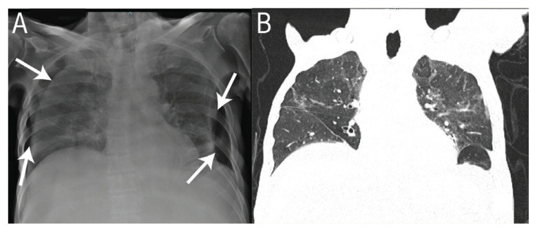
A 73-year-old male patient with unresectable, advanced dedifferentiated retroperitoneal liposarcoma being treated with palliative chemotherapy was admitted to a tertiary care hospital in Muscat, Oman, in 2020 with early satiety, poor oral intake, dehydration and ascites. A routine chest radiograph showed features of bilateral pneumothorax [Figure 1A]. However, at the time of the presumed radiological diagnosis, the patient was neither breathless nor desaturating and was haemodynamically stable. A computed tomography scan was done suspecting an extra thoracic shadow, such as a skin fold, which showed well expanded lungs without any evidence of pneumothorax [Figure 1B]. The patient and the relatives were reassured and no intervention was done. Consent was obtained from the patient to publish the details of his illness as well as his radiology images.
Figure 1.
A: Chest radiograph of a 73-year-old male patient showing features suggestive of pneumothorax bilaterally with curvilinear lines (arrows) mimicking collapsed lung borders. B: Computed tomography of the chest showing fully expanded lungs without any evidence of pneumothorax.
Comment
The curved shadow of skin folds can mimic the visceral pleural margin and can often be misinterpreted as a pneumothorax leading to unnecessary interventions.1 The area lateral to this sharp margin can often be perceived darker than the lung medial to it.2 Optical illusions such as Mach band effect are often helpful in demarcating the boundaries of anatomic structures with different optical densities on radiographs.3 However, they sometimes can also be mistaken for disease as was seen in the current case where a negative Mach band effect near a skin fold suggested pneumothorax. Pseudo-pneumothorax is usually seen when the film cassette is kept behind the patient with loose skin in a sitting or supine position. The features differentiating this artefact includes, a broader opaque density that fades medially, not following the expected border of the separated visceral pleura, terminating abruptly, extending beyond the pleural space over the chest wall and maybe more than one skin fold with two or more parallel lines.4 The absence of lung markings beyond this sharp curvilinear line as a differentiating feature may, at times, be limited as in the current case. Repeating the chest radiograph or a thoracic ultrasound are useful tools, but may not always resolve the dubious radiographic findings. The radiograph may still show the artefact and the ultrasound is accurate only when used by skilled operators.4 Computed tomography of the chest is the most sensitive and specific test for diagnosis of pneumothorax. In addition to the skin folds, the pleural line can also be mimicked by clothing or bed sheet folds, oxygen reservoir masks, elevated hemidiaphragm, rib or scapular borders, lung blebs or colonic interposition. These artefacts, when misinterpreted as pneumothorax, can lead to unnecessary and often catastrophic interventions and pneumothorax should be ruled out before any therapeutic procedure, especially when the clinical suspicion is low.
Footnotes
AUTHORS’ CONTRIBUTION
All authors were involved in patient care. JB wrote the initial manuscript draft and finalised the submission. AJ, SM and JA contributed to the literature review. All authors reviewed and approved the final version of the manuscript.
References
- 1.Niazi AK, Minko P, Nahrstedt CJ, Morris AR, Saha PJ, Elliott K, et al. A Case of Pseudo-pneumothorax with Complications. Cureus. 2018;10:e3263. doi: 10.7759/cureus.3263. [DOI] [PMC free article] [PubMed] [Google Scholar]
- 2.Kishimoto K, Watari T, Tokuda Y. Pseudo-pneumothorax: Skin fold is an excellent imitator. BMJ Case Rep. 2018;2018:bcr-2018226360. doi: 10.1136/bcr-2018-226360. [DOI] [PMC free article] [PubMed] [Google Scholar]
- 3.Buckle CE, Udawatta V, Straus CM. Now you see it, now you don't: Visual illusions in radiology. Radiographics. 2013;33:2087–102. doi: 10.1148/rg.337125204. [DOI] [PubMed] [Google Scholar]
- 4.Kattea MO, Lababede O. Differentiating Pneumothorax from the Common Radiographic Skinfold Artifact. Ann Am Thorac Soc. 2015;12:928–31. doi: 10.1513/AnnalsATS.201412-576AS. [DOI] [PubMed] [Google Scholar]


