Abstract
Objectives
This study aims to analyze whether the lateral posterior condylar offset (LPCO) and lateral posterior tibial slope (LPTS) values are associated with the presence of fabella by evaluating the frequency of fabella, its location, and whether it is bilateral and the relationship of the fabella with age, sex, and the presence of osteoarthritis.
Patients and methods
Between January 2016 and December 2020, computed tomography (CT) scans including 1,952 knee regions of 1,220 patients (861 males, 359 females; mean age: 54.5±19.7 years; range, 10 to 98 years) were retrospectively analyzed. Age, sex, and the presence of fabella whether unilateral (left or right) or bilateral were recorded. Of the patients with a bilateral knee CT, those with fabella on one side and without on the other were studied further to investigate the effect of fabella on the posterolateral corner (PLC). In these patients, the LPCO and LPTS values, presence of knee osteoarthritis, fabella-femoral distance, and sagittal anterior-posterior diameter of the fabella were evaluated.
Results
While there was no evidence of fabella in 867 (71.1%) patients, it was present in 353 (28.9%) patients. The linear correlation analysis revealed that the correlations between the right LPCO and the right LPTS (r=-0.295; p<0.001) and between the left LPCO and the left LPTS (r=-0.574; p<0.001) were significant. It was observed that LPTS decreased with increasing LPCO. According to the results of the point biserial correlation analysis, there was a significant correlation between the presence of fabella on the right side and the right LPCO value (r=-0.643; p<0.001) and between the presence of fabella on the left side and the left LPCO (r=-0.284; p=0.024). When the two knees were compared, fabella was less present in the knee whose LPCO was higher than that of the other knee, whereas it was more common in the knee whose LPCO was lower than that of the other knee. We found a significant correlation between each side's fabella and LPCO values and between the presence of fabella on the left side and the left LPTS.
Conclusion
The presence of fabella in the knee joint may be associated with LPCO and LPTS values of the knee. The comparison of the two knees of the same patient may reveal that if a fabella is present in a knee, the LPCO value of that knee is lower than that of the other knee. We believe that the reason for this is that the presence of fabella increases the distance to the center of rotation of the knee joint.
Keywords: Fabella, posterior condylar offset, posterior tibial slope, sesamoid bones.
Introduction
The posterolateral corner (PLC) consists of many different structures that act as static and dynamic stabilizers of the knee during motion.[1]
The posterolateral corner (PLC) consists of many different structures that act as static and dynamic stabilizers of the knee during motion.[2] The anatomy and function of PLC are still poorly understood.[3] The lateral posterior condylar offset (LPCO) and the lateral posterior tibial slope (LPTS) are important for understanding the function of bony structures in the anatomy of the PLC.[4] The fabella is thought to act as a stabilizer of the knee.[5] The fabella is a sesamoid bone located at the lateral head of the gastrocnemius muscle, plays a role in stabilizing the knee joint, and articulates directly with the lateral femoral condyle.[6] Together with the arcuate ligament, the popliteal oblique ligament, fabellofibular ligament, and fabella support the PLC of the knee.[7]
Studies on the fabella in the literature have focused on the prevalence and treatment of fabella syndrome.[8-10] Since there is no study examining the PLC bone morphology and its relationship with the fabella, the function of the fabella has not been fully elucidated, yet.
The fabella is the only bone in the human body whose prevalence has increased in the last century.[11] In general, the function of sesamoid bones is to reduce pressure and friction, redirect the direction of muscle movement, increase the mechanical strength of the muscle, or increase the strength of the tendon or ligament.[5,12] Several studies investigating the prevalence of the fabella in the general population have attempted to identify its prevalence using surgical/section reports, radiographs, and computed tomography (CT) and magnetic resonance imaging (MRI) scans.[8,11,13] The fabella occurs in 10 to 30% of the population and is mostly bilateral.[8,12]
The fabella is a sesamoid bone, and sesamoid bones usually act as amplifiers for the moment arm.[11] Due to this feature of sesamoid bones, we hypothesized that LPCO and LPTS values might be affected by the presence of the fabella. In the present study, we, therefore, aimed to analyze whether LPCO and LPTS values were related to the fabella and to present the prevalence rate of the fabella using a CT dataset. In addition, we aimed to investigate the normative values of LPCO and LPTS in the normal population, their differences between individuals and, most importantly, whether there was a relationship between LPCO and LPTS.
Patients and Methods
This single-center, retrospective study was conducted at Tokat Gaziosmanpaşa University, Department of Orthopedics and Traumatology between January 2016 and December 2020. All CT images containing the were collected from the electronic medical record system. The Picture Archiving and Communication Systems (PACS) software was used for all reconstructions and measurements (Sectra Workstation IDS7, version 21.2.11.6289, ©2019 Sectra AB).
There were a total of 1,445 patients with CT image of the knee region. In 225 patients, the knee region could not be assessed due to prostheses and various implants. The CTs including 1,952 knee regions of 1,220 patients (861 males, 359 females; mean age: 54.5±19.7 years; range, 10 to 98 years) were enrolled. Patients were excluded from the study, if they had an implant interfering with a clear assessment of the knee or if the quality of the image was poor. Data including age, sex, and the presence of the fabella were recorded. The patients were divided into six groups according to age (<25, 26-35, 36-45, 46-55, 56-65, >65 years). In the study, the CTs that were previously taken for routine follow-up and treatment of the patients were used. Since the CTs evaluated were not specifically taken for this study, the patients were exposed to no additional dose of radiation.
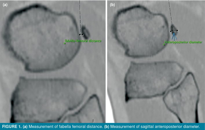
Of the patients with a bilateral knee CT, those with fabella on one knee, but not on the other were studied further to investigate the effect of the fabella on the PLC. In these patients, the LPCO and LPTS values, presence of knee osteoarthritis, fabellafemoral distance, and sagittal anteroposterior diameter of the fabella were determined (Figure 1).
Figure 1. (a) Measurement of fabella femoral distance. (b) Measurement of sagittal anteroposterior diameter.
The Kellgren-Lawrence classification was used for osteoarthritis classification; Grade 1: suspected narrowing of the joint space with possible osteophyte formation, Grade 2: possible narrowing of the joint space with definite osteophyte formation, Grade 3: definite joint space narrowing, moderate osteophyte formation, some sclerosis, and possible deformity of the bony ends and Grade 4: large osteophyte formation, severe narrowing of the joint space with significant sclerosis and definitive deformity of the bone ends.[14]
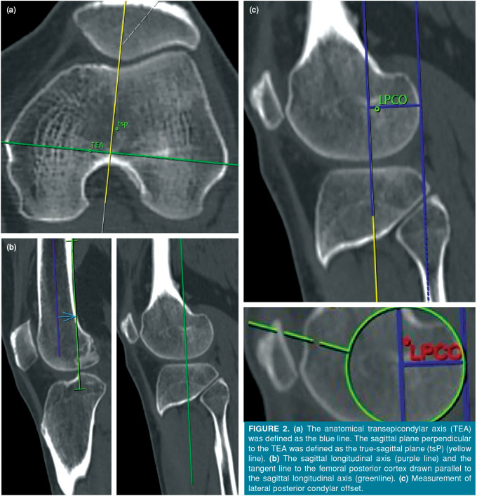
The LPCO measurements were performed according to previously published protocols.[14] The line connecting the most prominent points of the femoral epicondyles on the axial CT was defined as the transepicondylar axis (TEA). The true sagittal plane (tsP) was defined as the sagittal plane perpendicular to the TEA (Figure 2a).[15]
Figure 2. . (a) The anatomical transepicondylar axis (TEA) was defined as the blue line. The sagittal plane perpendicular to the TEA was defined as the true-sagittal plane (tsP) (yellow line). (b) The sagittal longitudinal axis (purple line) and the tangent line to the femoral posterior cortex drawn parallel to the sagittal longitudinal axis (greenline). (c) Measurement of lateral posterior condylar offset.
While the axis perpendicular to the line drawn along the TEA in the axial reformatted CT image was located at the center of the two condyles, the sagittal image was evaluated on CT. In the sagittal reformatted image, two separate points were detected on the distal femur 5-cm apart. The midpoint of the femur was found in these detected regions. By combining these two points, the sagittal line of the femur was determined (Figure 2b).[8] A new line was drawn parallel to the sagittal line of the femur and tangential to the posterior femoral cortex (Figure 2b). Then, in the axial reformatted CT image, the axis was shifted perpendicular to the line, along with the TEA, to the center of the lateral condyle. In the sagittal CT section, the circumferentially-located largest circle of the lateral posterior condyle was determined. The LPCO was determined by measuring the vertical distance between the line previously drawn tangential to the posterior cortex and the drawn circle (Figure 2c).
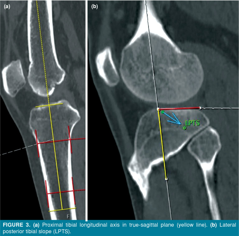
For LPTS measurement, two points of 5 cm and 15 cm distal to the knee joint were determined on the sagittal axis of the tibia (Figure 3a). The midpoint of the lines connecting the anteroposterior cortical lines was found (Figure 3a).[16] The angle between the line connecting these two points and the tangential line of the posterior tibial plateau was determined as the LPTS (Figure 3b).[10,11]
Figure 3. (a) Proximal tibial longitudinal axis in true-sagittal plane (yellow line). (b) Lateral posterior tibial slope (LPTS).
All measurements were performed by two experienced orthopedic surgeons at different times. One of the two orthopedists conducted the measurements again one month later. The results were obtained by averaging the measurements taken at different times.
Statistical analysis
Statistical analysis was performed using the IBM SPSS version 23.0 software (IBM Corp., Armonk, NY, USA). The Kolmogorov-Smirnov test was used to evaluate the distribution of the data. Descriptive data were presented in mean ± standard deviation (SD), median and interquartile range (IQR) or number and frequency, where applicable. The Student t-test was used to compare the parametric data, while the Mann-Whitney U test was performed to compare the non-parametric data. The Pearson and Spearman correlation analyses were conducted to identify the relationship between the condylar offset and tibial slope. The chi-square test, Fisher exact test, and Fisher-Freeman-Halton test were used to evaluate the categorical variables. A logistic regression model was established in which age and sex were considered possible predictors of the presence of the fabella. A p value of <0.05 was considered statistically significant.
Results
Bilateral CT were performed in 732 patients. Computed tomography was only performed on the right knee in 253 patients and on the left knee in 235 patients.
A total of 353 (28.9%) patients had fabella, while there was no evidence of fabella in 867 (71.1%) patients (Table I). Although fabella was more common in females (32.6%) than males (27.4%), this was not statistically significant (p=0.069). Considering all knee CT scans, fabella was absent in 1,361 (69.7%) knees, while it was observed in 591 knees (30.3%). The patients with bilateral fabellae were on average 65.7 years old, while those without bilateral fabella were on average 59.38 years old (p<0.001). The presence of the fabella among the age groups are given in Table II.
Table 1. Demographic and clinical characteristics of patients.
| n | % | Mean±SD | Range | 95% CI | |
| Age (year) | 54.5±19.7 | 10-98 | 53.4-55.7 | ||
| Sex | |||||
| Female | 359 | 29.4 | N/A | N/A | |
| Male | 861 | 70.6 | N/A | N/A | |
| Presence of the fabella in those who have bilateral knee CT | |||||
| Bilateral | 170 | 23.2 | N/A | N/A | |
| Right | 198 | 27.0 | N/A | N/A | |
| Left | 210 | 28.7 | N/A | N/A | |
| Presence of the fabella in those who have unilateral knee CT | |||||
| Right | 62 | 24.5 | N/A | N/A | |
| Left | 53 | 22.6 | N/A | N/A | |
| Presence of the fabella in all patients | 353 | 28.9 | N/A | N/A | |
| SD: Standard deviation; CI: Confidence interval; CT: Computed tomography. | |||||
Table 2. Presence of fabella among age groups.
| Unilateral CT | Bilateral CT | Total | ||||
| Right | Left | Right | Left | Right | Left | |
| Age groups (year) | ||||||
| <25 | 4 | 4 | 5 | 6 | 14 | 16 |
| 26-35 | 7 | 8 | 7 | 7 | 21 | 22 |
| 36-45 | 5 | 5 | 4 | 5 | 13 | 15 |
| 46-55 | 13 | 7 | 28 | 30 | 69 | 67 |
| 56-65 | 15 | 12 | 39 | 49 | 93 | 110 |
| >65 | 18 | 17 | 115 | 113 | 248 | 243 |
| CT: Computed tomography. | ||||||
The results of binary logistic regression analysis showed that fabella was observed more frequently with increasing age (hazard ratio [HR]: 1.015; 95% confidence interval [CI]: 1.000-1.032; p<0.001). Sex alone was not significant in this model. However, when we created a model in which age and sex were combined, more fabella were seen in older females (HR: 1.032; 95% CI: 1.001-1.033, p=0.032). Of 67 patients with bilateral knee CTs, one side with fabella, and other side without, four patients were excluded from the study, as healthy measurements of LPCO and LPTS could not be performed.
In the patients whose bilateral knee CTs were available, who were with fabella only on one side, there were 21 females and 42 males. There was no statistically significant difference between the patients with and without fabella in terms of the presence of gonarthrosis (Table III). Fabella was present on the right side in 27 patients and on the left side in 36 patients. The mean age was 61.2± 13.6 (range, 21 to 92) years, the mean sagittal anteroposterior diameter was 4.5±1.07 (range, 2 to 7.3) mm, the mean fabella-femoral distance was 2.3±0.55 (range, 1.2 to 5.1) mm, the mean right LPCO was 24.6±1.8 (range, 21.7 to 29.6) mm, the mean left LPCO was 24.3±1.7 (range, 19.4 to 28.9) mm, the mean right LPTS was 5.6±1.8 (range, 2.2 to 10.9), and the mean left LPTS was 5.7±1.8 (range, 2.1 to 10.8). Inter-observer and intra-observer compatibility of measurements were found to be excellent (Table IV).
Table 3. Comparison between unilateral fabella groups.
| Presence of Fabella | |||
| Right (n=27) | Left (n=36) | p | |
| Sex | 0.105 | ||
| Female | 12 | 9 | |
| Male | 15 | 27 | |
| Right gonarthrosis* | 0.456 | ||
| Grade 0 | 2 | 8 | |
| Grade 1 | 12 | 16 | |
| Grade 2 | 7 | 8 | |
| Grade 3 | 5 | 3 | |
| Grade 4 | 1 | 1 | |
| Left gonarthrosis* | 0.302 | ||
| Grade 0 | 2 | 8 | |
| Grade 1 | 17 | 16 | |
| Grade 2 | 5 | 9 | |
| Grade 3 | 2 | 3 | |
| Grade 4 | 1 | 0 | |
| * Fisher-Freeman-Halton test was used; Chi-square test was used; Kellgren- Lawrence classification was used. | |||
Table 4. Reliability analysis.
| Measurement | Intraobserver reliability | Interobserver reliability |
| Right LPCO | 0.97 | 0.94 |
| Left LPCO | 0.98 | 0.90 |
| Right LPTS | 0.98 | 0.94 |
| Left LPTS | 0.95 | 0.89 |
| LPCO: Lateral posterior condylar offset; LPTS: Lateral posterior tibial slope; All reliability data were assessed with intraclass correlation coefficient. | ||
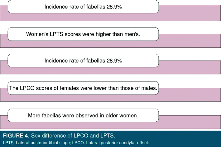
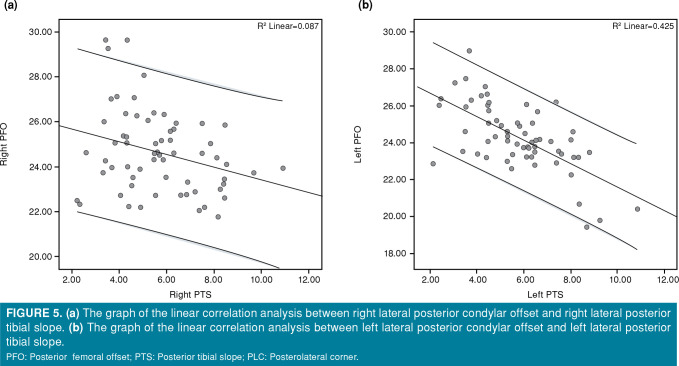
The LPTS values were higher in females than males. The LPCO values of females were lower than those of males (Table V, Figure 4). The linear correlation analysis results indicated that there was a negative correlation between the right LPCO and the right LPTS (r=-0.295; p<0.001) (Figure 5a) and between the left LPCO and the left LPTS (r=-0.574; p<0.001) (Figure 5b).
Table 5. Comparison of offset and slope between sex groups.
| Sex | p | ||||||
| Female (n=21) | Male (n=42) | ||||||
| Mean±SD | Range | 95% CI | Mean±SD | Range | 95% CI | ||
| Right LPCO | 23.7±1.3 | 21.7-27.1 | 23.1-24.3 | 25.0±1.8 | 22.1-29.6 | 24.4-25.6 | 0.006 |
| Left LPCO | 24.0±1.8 | 19.4-26.6 | 23.1-24.8 | 24.4±1.7 | 19.8-28.9 | 23.9-24.9 | 0.371 |
| Right LPTS | 6.5±1.9 | 3.3-10.9 | 5.7-7.4 | 5.2±1.7 | 2.2-9.6 | 4.6-5.7 | 0.006 |
| Left LPTS | 6.4±1.7 | 4.2-10.8 | 5.6-7.2 | 5.4±1.7 | 2.1-9.2 | 4.8-5.9 | 0.034 |
| CI: Confidence interval; LPCO: Lateral posterior condylar offset; LPTS: Lateral posterior tibial slope. | |||||||
Figure 4. Sex difference of LPCO and LPTS. LPTS: Lateral posterior tibial slope; LPCO: Lateral posterior condylar offset.
Figure 5. (a) The graph of the linear correlation analysis between right lateral posterior condylar offset and right lateral posterior tibial slope. (b) The graph of the linear correlation analysis between left lateral posterior condylar offset and left lateral posterior tibial slope. PFO: Posterior femoral offset; PTS: Posterior tibial slope; PLC: Posterolateral corner.
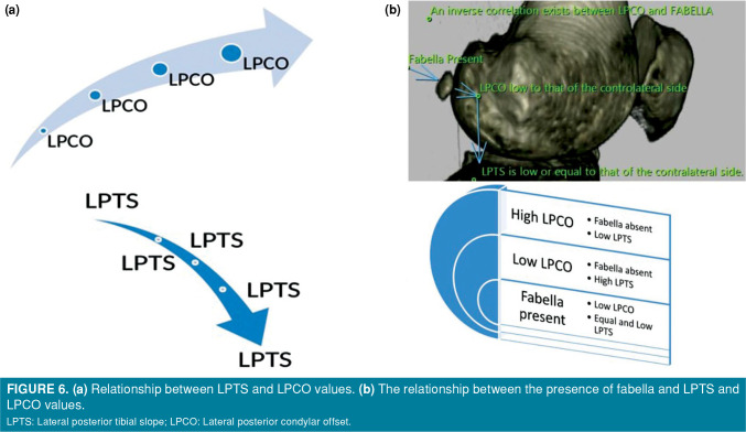
There was a significant correlation between the presence of fabella on the right side and the right LPCO (r=-0.643; p<0.001) and between the presence of fabella on the left side and the left LPCO (r=-0.284; p=0.024). The patients with fabella had lower LPCO values at the ipsilateral extremity. There was no significant correlation between the right fabella and the right LPTS (r=-0.149; p=0.244), while there was a significant correlation between the left fabella and the left LPTS (r=-0.273; p=0.031). The patients with left fabella had lower LPTS values on the left knee (Figure 6).
Figure 6. (a) Relationship between LPTS and LPCO values. (b) The relationship between the presence of fabella and LPTS and LPCO values. LPTS: Lateral posterior tibial slope; LPCO: Lateral posterior condylar offset.
Discussion
The prevalence of fabella varies according to age and sex.[16] The overall frequency of fabella in the patients evaluated in the present study was 28.9%. We found that the LPCO values showed more variability in the presence of fabella than the LPTS values. The knee with fabella had a lower LPCO; however, the LPTS value was lower or equal to that of the other side. When LPTS and LPCO were compared, an inverse correlation was found between the two parameters. There are few studies in the literature investigating normative data for posterior condylar offset (PCO) and posterior tibial slope (PTS).[4,17] Our study provides important normative data on LPCO, LPTS, and fabella.[18] These normal values not only explain the relationship between PCO and PTS, but also show the influence of fabella on this relationship.
In their study using CT scans, Hauser et al.[5] reported the prevalence of the fabella to be 30% in the Central European population. We found that the fabella was present in 28.9% of the patients. The prevalence of fabella varies according to ethnicity and geographic distribution.[19] Indeed, it even varies in different regions of the same country. Previous studies conducted in Turkey utilized radiographs and MRI of the knee to find fabella.[20] We, on the other hand, conducted our study using CT examinations, which best showed the bone structure. Unluturk et al.’s[13] study in which 1,000 patients were examined by MRI, the prevalence was 15.5%, and no sex difference was found. In our study, although no sex difference was found, the frequency of fabella increased in older females in the regression model including age and sex. Sari et al.[20] used radiographs in their studies and found that the overall prevalence of fabella was 24.3%, with a frequency of 25.8% in females and 20.6% in males. We can explain the reason for the high prevalence found in our study by the fact that the examination we used was CT, and that there were regional differences. Several studies have been conducted to identify the prevalence of fabella using imaging techniques. Some researchers have used radiographs in detecting the presence of fabella, while some others have utilized MRI.[8,13] The prevalence of fabella has been reported to be higher in cadaveric studies than in radiological studies.[21-23] Of note, no previous study has examined such a large population of knee CTs as ours. A systematic search of the literature revealed that there were few studies on the prevalence of fabella based on CT scans. One such study was conducted by Berthaume et al.[11] on 212 CT scans in which 94 fabellae were detected, with a prevalence of 44.34% for knees. Moreover, the authors reported a prevalence of 41.18% in males and 47.27% in females.
In our study, in which both knees of the patients were examined, we believe that the reason for low LPCO in knees with the presence of fabella is to compensate for the distance between tendons and ligaments, such as the popliteal posterolateral ligament, from the sagittal center of rotation of the knee. This is because sesamoid bones often act as amplifiers of the moment arm. This finding confirms the effects of increasing the mechanical muscle strength of sesamoid bones or increasing tendon or ligament strength.
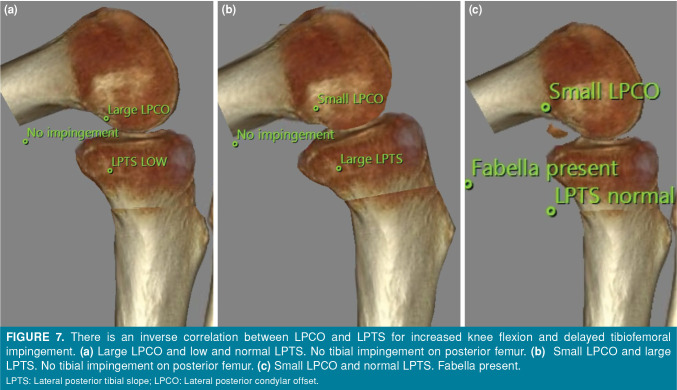
It has been reported in the literature that increased PCO and PTS values are associated with the increased flexion range of motion in prosthetic knees, while decreased PCO and PTS are associated with decreased range of motion.[2,24,25] Weinberg et al.[26] predicted an inverse correlation between PCO and PTS, and found that there was no impingement when the PCO was large and the PTS was normal, and that posterior tibial impingement occurred when the PCO was small and the PTS was normal. They also reported that when the PCO was small and the PTS was high, there was no impingement. However, these authors did not compare the relationship between the presence of fabella and PLC in their study.
Fabella is not observed in all individuals and its prevalence is 28.9%. Moreover, while fabella is present in one knee of an individual, it may not be seen in the other knee. In our study, among the patients with bilateral knee CT scans, the rate of those with fabella in only one knee was 9.1%. Since LPCO and LPTS values greatly vary in the population, the evaluation and comparison of both knees in the same patient is the main strength of our study. The sagittal geometry of the knee is designed to balance natural flexion range of motion with stability (Figure 7). Decreased LPCO and LPTS are probably associated with reduced motion and impingement due to mechanical impact in the posterior. Therefore, in most individuals, the inverse correlation between LPCO and LPTS, as we determined in our study, serves as a reconciliation of both functions. Regarding the effect of the presence of fabella on LPCO and LPTS, we detected that on the side where the fabella was present, LPCO was low and LPTS was normal or low (Figure 7). The presence of fabella in the posterolateral capsule helps to provide normal knee flexion range by reducing the adverse effect of decreased LPCO without posterolateral tibiofemoral contact and impingement. Fabella compensates for decreased LPCO in patients with low LPCO without the need for the tibial slope to excessively increase as a compensatory. Taken together, the presence of the fabella prevents tibial compression in the posterior femur by increasing the distance of the posterolateral soft tissue from the rotation center of the knee joint.
Figure 7. There is an inverse correlation between LPCO and LPTS for increased knee flexion and delayed tibiofemoral impingement. (a) Large LPCO and low and normal LPTS. No tibial impingement on posterior femur. (b) Small LPCO and large LPTS. No tibial impingement on posterior femur. (c) Small LPCO and normal LPTS. Fabella present. LPTS: Lateral posterior tibial slope; LPCO: Lateral posterior condylar offset.
Nonetheless, this study has some limitations. Additional regional populations can be included in future studies to compare regional differences. Also, the absence of knee range of motion values and the clinical evaluation of the PLC are the other limitations.
In conclusion, when the condylar offset decreases, LPTS increases to avoid impingement in flexion. Based on the data we obtained in our study, as a consequence of a low LPCO, tendons and ligaments in the PLC approach the center of rotation of the joint, which, in turn, results in higher energy requirements. We consider that the presence of fabella increases the distance of the tendons and ligaments to the center of rotation of the joint and allows the tendons and ligaments in the PLC to work with less energy rather than the higher energy produced by the joint. When LPCO is low and LPTS is normal/low, it is likely that tibia posteriorly compress to the posterior of the femur yielding impingement. When LPCO and LPTS are low, it can be speculated that the presence of fabella ensures no impingement during knee flexion. To the best of our knowledge, there are no studies in the literature assessing the relation of LPTS and LPCO with the presence or absence of fabella. Further prospective studies using knee MRI taken while the knee is in full flexion are needed to evaluate the relationship of the fabella, LPCO and LPTS with the PLC, and impingement.
Footnotes
Conflict of Interest: The authors declared no conflicts of interest with respect to the authorship and/or publication of this article.
Author Contributions: All authors declare that they have all participated in the design, execution, and analysis of the paper, and that they have approved the final version.
Financial Disclosure: The authors received no financial support for the research and/or authorship of this article.
References
- 1.Ranawat A, Baker CL 3rd, Henry S, Harner CD. Posterolateral corner injury of the knee: evaluation and management. J Am Acad Orthop Surg. 2008;16:506–518. [PubMed] [Google Scholar]
- 2.Bellemans J, Robijns F, Duerinckx J, Banks S, Vandenneucker H. The influence of tibial slope on maximal flexion after total knee arthroplasty. Knee Surg Sports Traumatol Arthrosc. 2005;13:193–196. doi: 10.1007/s00167-004-0557-x. [DOI] [PubMed] [Google Scholar]
- 3.Mehta N, Nayak T. From controversy to contemporary: A narrative review of the anatomy and biomechanics of the posterolateral corner of the knee. Arthroscopy and Orthopedic Sports Medicine. 2021;8:1–8. [Google Scholar]
- 4.Weinberg DS, Gebhart JJ, Wera GD. An anatomic investigation into the relationship between posterior condylar offset and posterior tibial slope of one thousand one hundred thirty-eight cadaveric knees. J Arthroplasty. 2017;32:1659–1664. doi: 10.1016/j.arth.2016.12.022. [DOI] [PubMed] [Google Scholar]
- 5.Hauser NH, Hoechel S, Toranelli M, Klaws J, Müller-Gerbl M. Functional and structural details about the fabella: What the important stabilizer looks like in the Central European population. Biomed Res Int. 2015;2015:343728–343728. doi: 10.1155/2015/343728. [DOI] [PMC free article] [PubMed] [Google Scholar]
- 6.Tabira Y, Saga T, Takahashi N, Watanabe K, Nakamura M, Yamaki K. Influence of a fabella in the gastrocnemius muscle on the common fibular nerve in Japanese subjects. Clin Anat. 2013;26:893–902. doi: 10.1002/ca.22153. [DOI] [PubMed] [Google Scholar]
- 7.Minowa T, Murakami G, Kura H, Suzuki D, Han SH, Yamashita T. Does the fabella contribute to the reinforcement of the posterolateral corner of the knee by inducing the development of associated ligaments. J Orthop Sci. 2004;9:59–65. doi: 10.1007/s00776-003-0739-2. [DOI] [PubMed] [Google Scholar]
- 8.Egerci OF, Kose O, Turan A, Kilicaslan OF, Sekerci R, Keles-Celik N. Prevalence and distribution of the fabella: A radiographic study in Turkish subjects. Folia Morphol (Warsz) 2017;76:478–483. doi: 10.5603/FM.a2016.0080. [DOI] [PubMed] [Google Scholar]
- 9.Robertson A, Jones SC, Paes R, Chakrabarty G. The fabella: A forgotten source of knee pain. Knee. 2004;11:243–245. doi: 10.1016/S0968-0160(03)00103-0. [DOI] [PubMed] [Google Scholar]
- 10.Weiner DS, Macnab I. The “abella syndrome”: An update. J Pediatr Orthop. 1982;2:405–408. [PubMed] [Google Scholar]
- 11.Berthaume MA, Di Federico E, Bull AMJ. Fabella prevalence rate increases over 150 years, and rates of other sesamoid bones remain constant: A systematic review. J Anat. 2019;235:67–79. doi: 10.1111/joa.12994. [DOI] [PMC free article] [PubMed] [Google Scholar]
- 12.Berthaume MA, Bull AMJ. Human biological variation in sesamoid bone prevalence: The curious case of the fabella. J Anat. 2020;236:228–242. doi: 10.1111/joa.13091. [DOI] [PMC free article] [PubMed] [Google Scholar]
- 13.Unluturk O, Duran S, Yasar Teke H. Prevalence of the fabella and its general characteristics in Turkish population with magnetic resonance imaging. Surg Radiol Anat. 2021;43:2047–2054. doi: 10.1007/s00276-021-02817-3. [DOI] [PubMed] [Google Scholar]
- 14.Cinotti G, Sessa P, Ripani FR, Postacchini R, Masciangelo R, Giannicola G. Correlation between posterior offset of femoral condyles and sagittal slope of the tibial plateau. J Anat. 2012;221:452–458. doi: 10.1111/j.1469-7580.2012.01563.x. [DOI] [PMC free article] [PubMed] [Google Scholar]
- 15.Balcarek P, Hosseini ASA, Streit U, Brodkorb TF, Walde TA. Sagittal magnetic resonance imaging-scan orientation significantly influences accuracy of femoral posterior condylar offset measurement. Arch Orthop Trauma Surg. 2018;138:267–272. doi: 10.1007/s00402-017-2838-0. [DOI] [PubMed] [Google Scholar]
- 16.Yoo JH, Chang CB, Shin KS, Seong SC, Kim TK. Anatomical references to assess the posterior tibial slope in total knee arthroplasty: A comparison of 5 anatomical axes. J Arthroplasty. 2008;23:586–592. doi: 10.1016/j.arth.2007.05.006. [DOI] [PubMed] [Google Scholar]
- 17.Bao L, Rong S, Shi Z, Wang J, Zhang Y. Measurement of femoral posterior condylar offset and posterior tibial slope in normal knees based on 3D reconstruction. BMC Musculoskelet Disord. 2021;22:486–486. doi: 10.1186/s12891-021-04367-6. [DOI] [PMC free article] [PubMed] [Google Scholar]
- 18.Atik OŞ. What are the expectations of an editor from a scientific article. Jt Dis Relat Surg. 2020;31:597–598. doi: 10.5606/ehc.2020.57896. [DOI] [PMC free article] [PubMed] [Google Scholar]
- 19.Asghar A, Naaz S, Chaudhary B. The ethnic and geographical distribution of fabella: A systematic review and meta-analysis of 34,733 Knees. e14743Cureus. 2021;13 doi: 10.7759/cureus.14743. [DOI] [PMC free article] [PubMed] [Google Scholar]
- 20.Sarı A, Dinçel YM, Çetin MÜ, Gunaydin B, Guney M. The prevalence of fabella in Turkish population and the association between the presence of fabella and osteoarthritis. SN Compr Clin Med. 2021;3:805–811. [Google Scholar]
- 21.Tatagari V, Brehman E, Adams CS. Evaluation of the gross anatomical incidence of fabellae in a north american cadaveric population. 2018. Available at: https://digitalcommons.pcom.edu/research_day/research_day_PA_2018/researchPA2018/49/2 .
- 22.Kawashima T, Takeishi H, Yoshitomi S, Ito M, Sasaki H. Anatomical study of the fabella, fabellar complex and its clinical implications. Surg Radiol Anat. 2007;29:611–616. doi: 10.1007/s00276-007-0259-4. [DOI] [PubMed] [Google Scholar]
- 23.Phukubye P, Oyedele O. The incidence and structure of the fabella in a South African cadaver sample. Clin Anat. 2011;24:84–90. doi: 10.1002/ca.21049. [DOI] [PubMed] [Google Scholar]
- 24.Antony J, Tetsworth K, Hohmann E. Influence of sagittal plane component alignment on kinematics after total knee arthroplasty. Knee Surg Sports Traumatol Arthrosc. 2017;25:1686–1691. doi: 10.1007/s00167-016-4098-x. [DOI] [PubMed] [Google Scholar]
- 25.Meric G, Gracitelli GC, Aram L, Swank M, Bugbee WD. Tibial slope is highly variable in patients undergoing primary total knee arthroplasty: Analysis of 13,546 computed tomography scans. J Arthroplasty. 2015;30:1228–1232. doi: 10.1016/j.arth.2015.02.012. [DOI] [PubMed] [Google Scholar]
- 26.Weinberg DS, Streit JJ, Gebhart JJ, Williamson DF, Goldberg VM. Important differences exist in posterior condylar offsets in an osteological collection of 1,058 femurs. J Arthroplasty. 2015;30:1434–1438. doi: 10.1016/j.arth.2015.02.027. [DOI] [PubMed] [Google Scholar]


