PLOS ONE logoLink to PLOS ONE
. 2022 Nov 10;17(11):e0276083. doi: 10.1371/journal.pone.0276083

Hyperbaric oxygen protects HT22 cells and PC12 cells from damage caused by oxygen-glucose deprivation/reperfusion via the inhibition of Nrf2/System Xc-/GPX4 axis-mediated ferroptosis

Chunxia Chen 1,*,#, Wan Chen 2,#, Xing Zhou 1,#, Yaoxuan Li 3, Xiaorong Pan 4,*, Xiaoyu Chen 1,*
Editor: Yoshiaki Tsuji5
PMCID: PMC9648730  PMID: 36355759

Abstract

This study was to investigate the protective effect of hyperbaric oxygen (HBO) on HT22 and PC12 cell damage caused by oxygen-glucose deprivation/reperfusion-induced ferroptosis. A 2-h oxygen-glucose deprivation and 24-h reperfusion model on HT22 and PC12 cells was used to simulate cerebral ischemia-reperfusion injury. Cell viabilities were detected by Cell Counting Kit-8 (CCK-8) method. The levels of reactive oxygen species (ROS) and lipid reactive oxygen species (Lipid ROS) were detected by fluorescent probes Dihydroethidium (DHE) and C11 BODIPY 581/591. Iron Colorimetric Assay Kit, malondialdehyde (MDA) and glutathione (GSH) activity assay kits were used to detect intracellular iron ion, MDA and GSHcontent. Cell ferroptosis-related ultrastructures were visualized using transmission electron microscopy (TEM). Furthermore, PCR and Western blot analyses were used to detect the expressions of ferroptosis-related genes and proteins. After receiving oxygen-glucose deprivation/reperfusion, the viabilities of HT22 and PC12 cells were significantly decreased; ROS, Lipid ROS, iron ions and MDA accumulation occurred in the cells; GSH contents decreased; TEM showed that cells were ruptured and blebbed, mitochondria atrophied and became smaller, mitochondrial ridges were reduced or even disappeared, and apoptotic bodies appeared. And the expressions of Nrf2, SLC7A11 and GPX4 genes were reduced; the expressions of p-Nrf2/Nrf2, xCT and GPX4 proteins were reduced. Notably, these parameters were significantly reversed by HBO, indicating that HBO can protect HT22 cells and PC12 cells from damage caused by oxygen-glucosedeprivation/reperfusion via the inhibition of Nrf2/System Xc-/GPX4 axis-mediated ferroptosis.

Introduction

Adequate cerebral blood perfusion is a key factor in maintaining normal brain function. chronic cerebral hypoperfusion (CCH) also known as chronic cerebral ischemia (CCI), prolonged ischemia of the brain can trigger neurodegeneration and eventually lead to progressive cognitive impairment. Currently, reperfusion is an effective treatment for patients with acute cerebral ischemia. However, reperfusion under severe cerebral ischemia is a double-edged sword, which often induces cerebral ischemia-reperfusion injury while restoring blood supply [1], which seriously threatens the life of patients. Recent studies have found that cell ferroptosis caused by abnormal iron metabolism may be an important pathophysiological mechanism of cerebral ischemia-reperfusion injury [2,3].

Ferroptosis was first proposed by Dixon et al. [4] in 2012. It is an iron-dependent, a new form of cell death caused by the formation of lipid peroxides. Ferroptosis is significantly different from apoptosis, necrosis, and autophagy in terms of morphology, biochemical characteristics, and gene expression [5,6]. In recent years, studies have found that ferroptosis is closely related to the occurrence and development of neurodegenerative diseases and traumatic nervous system injury diseases [710], and nerve cell death is the main pathological event of many neurological diseases [1113]. Brain tissue is prone to attack by hydroxyl radicals due to the presence of a large amount of unsaturated fatty acids and low levels of antioxidant enzymes, such as glutathione peroxidase (GPX4) [14]. Therefore, neurological diseases are closely related to ferroptosis.

Hyperbaric oxygen (HBO) therapy is a clinical use of pure oxygen in the environment of 2–3 times atmospheric pressure to treat ischemic and hypoxic diseases, such as carbon monoxide poisoning, cerebral infarction, decompression sickness and coronary heart disease, etc., and has obtained certain curative effects. According to the US FDA and the International Society of Underwater Hyperbaric Medicine, 13 clinical indications including decompression sickness, carbon monoxide poisoning, gas embolism, acute craniocerebral injury, and post-radiotherapy tissue damage can be treated with HBO [15]. HBO treatment has a strong effect of reducing oxidative stress, a strong anti-inflammatory effect, protecting cholinergic nerves, and delaying neuronal apoptosis [16,17]. Our previous study [18] also found that hyperbaric oxygen can protect PC12 cells from damage caused by oxygen-glucose deprivation/reperfusion. However, whether this effect is related to the inhibition of ferroptosis remains unknown. Therefore, this study used the oxygen-glucose deprivation/reperfusion injury model of HT22 cells and PC12 cells to simulate cerebral ischemia-reperfusion in vitro to explore the effect of HBO on the ferroptosis pathway.

Materials and methods

Culture and treatment of cells

HT22 cells were obtained from American Type Culture Collection (ATCC, Maryland, USA) and cultured in medium consisting of Dulbecco’s modified Eagle’s medium (DMEM, Gibco, USA), 10% fetal bovine serum (FBS, Gibco, USA) and 1% penicillin-streptomycin (Solarbio, Beijing). PC12 cells (ATCC, Maryland, USA) were maintained in RPMI 1640 medium (Gibco, USA) supplemented with 10% horse serum (Merck, USA), 5% FBS and 1% penicillin-streptomycin. Cells were divided into control group, model group, Hyperbaric oxygen group (HBO) group and HBO+Ferrostatin-1 (HBO+F)group. Cells of the control group were untreated. Cells of the model group were cultured in medium without sugar and serum and then put into a 37°C incubator with a mixture of 95% N2 and 5% CO2 gas for 2 h for oxygen sugar deprivation. Then, the cells were cultured with complete medium in a 37°C incubator with a mixture of 95% O2 and 5% CO2 for 24 h. Cells of the HBO group were placed in a hyperbaric chamber (Yantai Hongyuan CO., Ltd) and received pure oxygen (0.25 MPa, 60 min). Cells of the HBO+F group were received pure oxygen and then cultured in medium with Ferrostatin-1 (5 μmol/L, ferroptosis inhibitor, MedChemExpresss, USA) for 24h. This study was conducted in accordance with the Declaration of Helsinki and approved by Ethics Committee of People’s Hospital of Guangxi Zhuang Autonomous Region.

Cell viability assay

Cell viability was determined using a Cell Counting Kit-8 (CCK8, Dojindo, Japan) assay. Briefly, after cells in each group received treatment accordingly, 10 μL of CCK-8 solution was added to each well and then the cells were incubated at 37°C in a 95% air/5% CO2 atmosphere for 1 h. The optical density of the plate was then measured at 450 nm by using a microplate reader.

Detection of iron content in cells

Intracellular iron content was measured by Iron Colorimetric Assay Kit (APPLYGEN, Beijing). Cells in each group were received treatment accordingly and washed twice with cold PBS. 200 μl of Mixture A (prepared according to the instructions) was added to each well and the cells were incubated at a 60°C water bath for 1 h. 60 μl iron ion detection reagent was added to each well and the cells were incubated at room temperature for 30 min. The optical density of the plate was then measured at 570 nm by using a microplate reader.

Detection of reactive oxygen species (ROS) in cells

Intracellular reactive oxygen species (ROS) level was measured by superoxide anion fluorescent probe (Dihydroethidium, DHE, Beyotime). Cells in each group were received treatment accordingly and washed twice with PBS. 1 ml of DHE diluted in DMEM was added to each well and the cells were incubated at 37°C in the dark for 20 min and then were washed with DMEM 3 times. The red fluorescence intensity was observed under a fluorescent inverted microscope and was calculated by Image J software.

Measurement of lipid reactive oxygen species (Lip-ROS) in cells

Lipid ROS level was determined using BODIPYTM 581 /591 C11 dye (GLPBIO). Cells in each group were received treatment accordingly and washed twice with PBS. The cells were added with 10μmol /L BODIPYTM 581 /591 C11 and incubated at 37°C in the dark for 30 min and then were washed with PBS 3 times. The green fluorescence intensity was observed under a fluorescent inverted microscope and was calculated by Image J software.

Detection of MDA and GSH content in cells

Cells in each group were received treatment accordingly. Then, individual levels of MDA and GSH in cells were measured using malondialdehyde (MDA) and glutathione (GSH) activity assay kits (Beyotime, China) respectively according to the Kit Instructions. Individual contents of MDA and GSH were measured at 450 and 405 nm, respectively, with a microplate reader.

Observation of cell ultrastructure by transmission electron microscopy (TEM)

Cell ferroptosis-related ultrastructures were visualized using TEM. Briefly, cells were treated accordingly and fixed with cold 3% glutaraldehyde for 2 hours. Cells were then embedded in 1% osmium tetroxide and dehydrated. Subsequently, they were soaked in acetone and embedding medium overnight. Finally, they were stained with uranyl acetate and lead citrate. The ultrastructures of cells were examined using a transmission electron microscope (Hitachi H-7650, Japan).

The relative quantitative real-time PCR analysis

Real-time qPCR was performed to detect the levels of ferroptosis-related genes. Total RNA was extracted from cells using a total RNA rapid extraction kit (Beyotime, China). Next, cDNA was synthesized from the RNA with a cDNA synthesis kit (ES Science, China). The quantity of the mRNA was measured using a Super SYBR qPCR Master Mix kit (ES Science, China) and was performed in an ABI Prism 7300 real-time thermocycler (Applied Biosystems, Foster City, CA, USA). GADPH was used as an internal reference. Results were calculated using the 2−ΔΔCt method. The primers sequence are shown in Table 1.

Table 1. PCR primer sequences.

Gene Forward primer (5’-3’) Reverse primer (5’-3’)
Nrf2 GTGGTTTAGGGCAGAAGG TCTTTCTTACTCTGCCTCTA
SLC7A11 GCATTCCCAGGGGCTAACAT AATTTCTCCCATGCGGGTGT
GPX4 CCGCTTATTGAAGCCAGCAC TATCGGGCATGCAGATCGAC
GAPDH CGTGTTCCTACCCCCAATGT TGTCATCATACTTGGCAGGTTTCT

Western blot analysis

Western blot was used to detect the expressions of ferroptosis-related proteins. After the interventional cells were collected, a cell protein lysate (Solarbio, Beijing, China) containing PMSF (Solarbio, Beijing, China) and a phosphatase inhibitor (CWBIO, Beijing, China) was added into the cells to extract cellular proteins, and BCA protein detection kit (Beyotime, Shanghai, China) was used to detect protein concentration. The protein samples were mixed with 4 × loading buffer at a volume of 3:1, and denatured by boiling at 100°C for 8 min. After electrophoresis on SDS-PAGE gel (10%), the proteins was transferred to PVDF membranes (ISEQ00010; Millipore, Billerica, MA, USA). Subsequently, the membranes were blocked with 5% nonfat dry milk for 2 h and then incubated with p-Nrf2, Nrf2, xCT and GPX4 antibody (1: 1 000) at 4 ℃ overnight. After washed with TBST, the membranes were incubated with the anti-rabbit IgG (H+L) secondary antibodies (1: 5 000) at room temperature for 1 h. The membranes were detected using Infrared dual-color fluorescence imaging detection system (LI-COR Odyssey CLx). GADPH is used as an internal reference.

Statistical analysis

SPSS 20.0 Statistical software was used for data analysis. Measurement data were expressed as mean ± standard deviation (n = 3). One-way analysis of variance (ANOVA) was performed to compare the differences among multiple groups and P<0.05 was deemed a statistically significant difference.

Results

Effects of HBO on viability in HT22 cells and PC12 cells

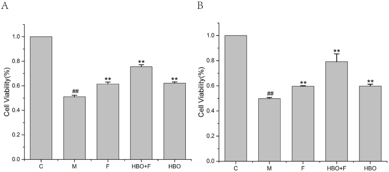
The CCK-8 assay showed that HBO enhances the viabilities of oxygen-glucose deprivation-reperfusion-injured HT22 cells and PC12 cells (Fig 1). Compared with the control group, the activities of HT22 cells and PC12 cells in the model group were significantly reduced. The activities of HT22 cells and PC12 cells in the HBO group and HBO+F group were significantly higher than the model group.

Fig 1. The effect of HBO on the viability of HT22 cells (A) and PC12 cells (B).

Fig 1

The results are presented as the mean ± SEM (N = 3). ##P < 0.01 vs. control group, **P < 0.01 vs. model group. C: Control group; M: Model group; HBO: Hyperbaric oxygen group; F: Ferrostatin-1 group; HBO+F: Hyperbaric oxygen + Ferrostatin-1 group.

Effects of HBO on iron content in HT22 cells and PC12 cells

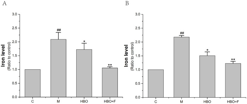
The iron ion detection experiment showed that compared with the control group, iron content accumulation occurred in HT22 cells and PC12 cells of the model group, while the iron ion content in the cells of the HBO group and HBO+F group was lower than that of the model group. The results showed that HBO could reverse oxygen-glucose deprivation-reperfusion-injured-induced increase in intracellular iron content (Fig 2).

Fig 2. The effect of HBO on iron content of HT22 cells (A) and PC12 cells (B).

Fig 2

The results are presented as the mean ± SEM (N = 3). ##P < 0.01 vs. control group, *P < 0.05, **P < 0.01 vs. model group. C: Control group; M: Model group; HBO: Hyperbaric oxygen group; HBO+F: Hyperbaric oxygen + Ferrostatin-1 group.

Effects of HBO on ROS and Lip-ROS in HT22 cells and PC12 cells

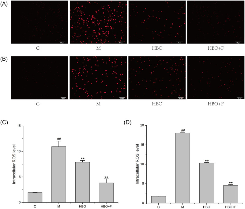
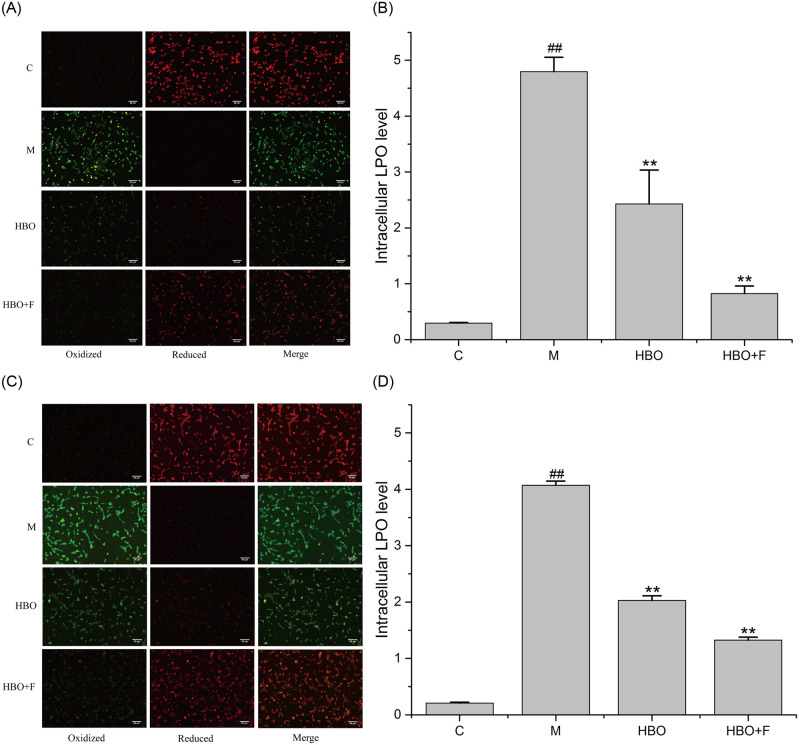
Fluorescent probe assay showed that compared with the control group, the ROS and Lipid ROS in the HT22 cells and PC12 cells in the model group were significantly increased, while the levels of ROS and Lipid ROS in the HBO group and HBO+F group were significantly lower than those in the model group. The result suggested that HBO can inhibit the generation of ROS and Lipid ROS in cells (Figs 3 and 4).

Fig 3. The effect of HBO on ROS of HT22 cells (A, C) and PC12 cells (B, D) (200×).

Fig 3

The results are presented as the mean ± SEM (N = 3). ##P < 0.01 vs. control group, **P < 0.01 vs. model group. C: Control group; M: Model group; HBO: Hyperbaric oxygen group; HBO+F: Hyperbaric oxygen + Ferrostatin-1 group. ROS: Reactive oxygen species.

Fig 4. The effect of HBO on Lipid ROS of HT22 cells (A, B) and PC12 cells (C, D) (200×).

Fig 4

The results are presented as the mean ± SEM (N = 3). ##P < 0.01 vs. control group, *P < 0.05, **P < 0.01 vs. model group. C: Control group; M: Model group; HBO: Hyperbaric oxygen group; HBO+F: Hyperbaric oxygen + Ferrostatin-1 group. LPO: Lipid reactive oxygen species.

Effects of HBO on MDA and GSH Content in HT22 cells and PC12 cells

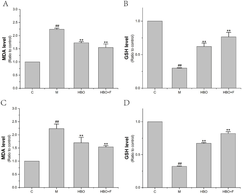
The experimental results showed that compared with the control group, the content of DMA in HT22 cells and PC12 cells in the model group was significantly increased, and the content of GSH in the cells was significantly decreased. After HBO treatment, compared with the model group, the content of MDA in the cells was significantly decreased, and the content of GSH in the cells was significantly increased (Fig 5).

Fig 5. The effect of MDA and GSH Content of HT22 cells (A, B) and PC12 cells (C, D).

Fig 5

The results are presented as the mean ± SEM (N = 3). ##P < 0.01 vs. control group, **P < 0.01 vs. model group. C: Control group; M: Model group; HBO: Hyperbaric oxygen group; HBO+F: Hyperbaric oxygen + Ferrostatin-1 group.

Effects of HBO on the ultrastructure of ferroptosis in HT22 cells and PC12 cells

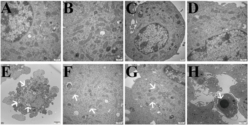
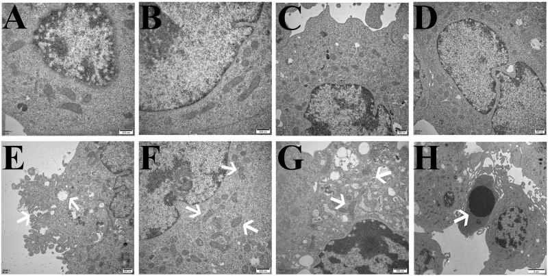
The TEM results showed that compared with the control group, the cell membranes of HT22 cells and PC12 cells in the model group were ruptured and blebbed, mitochondria atrophied and became smaller, mitochondrial ridges were reduced or even disappeared, and apoptotic bodies appeared. Compared with the model group, the HT22 cells and PC12 cells in the HBO group and HBO+F group were significantly improved (Figs 6 and 7).

Fig 6. Ultrastructural changes related to ferroptosis in HT22 cells observed by transmission electron microscopy.

Fig 6

A, B: Control group, with normal cell structure. C, D: HBO group, the cell structure was significantly improved compared with the model group. E,F,G,H: Model group, E: Cell membrane rupture and blebbing; F: Mitochondria become smaller; G: Mitochondrial degeneration; H: Apoptotic body.

Fig 7. Ultrastructural changes related to ferroptosis in PC12 cells observed by transmission electron microscopy.

Fig 7

A, B: Control group, with normal cell structure. C, D: HBO group, the cell structure was significantly improved compared with the model group. E,F,G,H: Model group, E: Cell membrane rupture and blebbing; F: Mitochondria become smaller; G: Mitochondrial degeneration; H: Apoptotic body.

Effects of HBO on the expression levels of ferroptosis-related genes

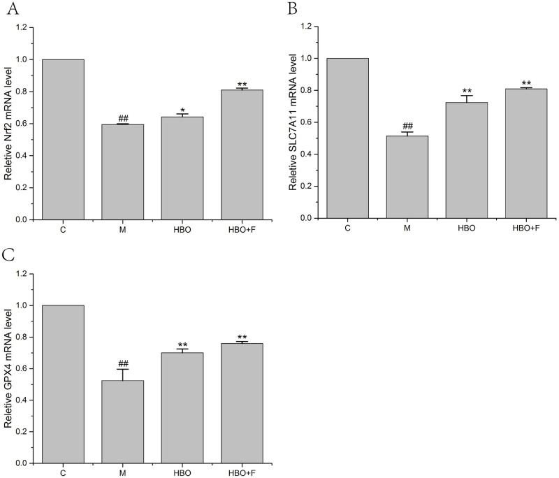
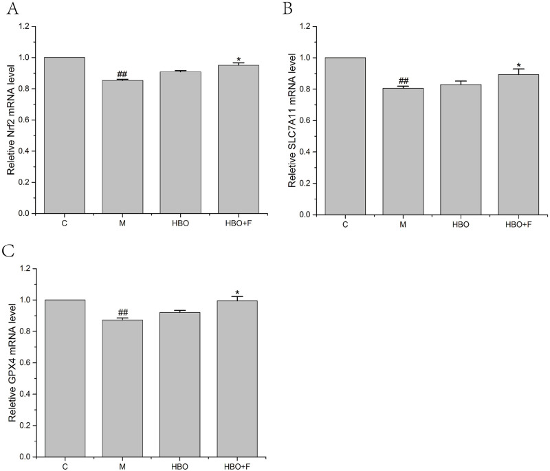
PCR results showed that compared with the control group, the expression levels of Nrf2, SLC7A11 and GPX4 genes in the HT22 cells and PC12 cells of the model group were significantly decreased; while the gene expressions of Nrf2, SLC7A11 and GPX4 genes in the cells of HBO group and HBO+F group were significantly higher than those in the model group (Figs 8 and 9).

Fig 8. The effect of HBO on the expression of Nrf2, SLC7A11 and GPX4 genes in HT22 cells.

Fig 8

The results are presented as the mean ± SEM (N = 3). Compared with the control group. ##P < 0.01 vs. control group, *P < 0.05, **P < 0.01 vs. model group. C: Control group; M: Model group; HBO: Hyperbaric oxygen group; HBO+F: Hyperbaric oxygen + Ferrostatin-1 group.

Fig 9. The effect of HBO on the expression of Nrf2, SLC7A11 and GPX4 genes in PC12 cells.

Fig 9

The results are presented as the mean ± SEM (N = 3). Compared with the control group. ##P < 0.01 vs. control group, *P < 0.05, **P < 0.01 vs. model group. C: Control group; M: Model group; HBO: Hyperbaric oxygen group; HBO+F: Hyperbaric oxygen + Ferrostatin-1 group.

Effects of HBO on the expression levels of ferroptosis-related proteins

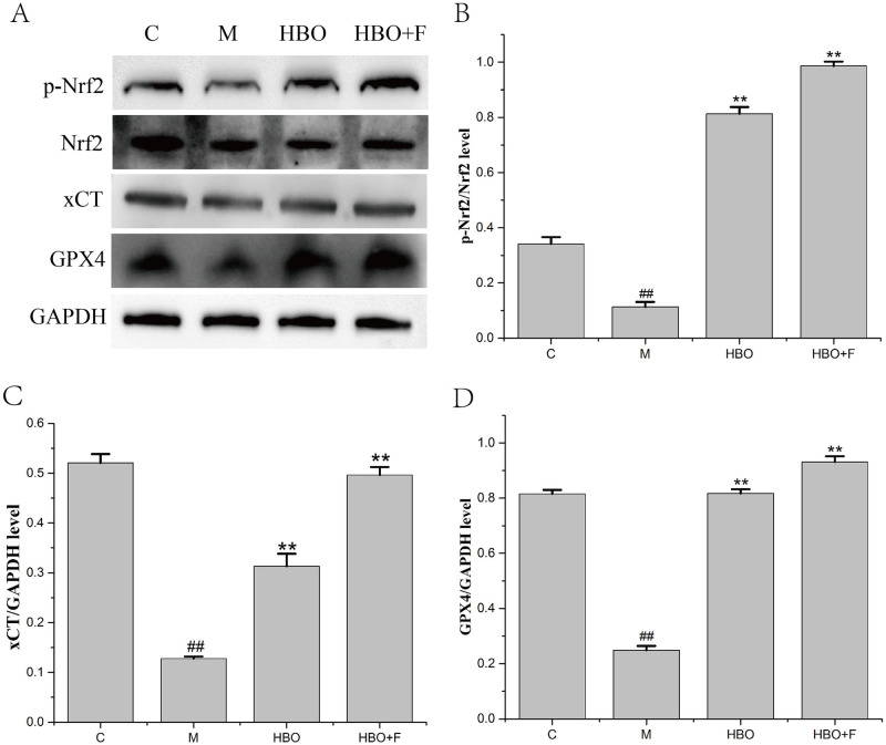
WB results showed that compared with the control group, the protein expression levels of p-Nrf2/Nrf2, xCT and GPX4 proteins in the HT22 cells of the model group were significantly decreased and the expression levels of p-Nrf2/Nrf2, xCT and GPX4 proteins in the cells of HBO group and HBO+F group were significantly higher than that in the model group (Fig 10).

Fig 10. The effect of HBO on the expression of p-Nrf2/Nrf2, xCT and GPX4 proteins in HT22 cells.

Fig 10

The results are presented as the mean ± SEM (N = 3). Compared with the control group. ##P < 0.01 vs. control group, **P < 0.01 vs. model group. C: Control group; M: Model group; HBO: Hyperbaric oxygen group; HBO+F: Hyperbaric oxygen + Ferrostatin-1 group.

Discussion

The process of ferroptosis is accompanied by the accumulation of a large amount of iron ions, and iron is an important cofactor. The balance of iron is essential for the proper functioning of the brain. Studies have found that iron accumulation exists in the brain of patients with many neurodegenerative diseases, and iron accumulation can lead to neurotoxicity through various mechanisms, including the production of oxygen free radicals that lead to oxidative stress, excitotoxicity, and promote inflammatory responses [19]. This study found that HBO can significantly reverse the oxygen-glucose deprivation-reperfusion-injury-induced decrease in cell viability and increase in intracellular iron content of HT22 cells and PC12 cells. It has been reported that cerebral ischemia-reperfusion can cause iron metabolism disorders in brain tissue, mainly manifested as increased iron content in brain tissue, resulting in iron overload. The increase of iron content in brain tissue can catalyze the production of free radicals to increase the force, thereby promoting lipid peroxidation and aggravating brain tissue damage [20,21].

Ferroptosis has its unique biochemical characteristics and morphology. Besides iron overload, accumulation of lipid ROS is also a major biochemical feature [22]. Morphological features include mitochondrial shrinkage, reduction or disappearance of mitochondrial cristae, increased mitochondrial membrane density, mitochondrial membrane rupture, and normal nuclear morphology, but lack of chromatin condensation [23]. This study showed that ROS, Lip-ROS and MDA were significantly increased, and GSH was significantly decreased in the cells with oxygen-glucose deprivation/reperfusion. TEM showed that the cell membrane was ruptured and blistered, mitochondria atrophied and became smaller, mitochondrial cristae were reduced or even disappeared, and apoptotic bodies appeared in the model groups. And HBO can reverse these changes. The results suggest that oxygen-glucose deprivation/reperfusion can induce ferroptosis in cells, and HBO can inhibit ferroptosis. Iron metabolism and lipid peroxidation are key factors mediating ferroptosis. Iron ions in the body are endocytosed into cells through the binding of TFR1 on the cell membrane. Iron ions are reduced to ferrous ions in the cells, and the excessive accumulation of ferrous ions in the cells will promote the generation of oxidative free radicals and lipid peroxidation product MDA through the Fenton reaction, thereby aggravating the damage of nerve cells [24,25].

Combined with our previous and current findings, we found that ferroptosis in the oxygen-glucose deprivation/reperfusion group was accompanied by apoptosis. Apoptosis is programmed series of events dependent on energy, as well as morphological features such as cell shrinkage, chromatin condensation, and presence of apoptotic bodies without inflammatory reactions [26,27]. Caspases are key molecules involved in the transduction of the apoptosis signal, and all of the pathways converge to the executioner caspase-3 [28]. The extrinsic pathway is initiated by the tumor necrosis factor (TNF) receptor family interacting with a ligand and then binds with procaspase-8 following ligand-receptor interaction to activation of caspase-3 which leads to execution of apoptosis [29,30]. The intrinsic pathway (mitochondrial pathway) employs alterations of inner mitochondrial membrane for induction of apoptosis. Apoptosis is triggered when the Bcl2-family proapoptotic proteins cause the opening of mitochondrial permeability transition pore and proapoptotic proteins into cytoplasm by interacting with apoptotic protease-activating factor 1 (Apaf-1) and procaspase-9 to constitute apoptosome [31]. An assembly of apoptosome leads to caspase-9 activation, which further activates caspase-3, for apoptotic execution [32]. TEM showed that apoptotic bodies appeared in the model groups. It suggested that apoptosis and ferroptosis coexist under certain conditions. Su et al. [33] found that reactive oxygen species can simultaneously induce apoptosis, autophagy and ferroptosis. Ye et al. [34] found that FBW7-NRA41-SCD1 axis synchronously regulates apoptosis and ferroptosis in pancreatic cancer cells.

As an important intracellular antioxidant molecule, cystine/glutamate transporter (System Xc-) is the upstream node molecule in the process of ferroptosis. Its main function is to maintain the balance of cystine (Cys) intake and glutamate (Glu) excretion. After being taken up by System Xc-, cystine is reduced to cysteine in cells and is involved in the synthesis of glutathione (GSH). Glutathione can reduce reactive oxygen species and reactive nitrogen species under the action of GPX4 [35]. When System Xc- is inhibited, the introduction of cystine (Cys) into cells is hindered and the cysteine necessary for the synthesis of GSH is reduced. Due to glutathione (GSH) depletion, the activity of glutathione peroxidase 4 (GPX4) is decreased or even inactivated. Intracellular lipid oxides (ROOH) cannot be metabolized to ROH and H2O2 without oxidative toxicity. The Fenton reaction then occurs, resulting in the production of a large amount of ROS, which severely disrupts the intracellular redox balance, causes cellular lipid peroxidative damage, and attacks biological macromolecules, thereby initiating ferroptosis [36,37]. SLC7A11 (also known as xCT) is the substrate-specific subunit that constitutes System Xc- and is responsible for the transport of cystine from the extracellular to the intracellular. When cells are under oxidative stress and cysteine deficiency, nuclear factor erythroid 2 like 2 (NRF2) and activating transcription factor 4 (ATF4) can induce SLC7A11 expression [38].

The System Xc-/GSH/GPX4 pathway, as one of the main regulatory axes of ferroptosis, plays an important role in cerebral ischemia-reperfusion injury [20,21]. PCR results showed that expressions of Nrf2, SLC7A11 and GPX4 genes were decreased, especially in HT22 cells. WB experiment showed that the expressions of p-Nrf2/Nrf2, xCT and GPX4 proteins in HT22 cells in the model group were significantly decreased. Nrf2-induced decreased SLC7A11 (xCT) expression, decreased glutamate-cysteine exchange, decreased GSH content, decreased GPX4 activity, and insufficient cellular ability to scavenge lipid peroxides resulted in the accumulation of a large number of oxidative products such as MDA. At the mitochondrial membrane, the mitochondrial membrane potential decreases, ferroptosis occurs, and nerve function is impaired. The expression of these genes and proteins can be reversed after HBO treatment. This suggested that HBO can inhibit ferroptosis through the System Xc-/GSH/GPX4 pathway.

In summary, our findings indicate that HBO can protect HT22 cells and PC12 cells from damage caused by oxygen-glucose deprivation/reperfusion via the inhibition of Nrf2/System Xc-/GPX4 axis-mediated ferroptosis. Our findings provide a basis for further research on the mechanism of HBO in cerebral ischemia-reperfusion.

Supporting information

S1 File

(7Z)

S2 File

(7Z)

Acknowledgments

The authors would like to thank Dr Weidong Li from Guangxi Medical University, China, for his technical support in immunohistochemistry.

Data Availability

All relevant data are within the manuscript and its Supporting information files.

Funding Statement

This study was supported by the National Natural Science Foundation of China (81960246 and 81701089), the Guangxi Natural Science Foundation (2020GXNSFAA238003 and 2017GXNSFBA198010), the Guangxi Sanitation and Family Planning Committee Project (No. Z20201096) and the Guangxi Medical and Health Appropriate Technology Research and Development Project (S2020076).

References

  • 1.Lu LQ, Tian J, Luo XJ, Peng J. Targeting the pathways of regulated necrosis: a potential strategy for alleviation of cardio-cerebrovascular injury. Cell Mol Life Sci. 2021. Jan;78(1):63–78. doi: 10.1007/s00018-020-03587-8 [DOI] [PMC free article] [PubMed] [Google Scholar]
  • 2.Guan X, Li X, Yang X, Yan J, Shi P, Ba L, et al. The neuroprotective effects of carvacrol on ischemia/reperfusion-induced hippocampal neuronal impairment by ferroptosis mitigation. Life Sci. 2019. Oct 15;235:116795. doi: 10.1016/j.lfs.2019.116795 [DOI] [PubMed] [Google Scholar]
  • 3.Lan B, Ge JW, Cheng SW, Zheng XL, Liao J, He C, et al. Extract of Naotaifang, a compound Chinese herbal medicine, protects neuron ferroptosis induced by acute cerebral ischemia in rats. J Integr Med. 2020. Jul;18(4):344–350. doi: 10.1016/j.joim.2020.01.008 [DOI] [PubMed] [Google Scholar]
  • 4.Dixon SJ, Lemberg KM, Lamprecht MR, Skouta R, Zaitsev EM, Gleason CE, et al. Ferroptosis: an iron-dependent form of nonapoptotic cell death. Cell. 2012. May 25;149(5):1060–72. doi: 10.1016/j.cell.2012.03.042 [DOI] [PMC free article] [PubMed] [Google Scholar]
  • 5.Hirschhorn T, Stockwell BR. The development of the concept of ferroptosis. Free Radic Biol Med. 2019. Mar;133:130–143. doi: 10.1016/j.freeradbiomed.2018.09.043 [DOI] [PMC free article] [PubMed] [Google Scholar]
  • 6.Yang JC, Ding L, Yu LD, et al. Nanomedicine enables autophagy-enhanced cancer-cell ferroptosis. Sci Bull, 2021, 66(5):464–477. [DOI] [PubMed] [Google Scholar]
  • 7.DeGregorio-Rocasolano N, Martí-Sistac O, Gasull T. Deciphering the Iron Side of Stroke: Neurodegeneration at the Crossroads Between Iron Dyshomeostasis, Excitotoxicity, and Ferroptosis. Front Neurosci. 2019. Feb 19;13:85. doi: 10.3389/fnins.2019.00085 [DOI] [PMC free article] [PubMed] [Google Scholar]
  • 8.Turchi R, Tortolici F, Guidobaldi G, Iacovelli F, Falconi M, Rufini S, et al. Frataxin deficiency induces lipid accumulation and affects thermogenesis in brown adipose tissue. Cell Death Dis. 2020. Jan 23;11(1):51. doi: 10.1038/s41419-020-2253-2 [DOI] [PMC free article] [PubMed] [Google Scholar]
  • 9.Gao X, Guo N, Xu H, Pan T, Lei H, Yan A, et al. Ibuprofen induces ferroptosis of glioblastoma cells via downregulation of nuclear factor erythroid 2-related factor 2 signaling pathway. Anticancer Drugs. 2020. Jan;31(1):27–34. doi: 10.1097/CAD.0000000000000825 [DOI] [PubMed] [Google Scholar]
  • 10.Cheng J, Fan YQ, Liu BH, Zhou H, Wang JM, Chen QX. ACSL4 suppresses glioma cells proliferation via activating ferroptosis. Oncol Rep. 2020. Jan;43(1):147–158. doi: 10.3892/or.2019.7419 [DOI] [PMC free article] [PubMed] [Google Scholar]
  • 11.Weiland A, Wang Y, Wu W, Lan X, Han X, Li Q, et al. Ferroptosis and Its Role in Diverse Brain Diseases. Mol Neurobiol. 2019. Jul;56(7):4880–4893. doi: 10.1007/s12035-018-1403-3 [DOI] [PMC free article] [PubMed] [Google Scholar]
  • 12.Han C, Liu Y, Dai R, Ismail N, Su W, Li B. Ferroptosis and Its Potential Role in Human Diseases. Front Pharmacol. 2020. Mar 17;11:239. doi: 10.3389/fphar.2020.00239 [DOI] [PMC free article] [PubMed] [Google Scholar]
  • 13.Magtanong L, Dixon SJ. Ferroptosis and Brain Injury. Dev Neurosci. 2018;40(5–6):382–395. doi: 10.1159/000496922 [DOI] [PMC free article] [PubMed] [Google Scholar]
  • 14.Mahoney-Sánchez L, Bouchaoui H, Ayton S, Devos D, Duce JA, Devedjian JC. Ferroptosis and its potential role in the physiopathology of Parkinson’s Disease. Prog Neurobiol. 2021. Jan;196:101890. doi: 10.1016/j.pneurobio.2020.101890 [DOI] [PubMed] [Google Scholar]
  • 15.Fife CE, Workmanm WT. Ethical issues, standards and quality control in practice of hyperbaric medicine. Springer, New York, 2016. [Google Scholar]
  • 16.Chen CX, Huang LY, Nong ZH, Li YX, Chen W, Huang JP, et al. Hyperbaric Oxygen Prevents Cognitive Impairments in Mice Induced by D-Galactose by Improving Cholinergic and Anti-apoptotic Functions. Neurochem Res. 2017, Jan 11. doi: 10.1007/s11064-016-2166-8 [DOI] [PubMed] [Google Scholar]
  • 17.Chen XY, Li YX, Chen W, Nong ZH, Huang JP, Chen CX. Protective effect of hyperbaric oxygen on cognitive impairment induced by D-Galactose in mice. Neurochem Res. 2016, 41:3032–41. doi: 10.1007/s11064-016-2022-x [DOI] [PubMed] [Google Scholar]
  • 18.Jianrong YANG, Wan CHEN, Xing ZHOU, Yaoxuan LI, Zhihuang NONG, Liyuan ZHOU, et al. Hyperbaric oxygen protects against PC12 and H9C2 cell damage caused by oxygen–glucose deprivation/reperfusion via the inhibition of cell apoptosis and autophagy. BIOCELL. 2022, 46(1):137–148. [Google Scholar]
  • 19.Raha AA, Vaishnav RA, Friedland RP, Bomford A, Raha-Chowdhury R. The systemic iron-regulatory proteins hepcidin and ferroportin are reduced in the brain in Alzheimer’s disease. Acta Neuropathol Commun. 2013. Sep 3;1:55. doi: 10.1186/2051-5960-1-55 [DOI] [PMC free article] [PubMed] [Google Scholar]
  • 20.Fang KM, Cheng FC, Huang YL, Chung SY, Jian ZY, Lin MC. Trace element, antioxidant activity, and lipid peroxidation levels in brain cortex of gerbils after cerebral ischemic injury. Biol Trace Elem Res. 2013. Apr;152(1):66–74. doi: 10.1007/s12011-012-9596-1 [DOI] [PubMed] [Google Scholar]
  • 21.Tuo QZ, Lei P, Jackman KA, Li XL, Xiong H, Li XL, et al. Tau-mediated iron export prevents ferroptotic damage after ischemic stroke. Mol Psychiatry. 2017. Nov;22(11):1520–1530. doi: 10.1038/mp.2017.171 [DOI] [PubMed] [Google Scholar]
  • 22.Latunde-Dada GO. Ferroptosis: role of lipid peroxidation, iron and ferritinophagy. BiochimBiophys Acta Gen Subi, 2017; 1861(8): 1893–1900. doi: 10.1016/j.bbagen.2017.05.019 [DOI] [PubMed] [Google Scholar]
  • 23.Xie Y, Hou W, Song X, Yu Y, Huang J, Sun X, et al. Ferroptosis: process and function. CellDeath Differ, 2016; 23(3): 369–379. doi: 10.1038/cdd.2015.158 [DOI] [PMC free article] [PubMed] [Google Scholar]
  • 24.Lu J, Xu F, Lu H. LncRNA PVT1 regulates ferroptosis through miR-214-mediated TFR1 and p53. Life Sci. 2020. Nov 1;260:118305. doi: 10.1016/j.lfs.2020.118305 [DOI] [PubMed] [Google Scholar]
  • 25.She X, Lan B, Tian H, Tang B. Cross Talk Between Ferroptosis and Cerebral Ischemia. Front Neurosci. 2020. Aug 6;14:776. doi: 10.3389/fnins.2020.00776 [DOI] [PMC free article] [PubMed] [Google Scholar]
  • 26.Zakeri Z, Bursch W, Tenniswood M, Lockshin RA. Cell death: programmed, apoptosis, necrosis, or other? Cell Death Differ. 1995. Apr;2(2):87–96. [PubMed] [Google Scholar]
  • 27.Elmore S. Apoptosis: a review of programmed cell death. Toxicol Pathol. 2007. Jun;35(4):495–516. doi: 10.1080/01926230701320337 [DOI] [PMC free article] [PubMed] [Google Scholar]
  • 28.Romero A, Novoa B, Figueras A. The complexity of apoptotic cell death in mollusks: An update. Fish Shellfish Immunol. 2015. Sep;46(1):79–87. doi: 10.1016/j.fsi.2015.03.038 [DOI] [PubMed] [Google Scholar]
  • 29.Ashkenazi A, Dixit VM. Death receptors: signaling and modulation. Science. 1998. Aug 28;281(5381):1305–8. doi: 10.1126/science.281.5381.1305 [DOI] [PubMed] [Google Scholar]
  • 30.Wajant H. The Fas signaling pathway: more than a paradigm. Science. 2002. May 31;296(5573):1635–6. doi: 10.1126/science.1071553 [DOI] [PubMed] [Google Scholar]
  • 31.Bao Q, Shi Y. Apoptosome: a platform for the activation of initiator caspases. Cell Death Differ. 2007. Jan;14(1):56–65. doi: 10.1038/sj.cdd.4402028 [DOI] [PubMed] [Google Scholar]
  • 32.Riedl SJ, Salvesen GS. The apoptosome: signalling platform of cell death. Nat Rev Mol Cell Biol. 2007. May;8(5):405–13. doi: 10.1038/nrm2153 [DOI] [PubMed] [Google Scholar]
  • 33.Su LJ, Zhang JH, Gomez H, Murugan R, Hong X, Xu D, et al. Reactive Oxygen Species-Induced Lipid Peroxidation in Apoptosis, Autophagy, and Ferroptosis. Oxid Med Cell Longev. 2019. Oct 13;2019:5080843. doi: 10.1155/2019/5080843 [DOI] [PMC free article] [PubMed] [Google Scholar]
  • 34.Ye Z, Zhuo Q, Hu Q, Xu X, Mengqi Liu, Zhang Z, et al. FBW7-NRA41-SCD1 axis synchronously regulates apoptosis and ferroptosis in pancreatic cancer cells. Redox Biol. 2021. Jan;38:101807. doi: 10.1016/j.redox.2020.101807 [DOI] [PMC free article] [PubMed] [Google Scholar]
  • 35.Florean C, Song S, Dicato M, Diederich M. Redox biology of regulated cell death in cancer: A focus on necroptosis and ferroptosis. Free Radic Biol Med, 2019, 134:177–189. doi: 10.1016/j.freeradbiomed.2019.01.008 [DOI] [PubMed] [Google Scholar]
  • 36.Ganguly U, Kaur U, Chakrabarti SS, Sharma P, Agrawal BK, Saso L, et al. Oxidative stress, neuroinflammation, andNADPH oxidase: implications in the pathogenesis and treatment of Alzheimer’s disease.Oxid Med Cell Longev, 2021, 2021:7086512. doi: 10.1155/2021/7086512 [DOI] [PMC free article] [PubMed] [Google Scholar]
  • 37.Li J, Cao F, Yin HL, Huang ZJ, Lin ZT, Mao N, et al. Ferroptosis: past, present and future. Cell Death Dis, 2020,11(2):88. doi: 10.1038/s41419-020-2298-2 [DOI] [PMC free article] [PubMed] [Google Scholar]
  • 38.Koppula P, Zhang Y, Zhuang L, Gan B. Amino acid trans- porter SLC7A11 /xCT at the crossroads of regulating redox homeostasis and nutrient dependency of cancer. Cancer Commun. 2018,38:12–26. [DOI] [PMC free article] [PubMed] [Google Scholar]

Decision Letter 0

Nukhet Aykin-Burns

18 May 2022

PONE-D-22-08821Hyperbaric oxygen protects HT22 cells and PC12 cells from damage caused by oxygen-glucose deprivation/reperfusion via the inhibition of Nrf2/System Xc-/GPX4 axis-mediated ferroptosisPLOS ONE

Dear Dr. Chen,

Thank you for submitting your manuscript to PLOS ONE. After careful consideration, we feel that it has merit but does not fully meet PLOS ONE’s publication criteria as it currently stands. Therefore, we invite you to submit a revised version of the manuscript that addresses the points raised during the review process.

Please submit your revised manuscript by Jul 02 2022 11:59PM. If you will need more time than this to complete your revisions, please reply to this message or contact the journal office at plosone@plos.org. When you're ready to submit your revision, log on to https://www.editorialmanager.com/pone/ and select the 'Submissions Needing Revision' folder to locate your manuscript file.

Please include the following items when submitting your revised manuscript:

  • A rebuttal letter that responds to each point raised by the academic editor and reviewer(s). You should upload this letter as a separate file labeled 'Response to Reviewers'.

  • A marked-up copy of your manuscript that highlights changes made to the original version. You should upload this as a separate file labeled 'Revised Manuscript with Track Changes'.

  • An unmarked version of your revised paper without tracked changes. You should upload this as a separate file labeled 'Manuscript'.

If you would like to make changes to your financial disclosure, please include your updated statement in your cover letter. Guidelines for resubmitting your figure files are available below the reviewer comments at the end of this letter.

If applicable, we recommend that you deposit your laboratory protocols in protocols.io to enhance the reproducibility of your results. Protocols.io assigns your protocol its own identifier (DOI) so that it can be cited independently in the future. For instructions see: https://journals.plos.org/plosone/s/submission-guidelines#loc-laboratory-protocols. Additionally, PLOS ONE offers an option for publishing peer-reviewed Lab Protocol articles, which describe protocols hosted on protocols.io. Read more information on sharing protocols at https://plos.org/protocols?utm_medium=editorial-email&utm_source=authorletters&utm_campaign=protocols.

We look forward to receiving your revised manuscript.

Kind regards,

Nukhet Aykin-Burns, PhD

Academic Editor

PLOS ONE

Journal Requirements:

When submitting your revision, we need you to address these additional requirements.

1. Please ensure that your manuscript meets PLOS ONE's style requirements, including those for file naming. The PLOS ONE style templates can be found at https://journals.plos.org/plosone/s/file?id=wjVg/PLOSOne_formatting_sample_main_body.pdf and 

https://journals.plos.org/plosone/s/file?id=ba62/PLOSOne_formatting_sample_title_authors_affiliations.pdf.

2. We note that the grant information you provided in the ‘Funding Information’ and ‘Financial Disclosure’ sections do not match. 

When you resubmit, please ensure that you provide the correct grant numbers for the awards you received for your study in the ‘Funding Information’ section.

3. PLOS requires an ORCID iD for the corresponding author in Editorial Manager on papers submitted after December 6th, 2016. Please ensure that you have an ORCID iD and that it is validated in Editorial Manager. To do this, go to ‘Update my Information’ (in the upper left-hand corner of the main menu), and click on the Fetch/Validate link next to the ORCID field. This will take you to the ORCID site and allow you to create a new iD or authenticate a pre-existing iD in Editorial Manager. Please see the following video for instructions on linking an ORCID iD to your Editorial Manager account: https://www.youtube.com/watch?v=_xcclfuvtxQ.

4. Your ethics statement should only appear in the Methods section of your manuscript. If your ethics statement is written in any section besides the Methods, please move it to the Methods section and delete it from any other section. Please ensure that your ethics statement is included in your manuscript, as the ethics statement entered into the online submission form will not be published alongside your manuscript. 

5. Please ensure that you refer to Figure 10 in your text as, if accepted, production will need this reference to link the reader to the figure.

Reviewers' comments:

Reviewer's Responses to Questions

Comments to the Author

1. Is the manuscript technically sound, and do the data support the conclusions?

The manuscript must describe a technically sound piece of scientific research with data that supports the conclusions. Experiments must have been conducted rigorously, with appropriate controls, replication, and sample sizes. The conclusions must be drawn appropriately based on the data presented.

Reviewer #1: Partly

Reviewer #2: Partly

**********

2. Has the statistical analysis been performed appropriately and rigorously?

Reviewer #1: N/A

Reviewer #2: N/A

**********

3. Have the authors made all data underlying the findings in their manuscript fully available?

The PLOS Data policy requires authors to make all data underlying the findings described in their manuscript fully available without restriction, with rare exception (please refer to the Data Availability Statement in the manuscript PDF file). The data should be provided as part of the manuscript or its supporting information, or deposited to a public repository. For example, in addition to summary statistics, the data points behind means, medians and variance measures should be available. If there are restrictions on publicly sharing data—e.g. participant privacy or use of data from a third party—those must be specified.

Reviewer #1: No

Reviewer #2: Yes

**********

4. Is the manuscript presented in an intelligible fashion and written in standard English?

PLOS ONE does not copyedit accepted manuscripts, so the language in submitted articles must be clear, correct, and unambiguous. Any typographical or grammatical errors should be corrected at revision, so please note any specific errors here.

Reviewer #1: No

Reviewer #2: Yes

**********

5. Review Comments to the Author

Please use the space provided to explain your answers to the questions above. You may also include additional comments for the author, including concerns about dual publication, research ethics, or publication ethics. (Please upload your review as an attachment if it exceeds 20,000 characters)

Reviewer #1: The author revealed the neuroprotective effects of hyperbaric oxygen (HBO) in HT22 and PC12 cells, while there are severe problems within the manuscript as listed below:

1. The morphology of PC12 cells is generally round or triangular, and the HT-22 has an elongated/polarized shape. However, the staining results (e.g. Fig 3) demonstrated their PC12 cells did not meet the standard morphology, as we can find that the PC12 here is more of a neuronal-like shape than HT-22. Detail has to be given.

2. The author did not use the standard complete medium (with horse serum) to culture the PC12 cells. Given reasons.

3. “SLC7ALL” (should be SLC7A11) is everywhere throughout the text, while it became correct in the Discussion and part of the figure (Fig 9), which infers the authors did not get the consensus regarding their research target.

4. The Ferrostatin-1 should serve as an individual group to treat the ferroptosis caused by O-G D/R, rather than being administered together with the HBO. Additional experimental results have to be given.

5. Lack of information: the number of repeats for individual experiments; scale bar for Fig3 and Fig4; abbreviations.

Reviewer #2: Major comment:

1. Please clarify whether HBOT protects PC 12 cells from damage caused by oxygen-glucose deprivation/reperfusion via the inhibition of ferroptosis or the inhibition of apoptosis and autophagy (previous publication in BIOCELL, 2022). The authors used the same condition of HBOT to treat PC 12 cells. The previous results showed the protection of HBOT for PC 12 cells from damage caused by oxygen–glucose deprivation/reperfusion via the inhibition of cell apoptosis and autophagy. However, in this study, they showed the protection of HBOT for PC 12 cells from damage caused by oxygen–glucose deprivation/reperfusion via the inhibition of cell ferroptosis. Is it possible that apoptosis, autophagy and ferroptosis can exist at the same condition or the same time, since the morphology, biochemical features and regulatory pathway for all are different?

2. Regarding the pressure and times of HBOT, the authors used 0.25 MPa and 60 min of HBOT to treat the cells. The pressure is higher than that used in clinical treatment. Why and how do the authors choose the proper pressure? If the authors were willing to mimic or simulate the clinical treatment, repetitive treatment of HBOT should be considered. Dose this effect persist after more than one time treatment? Furthermore, the PH value of culture medium may be changed in this condition that could influence the interpretation of protection mechanism.

3. Although the authors checked the morphology, related gene and protein expression of ferroptosis for HBOT treated PC cells directly, there was still some difference between HBO group and HBO+Ferrostatin-1 (HBO+F) group. Did that show statistic difference? It seemed that HBO and ferrostatin-1 had synergistic effect in ferroptosis which meant HBOT protection partially via the inhibition of ferroptosis. The authors may try to pretreat model group with ferrostatin-1 before HBOT to see how it goes. Otherwise, the authors need to justify the title based on the current results.

4. HBOT itself could produce ROS, especially while the higher pressure or the longer duration was used. Although some papers, as the authors cited, indicated HBOT might reduce the oxidative stress in the disease condition, it has been still arguable when HBOT was applied clinically in some neurological disorders, such as stroke. Please describe more to support your view in the discussion section.

Comments on Photos and Figures:

1. Please provide higher resolution images for Figures 3&4

2. Please present the figures differently, such as viability in the curve and intracellular ROS in columns, but combine images and levels of the same cell line with one Fraction.

**********

6. PLOS authors have the option to publish the peer review history of their article (what does this mean?). If published, this will include your full peer review and any attached files.

If you choose “no”, your identity will remain anonymous but your review may still be made public.

Do you want your identity to be public for this peer review? For information about this choice, including consent withdrawal, please see our Privacy Policy.

Reviewer #1: No

Reviewer #2: No

[NOTE: If reviewer comments were submitted as an attachment file, they will be attached to this email and accessible via the submission site. Please log into your account, locate the manuscript record, and check for the action link "View Attachments". If this link does not appear, there are no attachment files.]

While revising your submission, please upload your figure files to the Preflight Analysis and Conversion Engine (PACE) digital diagnostic tool, https://pacev2.apexcovantage.com/. PACE helps ensure that figures meet PLOS requirements. To use PACE, you must first register as a user. Registration is free. Then, login and navigate to the UPLOAD tab, where you will find detailed instructions on how to use the tool. If you encounter any issues or have any questions when using PACE, please email PLOS at figures@plos.org. Please note that Supporting Information files do not need this step.

PLoS One. 2022 Nov 10;17(11):e0276083. doi: 10.1371/journal.pone.0276083.r002

Author response to Decision Letter 0


16 Aug 2022

Journal Requirements:

1. Please ensure that your manuscript meets PLOS ONE's style requirements, including those for file naming. The PLOS ONE style templates can be found at https://journals.plos.org/plosone/s/file?id=wjVg/PLOSOne_formatting_sample_main_body.pdf and

https://journals.plos.org/plosone/s/file?id=ba62/PLOSOne_formatting_sample_title_authors_affiliations.pdf.

2. We note that the grant information you provided in the ‘Funding Information’ and ‘Financial Disclosure’ sections do not match.

When you resubmit, please ensure that you provide the correct grant numbers for the awards you received for your study in the ‘Funding Information’ section.

3. PLOS requires an ORCID iD for the corresponding author in Editorial Manager on papers submitted after December 6th, 2016. Please ensure that you have an ORCID iD and that it is validated in Editorial Manager. To do this, go to ‘Update my Information’ (in the upper left-hand corner of the main menu), and click on the Fetch/Validate link next to the ORCID field. This will take you to the ORCID site and allow you to create a new iD or authenticate a pre-existing iD in Editorial Manager. Please see the following video for instructions on linking an ORCID iD to your Editorial Manager account: https://www.youtube.com/watch?v=_xcclfuvtxQ.

4. Your ethics statement should only appear in the Methods section of your manuscript. If your ethics statement is written in any section besides the Methods, please move it to the Methods section and delete it from any other section. Please ensure that your ethics statement is included in your manuscript, as the ethics statement entered into the online submission form will not be published alongside your manuscript.

5. Please ensure that you refer to Figure 10 in your text as, if accepted, production will need this reference to link the reader to the figure.

Author Response: I have addressed the above additional requirements.

Reviewer #1: The author revealed the neuroprotective effects of hyperbaric oxygen (HBO) in HT22 and PC12 cells, while there are severe problems within the manuscript as listed below:

1. The morphology of PC12 cells is generally round or triangular, and the HT-22 has an elongated/polarized shape. However, the staining results (e.g. Fig 3) demonstrated their PC12 cells did not meet the standard morphology, as we can find that the PC12 here is more of a neuronal-like shape than HT-22. Detail has to be given.

Author Response: The PC12 cells in Fig 3 are more numerous, so it appears that the cells has an elongated/polarized shape. In order not to cause misunderstanding, the Fig 3B and Fig 3D have been replaced.

2. The author did not use the standard complete medium (with horse serum) to culture the PC12 cells. Given reasons.

Author Response: Sorry for the wrong description due to my carelessness. PC12 cells were maintained in RPMI 1640 medium supplemented with 10% horse serum, 5% fetal bovine serum and 1% penicillin-streptomycin. Modifications have been made in the Materials and methods section.

3. “SLC7ALL” (should be SLC7A11) is everywhere throughout the text, while it became correct in the Discussion and part of the figure (Fig 9), which infers the authors did not get the consensus regarding their research target.

Author Response: I'm very sorry for miswriting SLC7A11 as SLC7ALL. They have been modified in the manuscript.

4. The Ferrostatin-1 should serve as an individual group to treat the ferroptosis caused by O-G D/R, rather than being administered together with the HBO. Additional experimental results have to be given.

Author Response: A new experiment was added: the cell viability was analyzed when incubated the cells with Ferrostatin-1. As shown in Fig 1C and 1D, like the HBO group, the HT22 and PC12 cell activities of the Ferrostatin-1 groups were significantly higher than those of the model groups. Moreover, PC12 and H9C2 cell activities were higher than those of the HBO groups or the Ferrostatin-1 groups after being treated with Ferrostatin-1 and HBO at the same time. These results indicated that HBO may protect cells from damage by inhibiting ferroptosis.

5. Lack of information: the number of repeats for individual experiments; scale bar for Fig3 and Fig4; abbreviations.

Author Response: The missing information has been added in the manuscript.

Reviewer #2: Major comment:

1. Please clarify whether HBOT protects PC 12 cells from damage caused by oxygen-glucose deprivation/reperfusion via the inhibition of ferroptosis or the inhibition of apoptosis and autophagy (previous publication in BIOCELL, 2022). The authors used the same condition of HBOT to treat PC 12 cells. The previous results showed the protection of HBOT for PC 12 cells from damage caused by oxygen–glucose deprivation/reperfusion via the inhibition of cell apoptosis and autophagy. However, in this study, they showed the protection of HBOT for PC 12 cells from damage caused by oxygen–glucose deprivation/reperfusion via the inhibition of cell ferroptosis. Is it possible that apoptosis, autophagy and ferroptosis can exist at the same condition or the same time, since the morphology, biochemical features and regulatory pathway for all are different?

Author Response: Our findings and literature reports suggested that apoptosis, autophagy, and ferroptosis may coexist. During ferroptosis, the balance of the intracellular redox system is disrupted, leading to mitochondrial damage, which may promote the occurrence of autophagy, which may act as a feedback loop to further induce ferroptosis [1-3]. Autophagy is like a double-edged sword, which can induce apoptosis and inhibit apoptosis [4-6].

1. Li J, Liu J, Xu Y, Wu R, et al. Tumor heterogeneity in autophagy-dependent ferroptosis[J]. Autophagy,2021, 1-14.

2. Liu J, Yang M, Kang R, et al. Autophagic degradation of the circadian clock regulator promotes

ferroptosis[J]. Autophagy, 2019, 15(11): 2033-2035.

3. Munoz P, Casas J, Megias E, et al. The anti-cancer drug ABTL0812 induces ER stress-mediated cytotoxic autophagy by increasing dihydroceramide levels in cancer cells[J]. Autophagy, 2020, 1-18.

4. Maiuri MC, alckvar E, Kimchi A , Kroemer G. Self-eating and self-killing: crosstalk between autophagy and apoptosis. Nat Rev Mol Cell Biol. 2007 Sep;8(9):741-52.

5. Booth LA, Roberts JL, Dent P. The role of cell signaling in the crosstalk between autophagy and apoptosis in the regulation of tumor cell survival in response to sorafenib and neratinib. Semin Cancer Biol. 2020 Nov;66:129-139.

6. Pang H, Wu T, Peng Z, Tan Q, Peng X, Zhan Z, Song L, Wei B. Baicalin induces apoptosis and autophagy in human osteosarcoma cells by increasing ROS to inhibit PI3K/Akt/mTOR, ERK1/2 and β-catenin signaling pathways. J Bone Oncol. 2022 Feb 1;33:100415.

2. Regarding the pressure and times of HBOT, the authors used 0.25 MPa and 60 min of HBOT to treat the cells. The pressure is higher than that used in clinical treatment. Why and how do the authors choose the proper pressure? If the authors were willing to mimic or simulate the clinical treatment, repetitive treatment of HBOT should be considered. Dose this effect persist after more than one time treatment? Furthermore, the PH value of culture medium may be changed in this condition that could influence the interpretation of protection mechanism.

Author Response: Guidelines recommend using a pressure of 2 to 3 times standard atmospheric pressure for HBO therapy. Referring to previous studies, the pressure used for HBO treatment in this study was 2.5 times the atmospheric pressure, which did not exceed the recommended safe range. And, the cell viability was detected by CCK8 assay to determine the time of HBO treatment. All experiments in this study have been repeated three times with consistent results. After the pH test paper detection, the pH value of the culture medium basically did not change under the conditions of this study.

3. Although the authors checked the morphology, related gene and protein expression of ferroptosis for HBOT treated PC cells directly, there was still some difference between HBO group and HBO+Ferrostatin-1 (HBO+F) group. Did that show statistic difference? It seemed that HBO and ferrostatin-1 had synergistic effect in ferroptosis which meant HBOT protection partially via the inhibition of ferroptosis. The authors may try to pretreat model group with ferrostatin-1 before HBOT to see how it goes. Otherwise, the authors need to justify the title based on the current results.

Author Response: There was a statistical difference between the HBO group and the HBO+Ferrostatin-1 (HBO+F) group. HBO and ferrostatin-1 have a synergistic effect in inhibiting ferroptosis. A new experiment was added: the cell viability was analyzed when incubated the cells with Ferrostatin-1. As shown in Fig 1C and 1D, like the HBO group, the HT22 and PC12 cell activities of the Ferrostatin-1 groups were significantly higher than those of the model groups. Moreover, PC12 and H9C2 cell activities were higher than those of the HBO groups or the Ferrostatin-1 groups after being treated with Ferrostatin-1 and HBO at the same time. These results indicated that HBO may protect cells from damage by inhibiting ferroptosis.

4. HBOT itself could produce ROS, especially while the higher pressure or the longer duration was used. Although some papers, as the authors cited, indicated HBOT might reduce the oxidative stress in the disease condition, it has been still arguable when HBOT was applied clinically in some neurological disorders, such as stroke. Please describe more to support your view in the discussion section.

Author Response: Hyperbaric oxygen (HBO) therapy is a clinical use of pure oxygen in the environment of 2-3 times atmospheric pressure to treat ischemic and hypoxic diseases, such as carbon monoxide poisoning, cerebral infarction, decompression sickness and coronary heart disease, etc., and has obtained certain curative effects. According to the US FDA and the International Society of Underwater Hyperbaric Medicine, 13 clinical indications including decompression sickness, carbon monoxide poisoning, gas embolism, acute craniocerebral injury, and post-radiotherapy tissue damage can be treated with HBO [1-4]. This part is also highlighted in the introduction part.

1. O. Cheng, R.P. Ostrowski, B. Wu, W. Liu, J.H. Zhang, Cyclooxygenase-2 mediates

hyperbaric oxygen preconditioning in the rat model of transient global cerebral

ischemia, Stroke 42 (2) (2011) 484–490.

2. R.P. Ostrowski, G. Graupner, E. Titova, J. Zhang, J. Chiu, N. Dach, D. Corleone, J. Tang, J.H. Zhang, The hyperbaric oxygen preconditioning-induced brain protection is mediated by a reduction of early apoptosis after transient global cerebral ischemia, Neurobiol. Dis. 29 (1) (2008) 1–13.

3. R.E. Rosenthal, R. Silbergleit, P.R. Hof, Y. Haywood, G. Fiskum, Hyperbaric oxygen

reduces neuronal death and improves neurological outcome after canine cardiac

arrest, Stroke 34 (5) (2003) 1311–1316.

4. Z.N. Guo, L. Xu, Q. Hu, N. Matei, P. Yang, L.S. Tong, Y. He, Z. Guo, J. Tang, Y. Yang,

J.H. Zhang, Hyperbaric oxygen preconditioning attenuates hemorrhagic transformation through reactive oxygen species/thioredoxin-interacting protein/nod-like receptor protein 3 pathway in hyperglycemic middle cerebral artery occlusion rats, Crit. Care Med. 44 (6) (2016) e403–e411.

Comments on Photos and Figures:

1.Please provide higher resolution images for Figures 3&4.

Author Response: Higher resolution images for Figures 3&4 have been provided.

2.Please present the figures differently, such as viability in the curve and intracellular ROS in columns, but combine images and levels of the same cell line with one Fraction.

Author Response: The viability at different times was not researched in this study, so it is not suitable to present it as a curve.

While revising your submission, please upload your figure files to the Preflight Analysis and Conversion Engine (PACE) digital diagnostic tool, https://pacev2.apexcovantage.com/. PACE helps ensure that figures meet PLOS requirements. To use PACE, you must first register as a user. Registration is free. Then, login and navigate to the UPLOAD tab, where you will find detailed instructions on how to use the tool. If you encounter any issues or have any questions when using PACE, please email PLOS at figures@plos.org. Please note that Supporting Information files do not need this step.

Author Response: Before uploading,PACE have been used to verify the figures.

Attachment

Submitted filename: Response to Reviewers.doc

Decision Letter 1

Nukhet Aykin-Burns

12 Sep 2022

PONE-D-22-08821R1Hyperbaric oxygen protects HT22 cells and PC12 cells from damage caused by oxygen-glucose deprivation/reperfusion via the inhibition of Nrf2/System Xc-/GPX4 axis-mediated ferroptosisPLOS ONE

Dear Dr. Chen,

Thank you for submitting your manuscript to PLOS ONE. After careful consideration, we feel that it has merit but does not fully meet PLOS ONE’s publication criteria as it currently stands. Therefore, we invite you to submit a revised version of the manuscript that addresses the points raised during the review process. Make sure you answer the critique raised by both reviewers in your revised manuscript as well as in your rebuttal letter, including addition of more relevant references into the paper.

Please submit your revised manuscript by Oct 27 2022 11:59PM. If you will need more time than this to complete your revisions, please reply to this message or contact the journal office at plosone@plos.org. When you're ready to submit your revision, log on to https://www.editorialmanager.com/pone/ and select the 'Submissions Needing Revision' folder to locate your manuscript file.

Please include the following items when submitting your revised manuscript:

  • A rebuttal letter that responds to each point raised by the academic editor and reviewer(s). You should upload this letter as a separate file labeled 'Response to Reviewers'.

  • A marked-up copy of your manuscript that highlights changes made to the original version. You should upload this as a separate file labeled 'Revised Manuscript with Track Changes'.

  • An unmarked version of your revised paper without tracked changes. You should upload this as a separate file labeled 'Manuscript'.

If you would like to make changes to your financial disclosure, please include your updated statement in your cover letter. Guidelines for resubmitting your figure files are available below the reviewer comments at the end of this letter.

If applicable, we recommend that you deposit your laboratory protocols in protocols.io to enhance the reproducibility of your results. Protocols.io assigns your protocol its own identifier (DOI) so that it can be cited independently in the future. For instructions see: https://journals.plos.org/plosone/s/submission-guidelines#loc-laboratory-protocols. Additionally, PLOS ONE offers an option for publishing peer-reviewed Lab Protocol articles, which describe protocols hosted on protocols.io. Read more information on sharing protocols at https://plos.org/protocols?utm_medium=editorial-email&utm_source=authorletters&utm_campaign=protocols.

We look forward to receiving your revised manuscript.

Kind regards,

Nukhet Aykin-Burns, PhD

Academic Editor

PLOS ONE

[Note: HTML markup is below. Please do not edit.]

Reviewers' comments:

Reviewer's Responses to Questions

Comments to the Author

1. If the authors have adequately addressed your comments raised in a previous round of review and you feel that this manuscript is now acceptable for publication, you may indicate that here to bypass the “Comments to the Author” section, enter your conflict of interest statement in the “Confidential to Editor” section, and submit your "Accept" recommendation.

Reviewer #1: (No Response)

Reviewer #2: (No Response)

**********

2. Is the manuscript technically sound, and do the data support the conclusions?

The manuscript must describe a technically sound piece of scientific research with data that supports the conclusions. Experiments must have been conducted rigorously, with appropriate controls, replication, and sample sizes. The conclusions must be drawn appropriately based on the data presented.

Reviewer #1: Yes

Reviewer #2: Partly

**********

3. Has the statistical analysis been performed appropriately and rigorously?

Reviewer #1: Yes

Reviewer #2: No

**********

4. Have the authors made all data underlying the findings in their manuscript fully available?

The PLOS Data policy requires authors to make all data underlying the findings described in their manuscript fully available without restriction, with rare exception (please refer to the Data Availability Statement in the manuscript PDF file). The data should be provided as part of the manuscript or its supporting information, or deposited to a public repository. For example, in addition to summary statistics, the data points behind means, medians and variance measures should be available. If there are restrictions on publicly sharing data—e.g. participant privacy or use of data from a third party—those must be specified.

Reviewer #1: Yes

Reviewer #2: Yes

**********

5. Is the manuscript presented in an intelligible fashion and written in standard English?

PLOS ONE does not copyedit accepted manuscripts, so the language in submitted articles must be clear, correct, and unambiguous. Any typographical or grammatical errors should be corrected at revision, so please note any specific errors here.

Reviewer #1: Yes

Reviewer #2: Yes

**********

6. Review Comments to the Author

Please use the space provided to explain your answers to the questions above. You may also include additional comments for the author, including concerns about dual publication, research ethics, or publication ethics. (Please upload your review as an attachment if it exceeds 20,000 characters)

Reviewer #1: The author has addressed most concerns after this round revision, though there are minor problems needed to be corrected:

1. Fig 1. The author has added independent Ferrostatin-1 group as shown in Fig1C&D, so the Fig1A&B demonstrating the same experiment should be deleted.

2. “SLC7ALL” (should be SLC7A11) is still existing in Fig 8B.

Reviewer #2: According to the authors’ responses, some problems were not clarified clearly. Please provide more explanation with revised figures to support the rationale.

1) Although more literature suggests that apoptosis, autophagy, and ferroptosis may coexist under certain conditions, please list the percentage of cells undergoing ferroptosis and apoptosis or autophagy in the model (oxygen-glucose deprivation/reperfusion) group. How did you define the apoptotic body in Fig 6H? Was it under the same microscopic field where ferroptosis occurred? What proportion of apoptotic cells was found?

2) As the second comments on photos and figures, please present the figures separately, such as viability in the curve and intracellular ROS in columns, but combine images and levels of the same cell line with one fraction. The reason is that the previous research protocol demonstrated the HBOT protective effect against apoptosis and autophagy by using 0.25 MPa for 90 min/day, however, in the current study against ferroptosis HBOT with 0.25 MPa for 60 min/day was used. Were there the different results in cell viability and ROS levels? How did the authors design the two protocols?

3) In the discussion, please explain the potential differential signaling pathways triggering apoptosis or/and ferroptosis in the model (oxygen-glucose deprivation/reperfusion) group. Because some of listed references were not so relevant, please consider a rearrangement and provide more original discoveries on the coexistence of ferroptosis and apoptotic bodies under the same condition. It may further benefit and lighten the application of HBOT in neurological disorders.

4) The resolution for Figures 3 and 4 remained the same. Please try to treat cells at a proper density (2.5 x10⁵ cells/well) so that you may get a better picture. Some protocols recommend a short-term serum-free DMEM before ROS detection.

**********

7. PLOS authors have the option to publish the peer review history of their article (what does this mean?). If published, this will include your full peer review and any attached files.

If you choose “no”, your identity will remain anonymous but your review may still be made public.

Do you want your identity to be public for this peer review? For information about this choice, including consent withdrawal, please see our Privacy Policy.

Reviewer #1: No

Reviewer #2: No

**********

[NOTE: If reviewer comments were submitted as an attachment file, they will be attached to this email and accessible via the submission site. Please log into your account, locate the manuscript record, and check for the action link "View Attachments". If this link does not appear, there are no attachment files.]

While revising your submission, please upload your figure files to the Preflight Analysis and Conversion Engine (PACE) digital diagnostic tool, https://pacev2.apexcovantage.com/. PACE helps ensure that figures meet PLOS requirements. To use PACE, you must first register as a user. Registration is free. Then, login and navigate to the UPLOAD tab, where you will find detailed instructions on how to use the tool. If you encounter any issues or have any questions when using PACE, please email PLOS at figures@plos.org. Please note that Supporting Information files do not need this step.

PLoS One. 2022 Nov 10;17(11):e0276083. doi: 10.1371/journal.pone.0276083.r004

Author response to Decision Letter 1


15 Sep 2022

Reviewer #1: The author has addressed most concerns after this round revision, though there are minor problems needed to be corrected:

1. Fig 1. The author has added independent Ferrostatin-1 group as shown in Fig1C&D, so the Fig1A&B demonstrating the same experiment should be deleted.

Author Response: Fig1A&B have been deleted.

2. “SLC7ALL” (should be SLC7A11) is still existing in Fig 8B.

Author Response: It has been revised in Fig 8B.

Reviewer #2: According to the authors’ responses, some problems were not clarified clearly. Please provide more explanation with revised figures to support the rationale.

1) Although more literature suggests that apoptosis, autophagy, and ferroptosis may coexist under certain conditions, please list the percentage of cells undergoing ferroptosis and apoptosis or autophagy in the model (oxygen-glucose deprivation/reperfusion) group. How did you define the apoptotic body in Fig 6H? Was it under the same microscopic field where ferroptosis occurred? What proportion of apoptotic cells was found?

Author Response: Autophagy, apoptosis and ferroptosis exist in the process of cerebral ischemia-reperfusion injury. The formation of apoptotic bodies is because mitochondria, endoplasmic reticulum and other organelles in apoptotic cells are encapsulated by endoplasmic reticulum membrane together with other cytoplasmic components. After autophagosomes fuse with apoptotic cell membranes, autophagosomes are excreted out of cells to become apoptotic bodies. Our preliminary experimental results found that apoptosis and autophagy mainly occurred in oxygen-glucose deprivation for 0.5 h and reperfusion for 24h, and ferroptosis mainly occurred in oxygen-glucose deprivation for 2 h and reperfusion for 24h. This study focused on ferroptosis and did not observe the percentages of apoptosis and autophagy.

2) As the second comments on photos and figures, please present the figures separately, such as viability in the curve and intracellular ROS in columns, but combine images and levels of the same cell line with one fraction. The reason is that the previous research protocol demonstrated the HBOT protective effect against apoptosis and autophagy by using 0.25 MPa for 90 min/day, however, in the current study against ferroptosis HBOT with 0.25 MPa for 60 min/day was used. Were there the different results in cell viability and ROS levels? How did the authors design the two protocols?

Author Response: Literatures show that there is a time window for hyperbaric oxygen therapy, from 60-120min. In the pre-experiment for the current study, HBO with 0.25 MPa for 60, 90 min/day was used separately. It was found that the cell viability and ROS levels of 60 and 90 min/day were the same, so HBO with 0.25 MPa for 60 min/day was selected for this study.

3) In the discussion, please explain the potential differential signaling pathways triggering apoptosis or/and ferroptosis in the model (oxygen-glucose deprivation/reperfusion) group. Because some of listed references were not so relevant, please consider a rearrangement and provide more original discoveries on the coexistence of ferroptosis and apoptotic bodies under the same condition. It may further benefit and lighten the application of HBOT in neurological disorders.

Author Response: Combined with our previous and current findings, we found that ferroptosis in the oxygen-glucose deprivation/reperfusion group was accompanied by apoptosis. Potential differential signaling pathways triggering apoptosis and ferroptosis in the model (oxy-glucose deprivation/reperfusion) group have been explained in the discussion.

4) The resolution for Figures 3 and 4 remained the same. Please try to treat cells at a proper density (2.5 x10⁵ cells/well) so that you may get a better picture. Some protocols recommend a short-term serum-free DMEM before ROS detection.

Author Response: We tried to get the better pictures for Figures 3 and 4. Figure 3 with higher resolution has been re-uploaded. The re-uploaded Figure 4 is considered higher resolution than the first upload.

Attachment

Submitted filename: Response to Reviewers.docx

Decision Letter 2

Yoshiaki Tsuji

28 Sep 2022

Hyperbaric oxygen protects HT22 cells and PC12 cells from damage caused by oxygen-glucose deprivation/reperfusion via the inhibition of Nrf2/System Xc-/GPX4 axis-mediated ferroptosis

PONE-D-22-08821R2

Dear Dr. Chen,

We’re pleased to inform you that your manuscript has been judged scientifically suitable for publication and will be formally accepted for publication once it meets all outstanding technical requirements.

Within one week, you’ll receive an e-mail detailing the required amendments. When these have been addressed, you’ll receive a formal acceptance letter and your manuscript will be scheduled for publication.

An invoice for payment will follow shortly after the formal acceptance. To ensure an efficient process, please log into Editorial Manager at http://www.editorialmanager.com/pone/, click the 'Update My Information' link at the top of the page, and double check that your user information is up-to-date. If you have any billing related questions, please contact our Author Billing department directly at authorbilling@plos.org.

If your institution or institutions have a press office, please notify them about your upcoming paper to help maximize its impact. If they’ll be preparing press materials, please inform our press team as soon as possible -- no later than 48 hours after receiving the formal acceptance. Your manuscript will remain under strict press embargo until 2 pm Eastern Time on the date of publication. For more information, please contact onepress@plos.org.

Kind regards,

Yoshiaki Tsuji

Academic Editor

PLOS ONE

Additional Editor Comments (optional):

Reviewers' comments:

Reviewer's Responses to Questions

Comments to the Author

1. If the authors have adequately addressed your comments raised in a previous round of review and you feel that this manuscript is now acceptable for publication, you may indicate that here to bypass the “Comments to the Author” section, enter your conflict of interest statement in the “Confidential to Editor” section, and submit your "Accept" recommendation.

Reviewer #1: All comments have been addressed

Reviewer #2: All comments have been addressed

**********

2. Is the manuscript technically sound, and do the data support the conclusions?

The manuscript must describe a technically sound piece of scientific research with data that supports the conclusions. Experiments must have been conducted rigorously, with appropriate controls, replication, and sample sizes. The conclusions must be drawn appropriately based on the data presented.

Reviewer #1: Yes

Reviewer #2: Yes

**********

3. Has the statistical analysis been performed appropriately and rigorously?

Reviewer #1: (No Response)

Reviewer #2: Yes

**********

4. Have the authors made all data underlying the findings in their manuscript fully available?

The PLOS Data policy requires authors to make all data underlying the findings described in their manuscript fully available without restriction, with rare exception (please refer to the Data Availability Statement in the manuscript PDF file). The data should be provided as part of the manuscript or its supporting information, or deposited to a public repository. For example, in addition to summary statistics, the data points behind means, medians and variance measures should be available. If there are restrictions on publicly sharing data—e.g. participant privacy or use of data from a third party—those must be specified.

Reviewer #1: Yes

Reviewer #2: Yes

**********

5. Is the manuscript presented in an intelligible fashion and written in standard English?

PLOS ONE does not copyedit accepted manuscripts, so the language in submitted articles must be clear, correct, and unambiguous. Any typographical or grammatical errors should be corrected at revision, so please note any specific errors here.

Reviewer #1: (No Response)

Reviewer #2: Yes

**********

6. Review Comments to the Author

Please use the space provided to explain your answers to the questions above. You may also include additional comments for the author, including concerns about dual publication, research ethics, or publication ethics. (Please upload your review as an attachment if it exceeds 20,000 characters)

Reviewer #1: The author has addressed all the concerns after this round of revision.There is no further questions.

Reviewer #2: (No Response)

**********

7. PLOS authors have the option to publish the peer review history of their article (what does this mean?). If published, this will include your full peer review and any attached files.

If you choose “no”, your identity will remain anonymous but your review may still be made public.

Do you want your identity to be public for this peer review? For information about this choice, including consent withdrawal, please see our Privacy Policy.

Reviewer #1: No

Reviewer #2: No

**********

Acceptance letter

Yoshiaki Tsuji

26 Oct 2022

PONE-D-22-08821R2

Hyperbaric oxygen protects HT22 cells and PC12 cells from damage caused by oxygen-glucose deprivation/reperfusion via the inhibition of Nrf2/System Xc-/GPX4 axis-mediated ferroptosis

Dear Dr. Chen:

I'm pleased to inform you that your manuscript has been deemed suitable for publication in PLOS ONE. Congratulations! Your manuscript is now with our production department.

If your institution or institutions have a press office, please let them know about your upcoming paper now to help maximize its impact. If they'll be preparing press materials, please inform our press team within the next 48 hours. Your manuscript will remain under strict press embargo until 2 pm Eastern Time on the date of publication. For more information please contact onepress@plos.org.

If we can help with anything else, please email us at plosone@plos.org.

Thank you for submitting your work to PLOS ONE and supporting open access.

Kind regards,

PLOS ONE Editorial Office Staff

on behalf of

Dr. Yoshiaki Tsuji

Academic Editor

PLOS ONE

Associated Data

    This section collects any data citations, data availability statements, or supplementary materials included in this article.

    Supplementary Materials

    S1 File

    (7Z)

    S2 File

    (7Z)

    Attachment

    Submitted filename: Response to Reviewers.doc

    Attachment

    Submitted filename: Response to Reviewers.docx

    Data Availability Statement

    All relevant data are within the manuscript and its Supporting information files.


    Articles from PLOS ONE are provided here courtesy of PLOS

    RESOURCES