



Abstract
Chemical crosslinks known as advanced glycation end-products (AGEs) are associated with increased bone fracture risk and deteriorated bone mechanical properties. However, measurement of bone AGEs via ex vivo and in vitro methods has been limited to quantification of bulk fluorescent AGEs (fAGEs) and pentosidine only, which is a crosslinking fluorescent AGE. However, a non-crosslinking and non-fluorescent AGE such as carboxymethyl-lysine (CML) is found to be 40–100 times higher in quantity than pentosidine, but only one previous study has reported it in cortical bone, and one study reported it in trabecular bone. In our study, we wanted to investigate if accumulation of CML differs in cortical and trabecular compartments and if they are more strongly associated with bone mechanical properties than with fAGEs. We hypothesized that CML and fAGEs level would be higher in the trabecular compartment and show negative correlations to mechanical properties in cortical and trabecular bone. We obtained human cadaveric cortical and trabecular bone specimens, induced the formation of AGEs via the established in vitro ribosylation method, imaged specimens by microcomputed tomography to assess specimen geometry and microarchitecture, and mechanically tested cortical specimens by cyclic reference point indentation and fracture toughness tests and trabecular specimens by compression tests, followed by measurement of fAGEs and CML. fAGEs were 22 % higher in cortical bone (687 ± 44.8 ng Q/mg collagen) compared to trabecular bone (859 ± 317.1 ng Q/mg collagen), whereas CML levels were found to be 148 % higher in trabecular bone (6189.9 ± 866 ng/mg of protein) compared to cortical bone (924.6 ± 576.3 ng/mg of protein). Pooling the specimens from both the control and ribose groups, Spearman correlation analysis indicated that CML levels, but not fAGEs, are moderately associated with cortical porosity (r = +0.505, p ≤ 0.05) and mechanical properties such indentation depth (r = +0.460, p ≤ 0.05), total indentation depth (r = +0.440, p ≤ 0.05), and average energy dissipated (r = +0.465, p ≤ 0.05) in cortical bone. fAGEs showed a trend towards negative association with crack propagation toughness in cortical bone (r = −0.365, p = 0.055). No significant correlations were observed between CML and microarchitecture or mechanical properties in trabecular bone. CML levels were also associated with fAGEs in cortical bone (r = +0.596, p ≤ 0.05) but not in trabecular bone. Our preliminary findings indicate that CML, a non-crosslinking AGE, may affect bone material and mechanical properties differently than bulk fluorescent AGEs, given the higher accumulation of CML in each bone compartment. This study provides direction to future studies to quantify crosslinking and non-crosslinking AGEs separately as their effect on material and mechanical properties may be different and it would help identify better biomarkers for bone strength prediction.
Keywords: Bone, Carboxymethyl-lysine, Advanced glycation end-products, Trabecular, Cortical
1. Introduction
The accumulation of advanced glycation end products (AGEs), irreversible chemical crosslinks that form on proteins and lipids, negatively affects the biophysical properties and functions of different tissues, including skin, bone, and blood vessels (Ahmed and Thornalley, 2007; Gkogkolou and Böhm, 2012; Genuth et al., 2005; Meng et al., 2001). AGEs result from a series of reactions, many driven by oxidation, following the spontaneous post-translational addition of sugar to a protein. This set of reactions, collectively referred to as the Maillard reaction, occurs by a process known as the non-enzymatic glycation (NEG) (Dyer et al., 1993; Hein et al., 2003; Shiraki et al., 2008; Singh et al., 2001). In bone, NEG increases with age (Tang and Vashishth, 2011). Studies with in vitro incubation with ribose indicate that accumulation of AGEs is associated with deteriorated bone mechanical properties (Tang et al., 2007; Vashishth et al., 2001), in vivo studies in mice report that bone AGEs are associated with reduced bone turnover (Rubin et al., 2016; Illien-Jünger et al., 2018), and ex-vivo studies using bones from patients with diabetes indicate decreased post-yield deformation associated with increased AGEs (Rubin et al., 2016; Seeman and Delmas, 2006; Ural et al., 2015).
The rate of NEG modification increases with hyperglycemia and higher oxidative stress (Shiraki et al., 2008; Su et al., 2008; Moldogazieva, 2019). Thus, AGEs likely play a critical role in deteriorating bone health in patients with hyperglycemia (i.e., diabetics). Total fluorescent AGEs (fAGEs) can be measured, but bone AGEs are often represented by a single fAGE marker, pentosidine (Shiraki et al., 2008; Grandhee and Monnier, 1991; Schwartz et al., 2009; Miura et al., 2003; Neumann et al., 2014). Increased amounts of the AGE pentosidine detected in urine have been associated with bone fragility in patients with type 2 diabetes (T2D) (Schwartz et al., 2009). Recent studies indicate that there are higher fAGEs in T2D bone compared to non-T2D bone (Karim et al., 2019a; Moseley et al., 2021; Hunt et al., 2019). Importantly, pentosidine is one of the numerous fAGEs and represents only a fraction of all AGEs (Viguet-Carrin et al., 2006). Depending on the type of bone, pentosidine is present in varying ratios to fAGEs and is weakly correlated to the level of fAGEs (Karim and Vashishth, 2012). This suggests that pentosidine alone may not be an adequate assessment of the AGE content of bone, and thus, it is vital to identify and measure other AGEs in bone. Further, unlike other tissues, there has been limited assessment of non-fluorescent AGEs in bone.
Carboxymethyl-lysine (CML) is an abundant and well-characterized non-crosslinking, non-fluorescent AGE that is considered a biomarker of oxidative stress and long-term protein damage (Barzilay et al., 2014; Thomas et al., 2018; Huebschmann et al., 2006). CML accumulates in serum, skin, heart, arteries, and intervertebral discs, and CML content is increased in cases of uremia or diabetes (Dyer et al., 1993; Schleicher et al., 1997; Schalkwijk et al., 2004). Assessment of AGE content, including CML, to gauge biomechanical deterioration is common in numerous tissues, and the levels of CML detected are higher than that of the pentosidine (Verzijl et al., 2002). The formation of CML via ribose is confirmed by previous studies (Chen et al., 2019; Ban et al., 2022; Alsamad et al., 2021). Evidence of a possible relationship between CML and mechanical deterioration of bone is implied by studies that show increased levels of circulating CML that correlate with increased hip fracture risk in older patients independent of bone mineral density, and an increase in circulating CML levels in osteoporotic patients. (Hein et al., 2003; Barzilay et al., 2014). Thus, it is necessary to determine the levels of CML in bone and whether it is related to bone's mechanical properties.
A previous study shows that CML is present in cortical bone in orders of magnitude higher than pentosidine and is correlated with crack propagation toughness, suggesting CML as a valuable representative marker of AGEs in bone (Thomas et al., 2018). Another recent study has reported higher levels of CML compared to pentosidine in human cancellous bone, but no data was reported on the effect of CML on bone mechanical properties (Arakawa et al., 2020). Our goal was to quantify CML in both trabecular and cortical human bone to determine if it differs between the two bone types and to understand if it relates to bone mechanical properties similar to measures of fAGEs. We hypothesized that CML and fAGEs will be positively correlated with each other and that they will be negatively correlated with mechanical properties.
2. Methods
2.1. Bone specimen collection
One tibia each from 10 female human donors (ages 57–96 years old) with no history of bone metabolic disorders were acquired from a donor bank (Anatomy Gifts Registry, Hanover, MD). All donor specimens were free of fixatives, shipped frozen on dry ice, and stored immediately at −20 °C until use. Cortical bone beams were cut from the proximal midshaft parallel to the long axis of the bone using an Isomet 1000 low-speed diamond blade saw (Buehler, Lake Bluff, IL) and polished to exact dimensions (2 × 2 × 20 mm) using a Model 900 polishing machine (South Bay Technology, San Clemente, CA) (Fig. 1). Four polished beams were extracted from each donor (n = 40) (Fig. 1). Beams were randomly assigned to either control (n = 20) or ribose (n = 20) groups such that each experimental group had an even but randomized distribution of beams from all donors. All specimens were wrapped in saline-soaked gauze and stored at −20 °C when not in use.
Fig. 1.
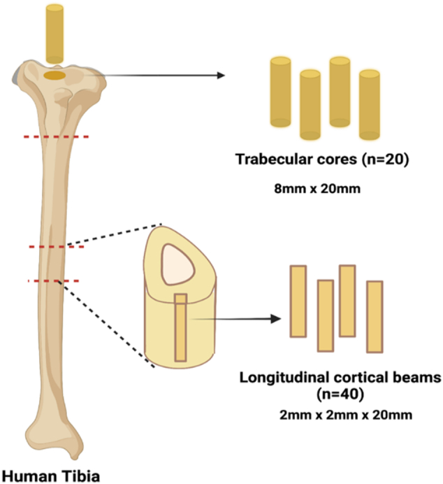
Schematic illustrating location and orientation of bone specimens excised from human tibias.
From the donors, the tibial plateau was secured in a wood vice and placed on a drill press (WEN 12” Variable Speed Drill Press). A diamond-tipped coring drill (Starlite Industries) was used to extract cylindrical cores of trabecular bone with dimensions of 8 mm × 20 mm. 3–4 cores were extracted from each donor, of which 2 intact cores/donor were used, yielding 20 intact cores (n = 20) (Fig. 1). Two cores were randomly assigned to a group: control (n = 10) or ribose (n = 10), such that each experimental group had even but randomized distribution of cores from all donors.
All specimens were wrapped in saline-soaked gauze and stored at −20 °C when not in use.
2.2. In vitro incubation
A buffer solution was created with 25 mM amino-n-caproic acid, 5 mM benzamidine, 10 mM n-ethylmaleimide, and 30 mM HEPES in Hanks' Balanced Salt Solution as done in previous studies (Tang and Vashishth, 2011; Vashishth et al., 2001; Viguet-Carrin et al., 2008a). Half of this solution was designated as the control solution, while the other half was then mixed with d-ribose to create a 0.6 M ribose solution. Bone specimens were submerged in corresponding solutions with 0.1875 L of solution per bone sample based on pilot studies conducted in our laboratory. Specimens were incubated for 10 days at 37 °C, with pH maintained daily between 7.2 and 7.6. Bone specimens of 2 × 2 × 2 mm dimensions were cut from the ends of incubated beams and 2 mm thick core specimens were cut and stored at −80 °C after incubation for crosslink analysis.
2.3. Micro-computed tomography imaging
All samples were scanned using a high-resolution microcomputed tomography imaging system (μCT40, Scanco Medical AG, Brüttisellen, Switzerland). Trabecular cores were scanned with a 15 μm isotropic voxel size, 70 kVp peak x-ray tube potential, 114 uA x-ray tube intensity, and 300 ms integration time. Trabecular microarchitecture was evaluated with the standard trabecular morphology analysis script in the Scanco Evaluation program. A threshold of 375 mgHA/cm3 was used to segment trabecular bone during analysis. Cortical beams were scanned with a 12 μm isotropic voxel size, 70 kVp peak x-ray tube potential, 114 uA x-ray tube intensity, and 200 ms integration time. A threshold of 700 mgHA/cm3 was used to segment cortical bone during analysis.
2.4. Mechanical testing
2.4.1. Cyclic reference point indentation
Tissue level cortical bone mechanical properties were assessed by cyclic reference point indentation (cRPI) via 3 indents per beam for 20 cycles at a maximum force of 6 N using a Biodent Hfc (Active Life Scientific, Santa Barbara, CA).
2.4.2. Fracture toughness testing
Cortical beams were tested via notched fracture toughness tests in four-point bending configuration with notched side in tension following protocols in our prior work (Merlo et al., 2020). We use a four-point bending test configuration, which generates a pure mode-I (opening loading conditions) around the crack tip as the shear stress is zero between the loading points, which is not induced by the three-point bending configuration.
Beams were thawed at room temperature for 30 min and notched on the periosteal surface with a diamond saw blade to a depth of ~250 μm. Then a razor blade was inserted in the notch to create a crack by dropping a 50 g weight from 50 mm height onto the razor blade (average initial crack depth: 540 μm). Beams were then tested in random order and loaded at a rate of 0.001 mm/s until failure using a 5 kN load cell. Upper and lower spans of the bending fixture were set to 8 mm and 16 mm, respectively. The specimens were kept hydrated with saline.
A dot was marked on beams before testing to use as an indicator in images. An Allied Vision Prosilica CCD camera (Stadtroda, Germany) with a view field of ~5 × 5 mm was used to observe the fracture to determine the instant of crack initiation, which was used with the corresponding load data to calculate the fracture toughness, KIC using the following equations:
| (1) |
| (2) |
In these equations, K represents fracture toughness, c is the distance between the upper and lower supports divided by 2, P is the applied load, a is the initial crack depth, h is the height of the sample, and w is the width of the sample (Munz and Fett, 1999). is a geometric correction factor. An a/W of ¼ was considered here to provide enough duration for crack growth upon initiation. ASTM standards were used as guideline and followed as closely as possible.
As human bone exhibits a non-linear load-displacement behavior (Fig. 2), a non-linear fracture mechanics based J-fracture toughness is determined (Willett et al., 2019; Granke et al., 2016; Granke et al., 2015). This approach provides the fracture toughness from both elastic and plastic behavior. The J-value is determined using ASTM Standard E1820-15a, with the equation used for plane stress conditions as follows.
| (3) |
Where KIC is determined from Eq. (1), and
| (4) |
Where, Apl = area under force displacement curve, ηpl = 1.9 if the load-line displacement is used for Apl, BN = net specimen thickness and b0 = W – a (W = specimen width, a = crack length).
Fig. 2.

Load-displacement diagrams for (a) vehicle and (b) Ribose. Lower peak loads were observed in the ribose group compared to the vehicle group.
The elastic modulus (E) was obtained from traditional microindentation tests performed on these specimens, using a method described previously by our group (Merlo et al., 2019).
2.4.3. Compression testing
Compression experiments on twenty core specimens were conducted using a material testing machine (Shimadzu AGS-X Mechanical Tester). The quasi-static compression load was measured with a commercial load cell applied at a constant velocity rate of 0.001 mm/s. Axial displacement and strain were measured via a 5000 N load cell. Force-displacement data and geometry data obtained from microCT were used to calculate yield stress and strain, ultimate stress and strain, total strain energy, pre-and post-yield strain energy, ultimate toughness, and stiffness.
2.5. Measurement of total fluorescent advanced glycation end-products
Bone specimens saved for AGE quantification following incubation were defatted by three 15-min washes in 250 μL cold isopropyl ether while undergoing constant agitation. These samples were then lyophilized for 8 h using a FreeZone 2.5 Liter freeze dry system (Labconco, Kansas City, MO) and hydrolyzed based on the amount of dry mass in 6 N HCl (10 μL/mg bone) for 20 h at 110 °C. The resulting hydrolysates were centrifuged at 13,000 RPM at 4 °C to allow for the removal of unwanted debris. Centrifuged hydrolysates were stored at −80 °C in complete darkness before quantifying AGEs by a fluorometric assay. Total fluorescent AGEs were quantified and normalized to collagen content using previously published protocols (Vashishth et al., 2001; Viguet-Carrin et al., 2008a). Fluorescence for quinine standards and 150× diluted hydrolysates were measured at 360/460 nm excitation/emission using a Synergy HTX Multi-Mode Reader (BioTek, Winooski, VT). A chloramine-T solution was added to hydroxyproline standards (stock: 2000 μg/mL L-hydroxyproline per 0.001 N HCl) and the diluted hydrolysates. These solutions were incubated at room temperature to allow for the oxidation of hydroxyproline. Perchloric acid (3.15 M) was added to quench residual chloramine-T and then incubated at room temperature. A p-dimethylaminobenzaldehyde solution was also added and incubated at 60 °C. All standards were left to cool at room temperature in complete darkness. Absorbance was measured at 570 nm using a microplate reader. Collagen content was measured based on the quantity of hydroxyproline quantity (Ignat’eva et al., 2007). Total fluorescent AGEs were quantified in terms of ng quinine/mg collagen.
2.6. Measurement of carboxymethyl-lysine
CML was quantified in all cores (n = 20) and beams (n = 40). The samples were defatted by three 15-min washes in 250 μL cold isopropyl ether while undergoing constant agitation. These samples were then lyophilized for 8 h using a FreeZone 2.5 Liter freeze dry system (Labconco, Kansas City, MO) and cut into 50–80 mg pieces for protein extraction. The bone pieces were homogenized in 0.5 M EDTA solution using a bead mill homogenizer (Omnibead Ruptor 6) for 90–100 cycles at a speed of 4350 rpm, with 45-s intervals between the cycles. 2 mL pre-filled bead mill tubes with stainless steel beads were used for homogenizing the cubes. The homogenized bone samples were then demineralized in 1 mL of 0.5 M EDTA solution at 4 °C for 6 days, with EDTA changes alternate days, by centrifugation at 4000 g. The insoluble crosslinked organic matrix, leftover after demineralization, was separated by centrifugation at 13000 rpm for 15 min. The supernatant was discarded, and the pellet that consists of crosslinked collagen was reserved. This collagen is then solubilized by heat denaturation and trypsin digestion. The pellet was dissolved in 100 μL Hank's buffer and heated at 85 °C for 10 min, followed by trypsin digestion using100 μL of 1 mg/mL trypsin solution for 16 h at 37 °C. The samples were heated again at 85 °C for 10 mins and trypsinized with 50 μL of 1 mg/mL trypsin to digest any remaining collagen for 3 h at 37 °C (Thomas et al., 2018).
All CML content was measured for all demineralized samples with a commercially available CML Competitive ELISA kit (catalog # STA-816, Cell BioLabs, San Diego, CA) following the manufacturer's protocol. The bone protein extract was diluted with kit-provided diluent to fall within the measurable concentration range of the kit and measured in triplicates. The protein content of the final sample was assessed using a Bradford assay with a BSA standard, and all samples were diluted to have a final protein concentration of 1 mg. CML levels are reported as ng of CML per mg of protein (Thomas et al., 2018).
2.7. Statistical analysis
With post-hoc power analysis, the sample size was found to be sufficient for our primary outcomes i.e., fAGEs (d = 2.4 beams, 1.31 cores, for α = 0.05,1-β = 0.95) and CML (d = 1.6beams, d = 6.01cores, for α = 0.05,1-β = 0.95). Outliers were determined using boxplots as values beyond two standard deviations from the mean of each group. An analysis of covariance (ANCOVA) was conducted to determine differences between groups for all variables, with donor age considered as a confounding variable. Data were tested for normality prior to linear regression and statistical analysis of correlations. Spearman correlations were run between total fluorescent AGEs and mechanical properties for both trabecular cores and cortical beams. Differences in fAGEs and CML between trabecular and cortical specimens in the control and ribose-incubated groups were determined using t-tests. All statistical analyses were performed using SPSS and GraphPad Prism 9.
3. Results
3.1. Comparison of control and ribose-incubated groups
In cortical bone, fAGEs and CML levels were significantly higher in ribose-incubated bone compared to control by +217.3 % (p < 0.0001) and +390.8 % (p < 0.0001), respectively (Table 1, Fig. 3-A, B). In trabecular bone, fAGEs and CML levels were also significantly higher in ribose-incubated bone compared to control by +102.6 % (p < 0.001) and + 600.1 % (p < 0.0001), respectively (Table 2, Fig. 3-C, D).
Table 1.
Mean and standard deviation of microCT properties, AGE measurements, and mechanical properties per treatment group in human cortical beams. The sample size was same for all measurements assessed.
| Control (n = 20) | Ribose (n = 20) | % Change | |
|---|---|---|---|
| MicroCT properties | |||
| Ct.TMD (mg HA/ccm) | 1095.9 ± 25.7 | 1093.2 ± 23.5 | – |
| Ct.Po (%) | 14.2 ± 11.4 | 17.9 ± 12.3 | – |
| AGE measurements | |||
| fAGEs (ng quinine/mg collagen) | 270.7 ± 138.0 | ⁎⁎⁎859.0 ± 317.1 | +217.3 % |
| CML (ng/mg of protein) | 188.4 ± 258.3 | ⁎⁎⁎924.6 ± 576.3 | +390.8 % |
| Mechanical properties | |||
| Stress intensity factor (KIC) | 5.43 ± 1.98 | 4.40 ± 0.55 | – |
| J-fracture toughness (kJ/m2) | 17.74 ± 7.8 | 17.15 ± 8,2 | – |
| Indentation Distance (ID, μm) | 65.1 ± 11.3 | 67.0 ± 8.1 | – |
| Creep Indentation Distance (CID, μm) | 5.2 ± 1.0 | 5.7 ± 1.2 | – |
| Total Indentation Distance (TID, μm) | 71.6 ± 12.6 | 73.9 ± 9.3 | – |
| Indentation Distance Increase (IDI, μm) | 11.0 ± 2.8 | 11.8 ± 2.5 | – |
| Average Energy Dissipated (Avg ED, μJ) | 17.1 ± 2.9 | 17.3 ± 3.0 | – |
| Average Unloading Slope (Avg US, N/μm) | 0.45 ± 0.03 | 0.45 ± 0.04 | – |
| Average Loading Slope (Avg LS, N/μm) | 0.33 ± 0.03 | 0.33 ± 0.03 | |
Differences were determined by ANCOVA with age as a confounding variable.
⁎ p ≤ 0.05.
⁎⁎ p ≤ 0.01.
p ≤ 0.0001.
Fig. 3.

CML and fAGE levels in ribose incubated bone vs control in cortical beams (A, C) and trabecular cores (B, D).
Table 2.
Mean and standard deviation of microCT properties, AGE measurements, and mechanical properties per treatment group in human trabecular cores. The sample size was same for all measurements assessed.
| Control (n = 20) | Ribose (n = 20) | % Change | |
|---|---|---|---|
| MicroCT properties | |||
| BV/TV (%) | 11.7 ± 3.3 | 12.9 ± 4.6 | – |
| Conn.D (1/mm3) | 6.4 ± 1.5 | 5.6 ± 1.5 | – |
| SMI | 2.0 ± 0.2 | 2.0 ± 0.6 | – |
| Tb.N (1/mm) | 1.44 ± 0.15 | 1.38 ± 0.11 | – |
| Tb.Th (mm) | 0.13 ± 0.01 | 0.14 ± 0.02 | – |
| Tb.Sp (mm) | 0.68 ± 0.07 | 0.69 ± 0.07 | – |
| AGE measurements | |||
| fAGEs (ng quinine/mg collagen) | 260.4 ± 110.4 | ⁎⁎687.7 ± 444.8 | +164 % |
| CML (ng/mg of protein) | 884.2 ± 896.9 | ⁎⁎⁎6189.9 ± 866.0 | +600.1 % |
| Mechanical properties | |||
| Ultimate stress (MPa) | 1.8 ± 1.2 | 1.8 ± 1.0 | – |
| Ultimate strain | 0.06 ± 0.03 | 0.05 ± 0.02 | – |
| Pre-yield energy (mJ) | 0.005 ± 0.005 | 0.010 ± 0.018 | – |
| Post-yield energy (mJ) | 0.070 ± 0.053 | 0.041 ± 0.039 | – |
| Total energy (mJ) | 0.076 ± 0.058 | 0.057 ± 0.037 | – |
Differences were determined by ANCOVA with age as a confounding variable.
⁎p ≤ 0.05.
p ≤ 0.01.
p ≤ 0.0001.
In cortical bone, we observed no significant differences in variables assessed by microCT such as cortical tissue mineral density (Ct. TMD) or cortical porosity (Ct. Po) or any mechanical properties assessed by cRPI. We found no significant differences between crack initiation toughness (KIC) and crack propagation toughness (J-int) (Table 1, Fig. 4). Similarly, in trabecular bone, no differences were observed in microarchitectural variables or mechanical properties (Table 2).
Fig. 4.
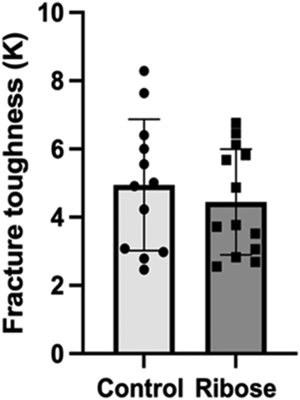
Fracture toughness (KIC) measurements were not different between control and ribose incubated groups (p = 0.645).
We found fAGEs were higher in cortical bone compared to trabecular bone in both control (3.9 %) and ribose groups (25 %). On the other hand, CML levels were higher in trabecular bone compared to cortical bone in both control (4.7 %) and ribose groups (6.7 %) (Table 1, Table 2).
3.2. Correlations between fluorescent advanced glycation end-products, carboxymethyl-lysine, microarchitecture, and mechanical properties
In cortical bone, we observed no significant correlations between fAGEs, CML, microarchitectural and mechanical properties in control or ribose incubated groups. However, when the two groups were pooled together, we found CML was positively correlated to fAGEs (r = +0.598, p ≤ 0.05) (Fig. 5), cRPI variables such as average energy dissipated (r = +0.465, p ≤ 0.05), indentation depth (r = +0.460, p ≤ 0.05), total indentation depth (r = +0.440, p ≤ 0.05) (Fig. 6) and microCT variable such as Ct. Po (r = +0.505, p ≤ 0.05) (Fig. 7). fAGEs showed a trend towards negative association with crack propagation toughness(J-int) (r = −0.365, p = 0.055).
Fig. 5.

Spearman's correlation between CML and fAGEs in cortical bone (A) and trabecular bone (B).
Fig. 6.

Spearman's correlation between CML and variables assessed by cyclic reference point indentation (A) CML and total indentation depth, (B) CML and indentation depth and (C) CML and average energy dissipated.
Fig. 7.
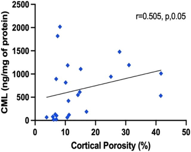
Spearman's correlation between CML and cortical porosity as assessed by microCT scan.
In trabecular bone, we found fAGEs were correlated to Tb. N (r = +0.833, p ≤ 0.005) in the control group, but no other correlation was observed between fAGEs and other parameters in control, ribose, or pooled groups. We found no correlations between CML and other parameters in the control, ribose, or pooled group.
4. Discussion
A growing body of literature indicates advanced glycation end-products (AGEs) correlate with deteriorated bone mechanical properties; however, individual AGEs may serve as better markers than bulk measurement of fluorescent AGEs (fAGEs) (Tang and Vashishth, 2011; Tang et al., 2007; Ural et al., 2015; Hunt et al., 2019; Karim and Vashishth, 2012; Thomas et al., 2018). Our goal was to understand how the individual non-crosslinking AGE such as carboxymethyl-lysine (CML) relates to fAGEs and bone's mechanical properties and how it differs between trabecular and cortical bone types. This study reports that CML levels are significantly higher in trabecular bone than cortical bone, and in cortical bone are positively correlated to fAGE levels and deteriorated mechanical properties.
In this study, we use an established method of in vitro incubation with ribose sugar to induce CML formation and other AGEs (Thomas et al., 2018; Chen et al., 2019; Viguet-Carrin et al., 2008b). The CML levels we observed in cortical bone (200–1000 ng/mg of protein) were comparable to the CML levels reported in the literature (100–1000 ng/mg of protein) (Thomas et al., 2018), indicating that our in-vitro incubation protocol effectively induced a high level of AGEs. Firstly, we observed that CML levels are much higher in both cortical and trabecular bone compared to fAGEs. This is likely due to CML being a non-crosslinking AGE and forming much faster than other crosslinking AGEs. Secondly, we observed that CML levels are 6.7 % higher in the ribose group and 4.7 % higher in the control group in trabecular bone compared to cortical bone. This suggests that the porous nature of trabecular bone allows for better access of free-floating sugars to the amino acid residues within the collagen network, resulting in a higher accumulation of AGEs in trabecular bone than in cortical bone (Karim and Vashishth, 2012; Karim et al., 2013). Our findings are in line with a previous studies that have reported higher levels of CML in cortical (Thomas et al., 2018) and trabecular bone (Arakawa et al., 2020). Our study provides new information by showing the difference in accumulation of CML in cortical vs trabecular bone and preliminary evidence of its association with bone mechanical properties, which are not reported in earlier studies.
The CML content in cortical bone shows a moderate and significant positive correlation with fAGEs, suggesting that it may serve as a useful marker for glycation in bone along with fAGEs. CML but not fAGEs is positively correlated with microarchitecture and mechanical properties. Regarding microarchitecture, there was specifically a positive association between CML and cortical porosity in cortical bone. As cortical porosity is intimately linked to the bone remodeling and fracture toughness of bone (Tang and Vashishth, 2011; Granke et al., 2016), elevated levels of CML might play a role in altering bone remodeling with a consequent effect on cortical porosity, however since the cortical porosity in our ribose and control group was not significantly different, we don't observe much differences in the fracture toughness between these groups. Studies have reported changes in the function of osteoblasts and osteocytes in the presence of AGEs to indicate an altered bone remodeling (Vaidya et al., 2021; Tanaka et al., 2015; Suzuki et al., 2020), yet the role of CML in bone remodeling remains unknown. As we utilized cadaver bones devoid of live cells, we could not assess CML's role in turnover, and further work assessing CML's impact directly on the turnover process is needed to evaluate this scenario.
CML is also significantly correlated with bone mechanical properties assessed by cRPI. Higher indentation distances indicate that the indentation test probe travels deeper into the bone's surface, suggesting weaker material strength. Bulk AGEs have been previously shown to be correlated to bone material strength assessed using cRPI (Granke et al., 2015; Karim et al., 2018; Furst et al., 2016). Our results indicate that bones with higher levels of CML are easier to indent, indicating inferior bone tissue. Interestingly, CML is positively correlated with cRPI-derived energy dissipation, which is a measure of plasticity. We did not assess collagen denaturation or the amount of microcracking, which would need to be done in the future to offer insight into why energy dissipation is altered with higher levels of CML, but our study provides direction for future studies. No significant correlation was observed between CML and fracture toughness as observed in previous work (Thomas et al., 2018), however the authors used crack propagation toughness calculated elsewhere (Poundarik et al., 2015) and did not test it themselves. Post-hoc power analysis indicated our study was underpowered for fracture toughness data, thus analysis on a larger sample size is necessary to clarify this finding. Unlike CML, fAGEs did not correlate with microarchitectural and mechanical properties in cortical bone. Several reports in literature of fAGEs deteriorating mechanical and material properties of cortical bone suggest that using a broader age range of specimens than that used in our present study may be needed to determine the relationship between fAGEs and bone's mechanical behavior (Merlo et al., 2019; Furst et al., 2016; Karim et al., 2019b; Yamamoto and Sugimoto, 2016). CML levels were orders of magnitude higher in our ribose incubated cortical bones and clearly indicated a relationship with mechanical properties even within our limited age range, further emphasizing its potential to serve as a useful biomarker in cortical bone.
Unlike cortical bone, CML did not relate with fAGEs, or any mechanical properties assessed by compression tests in trabecular bone. Trabecular bone is highly porous, heterogeneous, has a high surface area to volume ratio, and thus has more active bone remodeling than cortical bone (Vaidya et al., 2021; Eriksen, 2010). Since the effect of CML on active remodeling cannot be tested on cadaver bones, future studies on bone cells and organ culture could help elucidate the effect of CML on bone's mechanical behavior. Additional tests with a larger sample size might be needed to identify any relationships with CML in trabecular bone.
Our results should be considered in light of a few limitations. Our study uses cadaveric bone from females over 50 years old. This age group is clinically relevant due to the prevalence of fractures in older women, but our results only represent this group, and findings may not be the same for bone acquired from younger and male donors. Although these donors were not identified as having diabetes, there is no record of their blood sugar levels, which could impact the results of CML and fAGE accumulation. Further, although our study was well powered for fAGE and CML measurement, post-hoc power analysis indicates that the study was underpowered to detect differences in mechanical and microarchitecture properties. This may help explain why there were no significant differences detected in these variables between groups despite the difference in AGE content. Future work must include a larger sample size to further clarify our findings. It should be noted that we report CML as ng/ml of protein as quantified by Bradfords assay. Thus, we do not compare the CML levels directly to fAGE (ng/mg collagen) levels. We use a ribosylation method to amplify AGEs in the bone samples, however, it should be noted that ribose favors formation of pentosidine (pentose-derived) over other fAGEs (Chen et al., 2019; Alsamad et al., 2021). The fAGEs induced by ribosylation does not allow us to distinguish between non-crosslinking and crosslinking AGEs and quantifies only a small portion of fAGEs, but ribosylation is favored due to its ability to amplify AGEs in fewer days compared to glucose incubation. Although CML is one of the many AGEs that might serve as an important biomarker for glycation and other AGEs, this study helps direct our future work to identify and quantify other critical AGEs. It is also unknown whether non-crosslinking and crosslinking AGEs affect the mechanical properties of bone differently, which may be important to consider in studies hereafter. Further, it should be noted that although our study normalizes fAGEs to collagen content, there is no distinction between fAGEs formed on collagen vs non-collagenous proteins. Lastly, during fracture toughness testing, the crack propagates so suddenly after crack initiation, that our digital camera did not have enough framing speed to capture the event, thus we have no record of crack length vs time information during the propagation, and we are unable to generate R-curves in this study. To compensate for it, we employed ASTM Standard E1820-15a and determined J-based fracture toughness that includes both Jelastic (based on crack initiation conditions) and Jplastic (based on crack propagation conditions). Further work in larger sample sizes in a broader age range with additional mechanical characterization is needed to confirm these results. Additionally, we need to understand the pathways through which CML may have this kind of impact on bone mechanics. One such pathway is through changes in bone turnover. As CML is involved in the receptor for AGE (RAGE) signaling (Gaens et al., 2014), more studies should aim to assess the effect of CML on bone cells to determine if it alters turnover through RAGE signaling changes.
Despite these limitations, we report differences in CML levels in human cortical and trabecular bone, and show preliminary evidence of its association with bone material and mechanical properteis. Altogether, our results show that CML may impact the mechanical behavior of bone and could be a useful marker for non-crosslinking glycation or early glycation changes and warrants further investigation.
CRediT authorship contribution statement
Rachana Vaidya: Methodology, Formal analysis, Investigation, Writing – original draft, Writing – review & editing, Visualization. Taraneh Rezaee: Methodology, Formal analysis, Investigation, Writing – original draft, Writing – review & editing, Visualization. Tianna Edwards: Methodology, Formal analysis, Investigation, Writing – original draft, Writing – review & editing, Visualization. Richard Bender: Methodology, Investigation, Writing – review & editing. Arune Vickneswaran: Methodology, Formal analysis, Investigation, Writing – review & editing. Vijaya Chalivendra: Methodology, Formal analysis, Writing – review & editing, Visualization, Supervision. Lamya Karim: Methodology, Formal analysis, Writing – original draft, Writing – review & editing, Visualization, Resources, Supervision, Project administration, Funding acquisition.
Declaration of competing interest
None of the authors have any other conflicts of interest to declare.
Acknowledgments
Donor bones were acquired with funding by the Multidisciplinary Seed Funding Program from the Office of the Provost at the University of Massachusetts Dartmouth. Lamya Karim is currently supported by the National Institutes of Health (award # K01AR069685). The content is solely the responsibility of the authors and does not necessarily represent the official views of the funding sources.
We would like to thank Mohammed Shonar for his help with the MATLAB code.
Contributor Information
Rachana Vaidya, Email: vaidya@wustl.edu.
Taraneh Rezaee, Email: trezaee@ccny.cuny.edu.
Tianna Edwards, Email: tedwards1896@gmail.com.
Richard Bender, Email: rbender264@gmail.com.
Arune Vickneswaran, Email: avickneswaran@umassd.edu.
Vijaya Chalivendra, Email: vchalivendra@umassd.edu.
Lamya Karim, Email: lkarim@umassd.edu.
Data availability
Data will be made available on request.
References
- Ahmed N., Thornalley P. Advanced glycation endproducts: what is their relevance to diabetic complications? Diabetes. Obes. Metab. 2007;9(3):233–245. doi: 10.1111/j.1463-1326.2006.00595.x. [DOI] [PubMed] [Google Scholar]
- Alsamad F., et al. In depth investigation of collagen non-enzymatic glycation by raman spectroscopy. Spectrochim. Acta A Mol. Biomol. Spectrosc. 2021;251 doi: 10.1016/j.saa.2020.119382. [DOI] [PubMed] [Google Scholar]
- Arakawa S., et al. Mass spectrometric quantitation of AGEs and enzymatic crosslinks in human cancellous bone. Sci. Rep. 2020;10(1):18774. doi: 10.1038/s41598-020-75923-8. [DOI] [PMC free article] [PubMed] [Google Scholar]
- Ban I., Sugawa H., Nagai R. Protein modification with ribose generates N δ-(5-hydro-5-methyl-4-imidazolone-2-yl)-ornithine. Int. J. Mol. Sci. 2022;23(3):1224. doi: 10.3390/ijms23031224. [DOI] [PMC free article] [PubMed] [Google Scholar]
- Barzilay J.I., et al. Circulating levels of carboxy-methyl-lysine (CML) are associated with hip fracture risk: the cardiovascular health study. J. Bone Miner. Res. Off. J. Am. Soc. Bone Miner. Res. 2014;29(5):1061–1066. doi: 10.1002/jbmr.2123. [DOI] [PMC free article] [PubMed] [Google Scholar]
- Chen Y., et al. D-ribose contributes to the glycation of serum protein. Biochim. Biophys. Acta (BBA) - Mol. Basis Dis. 2019;1865(9):2285–2292. doi: 10.1016/j.bbadis.2019.05.005. [DOI] [PubMed] [Google Scholar]
- Dyer D.G., et al. Accumulation of maillard reaction products in skin collagen in diabetes and aging. J. Clin. Invest. 1993;91(6):2463–2469. doi: 10.1172/JCI116481. [DOI] [PMC free article] [PubMed] [Google Scholar]
- Eriksen E.F. Cellular mechanisms of bone remodeling. Rev. Endocr. Metab. Disord. 2010;11(4):219–227. doi: 10.1007/s11154-010-9153-1. [DOI] [PMC free article] [PubMed] [Google Scholar]
- Furst J.R., et al. Advanced glycation endproducts and bone material strength in type 2 diabetes. J. Clin. Endocrinol. Metab. 2016;101(6):2502–2510. doi: 10.1210/jc.2016-1437. [DOI] [PMC free article] [PubMed] [Google Scholar]
- Gaens K.H., et al. Nε-(carboxymethyl)lysine-receptor for advanced glycation end product axis is a key modulator of obesity-induced dysregulation of adipokine expression and insulin resistance. Arterioscler. Thromb. Vasc. Biol. 2014;34(6):1199–1208. doi: 10.1161/ATVBAHA.113.302281. [DOI] [PubMed] [Google Scholar]
- Genuth S., et al. Glycation and carboxymethyllysine levels in skin collagen predict the risk of future 10-year progression of diabetic retinopathy and nephropathy in the diabetes control and complications trial and epidemiology of diabetes interventions and complications participants with type 1 diabetes. Diabetes. 2005;54(11):3103–3111. doi: 10.2337/diabetes.54.11.3103. [DOI] [PMC free article] [PubMed] [Google Scholar]
- Gkogkolou P., Böhm M. Advanced glycation end products: key players in skin aging? Dermato-endocrinology. 2012;4(3):259–270. doi: 10.4161/derm.22028. [DOI] [PMC free article] [PubMed] [Google Scholar]
- Grandhee S.K., Monnier V.M. Mechanism of formation of the maillard protein cross-link pentosidine. Glucose, fructose, and ascorbate as pentosidine precursors. J. Biol. Chem. 1991;266(18):11649–11653. [PubMed] [Google Scholar]
- Granke M., et al. Identifying novel clinical surrogates to assess human bone fracture toughness. J. Bone Miner. Res. 2015;30(7):1290–1300. doi: 10.1002/jbmr.2452. [DOI] [PMC free article] [PubMed] [Google Scholar]
- Granke M., et al. Prevalent role of porosity and osteonal area over mineralization heterogeneity in the fracture toughness of human cortical bone. J. Biomech. 2016;49(13):2748–2755. doi: 10.1016/j.jbiomech.2016.06.009. [DOI] [PMC free article] [PubMed] [Google Scholar]
- Hein G., et al. Advanced glycation end-products pentosidine and N epsilon-carboxymethyllysine are elevated in serum of patients with osteoporosis. Rheumatology (Oxford) 2003;42(10):1242–1246. doi: 10.1093/rheumatology/keg324. [DOI] [PubMed] [Google Scholar]
- Huebschmann A.G., et al. Diabetes and advanced glycoxidation end products. Diabetes Care. 2006;29(6):1420–1432. doi: 10.2337/dc05-2096. [DOI] [PubMed] [Google Scholar]
- Hunt H.B., et al. Altered tissue composition, microarchitecture, and mechanical performance in cancellous bone from men with type 2 diabetes mellitus. J. Bone Miner. Res. 2019;34(7):1191–1206. doi: 10.1002/jbmr.3711. [DOI] [PMC free article] [PubMed] [Google Scholar]
- Ignat’eva N.Y., et al. Determination of hydroxyproline in tissues and the evaluation of the collagen content of the tissues. Journal of Analytical Chemistry. 2007;62(1):51–57. [Google Scholar]
- Illien-Jünger S., et al. Dietary advanced glycation end products have sex-and age-dependent effects on vertebral bone microstructure and mechanical function in mice. J. Bone Miner. Res. 2018;33(3):437–448. doi: 10.1002/jbmr.3321. [DOI] [PMC free article] [PubMed] [Google Scholar]
- Karim L., Vashishth D. Heterogeneous glycation of cancellous bone and its association with bone quality and fragility. PLoS One. 2012;7(4) doi: 10.1371/journal.pone.0035047. [DOI] [PMC free article] [PubMed] [Google Scholar]
- Karim L., et al. Differences in non-enzymatic glycation and collagen cross-links between human cortical and cancellous bone. Osteoporos. Int. 2013;24(9):2441–2447. doi: 10.1007/s00198-013-2319-4. [DOI] [PMC free article] [PubMed] [Google Scholar]
- Karim L., et al. Bone microarchitecture, biomechanical properties, and advanced glycation end-products in the proximal femur of adults with type 2 diabetes. Bone. 2018;114:32–39. doi: 10.1016/j.bone.2018.05.030. [DOI] [PMC free article] [PubMed] [Google Scholar]
- Karim L., Rezaee T., Vaidya R. The effect of type 2 diabetes on bone biomechanics. Curr. Osteoporos. Rep. 2019;17(5):291–300. doi: 10.1007/s11914-019-00526-w. [DOI] [PMC free article] [PubMed] [Google Scholar]
- Karim L., Rezaee T., Vaidya R. The effect of type 2 diabetes on bone biomechanics. Curr. Osteoporos. Rep. 2019;17(5):291–300. doi: 10.1007/s11914-019-00526-w. [DOI] [PMC free article] [PubMed] [Google Scholar]
- Meng J., et al. Carboxymethyllysine in dermal tissues of diabetic and nondiabetic patients with chronic renal failure: relevance to glycoxidation damage. Nephron. 2001;88(1):30–35. doi: 10.1159/000045955. [DOI] [PubMed] [Google Scholar]
- Merlo K., et al. In vitro-induced high sugar environments deteriorate human cortical bone elastic modulus and fracture toughness. J. Orthop. Res. 2019;38 doi: 10.1002/jor.24543. [DOI] [PMC free article] [PubMed] [Google Scholar]
- Merlo K., et al. In vitro-induced high sugar environments deteriorate human cortical bone elastic modulus and fracture toughness. J. Orthop. Res. 2020;38(5):972–983. doi: 10.1002/jor.24543. [DOI] [PMC free article] [PubMed] [Google Scholar]
- Miura J., et al. Serum levels of non-carboxymethyllysine advanced glycation endproducts are correlated to severity of microvascular complications in patients with type 1 diabetes. J. Diabetes Complicat. 2003;17(1):16–21. doi: 10.1016/s1056-8727(02)00183-6. [DOI] [PubMed] [Google Scholar]
- Moldogazieva N.T., et al. Oxidative stress and advanced lipoxidation and glycation end products (ALEs and AGEs) in aging and age-related diseases. Oxidative Med. Cell. Longev. 2019;2019 doi: 10.1155/2019/3085756. [DOI] [PMC free article] [PubMed] [Google Scholar]
- Moseley K.F., et al. Advanced glycation endproducts and bone quality: practical implications for people with type 2 diabetes. Curr. Opin. Endocrinol. Diabetes Obes. 2021;28(4):360–370. doi: 10.1097/MED.0000000000000641. [DOI] [PubMed] [Google Scholar]
- Munz D., Fett T. Springer; Berlin, New York, NY: 1999. Ceramics: Mechanical Properties, Failure Behaviour, Materials Selection. [Google Scholar]
- Neumann T., et al. High serum pentosidine but not esRAGE is associated with prevalent fractures in type 1 diabetes independent of bone mineral density and glycaemic control. Osteoporosis Int. 2014;25(5):1527–1533. doi: 10.1007/s00198-014-2631-7. [DOI] [PubMed] [Google Scholar]
- Poundarik A.A., et al. A direct role of collagen glycation in bone fracture. J. Mech. Behav. Biomed. Mater. 2015;52:120–130. doi: 10.1016/j.jmbbm.2015.08.012. [DOI] [PMC free article] [PubMed] [Google Scholar]
- Rubin M.R., et al. Advanced glycation endproducts and bone material properties in type 1 diabetic mice. PLoS ONE. 2016;11(5) doi: 10.1371/journal.pone.0154700. [DOI] [PMC free article] [PubMed] [Google Scholar]
- Schalkwijk C.G., et al. Increased accumulation of the glycoxidation product nepsilon-(carboxymethyl)lysine in hearts of diabetic patients: generation and characterisation of a monoclonal anti-CML antibody. Biochim. Biophys. Acta. 2004;1636(2–3):82–89. doi: 10.1016/j.bbalip.2003.07.002. [DOI] [PubMed] [Google Scholar]
- Schleicher E.D., Wagner E., Nerlich A.G. Increased accumulation of the glycoxidation product N(epsilon)-(carboxymethyl)lysine in human tissues in diabetes and aging. J. Clin. Invest. 1997;99(3):457–468. doi: 10.1172/JCI119180. [DOI] [PMC free article] [PubMed] [Google Scholar]
- Schwartz A.V., et al. Pentosidine and increased fracture risk in older adults with type 2 diabetes. J. Clin. Endocrinol. Metab. 2009;94(7):2380–2386. doi: 10.1210/jc.2008-2498. [DOI] [PMC free article] [PubMed] [Google Scholar]
- Seeman E., Delmas P.D. Bone quality–the material and structural basis of bone strength and fragility. N. Engl. J. Med. 2006;354(21):2250–2261. doi: 10.1056/NEJMra053077. [DOI] [PubMed] [Google Scholar]
- Shiraki M., et al. Nonenzymatic collagen cross-links induced by glycoxidation (pentosidine) predicts vertebral fractures. J. Bone Miner. Metab. 2008;26(1):93–100. doi: 10.1007/s00774-007-0784-6. [DOI] [PubMed] [Google Scholar]
- Singh R., et al. Advanced glycation end-products: a review. Diabetologia. 2001;44(2):129–146. doi: 10.1007/s001250051591. [DOI] [PubMed] [Google Scholar]
- Su J., et al. Role of advanced glycation end products with oxidative stress in resistance artery dysfunction in type 2 diabetic mice. Arterioscler. Thromb. Vasc. Biol. 2008;28(8):1432–1438. doi: 10.1161/ATVBAHA.108.167205. [DOI] [PMC free article] [PubMed] [Google Scholar]
- Suzuki R., et al. Intracellular accumulation of advanced glycation end products induces osteoblast apoptosis via endoplasmic reticulum stress. J. Bone Miner. Res. 2020;35(10):1992–2003. doi: 10.1002/jbmr.4053. [DOI] [PubMed] [Google Scholar]
- Tanaka K., et al. Effects of high glucose and advanced glycation end products on the expressions of sclerostin and RANKL as well as apoptosis in osteocyte-like MLO-Y4-A2 cells. Biochem. Biophys. Res. Commun. 2015;461(2):193–199. doi: 10.1016/j.bbrc.2015.02.091. [DOI] [PubMed] [Google Scholar]
- Tang S., Vashishth D. The relative contributions of non-enzymatic glycation and cortical porosity on the fracture toughness of aging bone. J. Biomech. 2011;44(2):330–336. doi: 10.1016/j.jbiomech.2010.10.016. [DOI] [PMC free article] [PubMed] [Google Scholar]
- Tang S.Y., Zeenath U., Vashishth D. Effects of non-enzymatic glycation on cancellous bone fragility. Bone. 2007;40(4):1144–1151. doi: 10.1016/j.bone.2006.12.056. [DOI] [PMC free article] [PubMed] [Google Scholar]
- Thomas C.J., et al. Accumulation of carboxymethyl-lysine (CML) in human cortical bone. Bone. 2018;110:128–133. doi: 10.1016/j.bone.2018.01.028. [DOI] [PMC free article] [PubMed] [Google Scholar]
- Ural A., et al. Association between non-enzymatic glycation, resorption, and microdamage in human tibial cortices. Osteoporos. Int. 2015;26(3):865–873. doi: 10.1007/s00198-014-2938-4. [DOI] [PMC free article] [PubMed] [Google Scholar]
- Vaidya R., Church A., Karim L. In: The Science, Etiology and Mechanobiology of Diabetes and its Complications. Gefen A., editor. Academic Press; 2021. Chapter 18 - effect of type 2 diabetes on bone cell behavior; pp. 313–326. [Google Scholar]
- Vashishth D., et al. Influence of nonenzymatic glycation on biomechanical properties of cortical bone. Bone. 2001;28(2):195–201. doi: 10.1016/s8756-3282(00)00434-8. [DOI] [PubMed] [Google Scholar]
- Verzijl N., et al. Crosslinking by advanced glycation end products increases the stiffness of the collagen network in human articular cartilage: a possible mechanism through which age is a risk factor for osteoarthritis. Arthritis Rheum. 2002;46(1):114–123. doi: 10.1002/1529-0131(200201)46:1<114::AID-ART10025>3.0.CO;2-P. [DOI] [PubMed] [Google Scholar]
- Viguet-Carrin S., et al. Contribution of the advanced glycation end product pentosidine and of maturation of type I collagen to compressive biomechanical properties of human lumbar vertebrae. Bone. 2006;39(5):1073–1079. doi: 10.1016/j.bone.2006.05.013. [DOI] [PubMed] [Google Scholar]
- Viguet-Carrin S., et al. An in vitro model to test the contribution of advanced glycation end products to bone biomechanical properties. Bone. 2008;42(1):139–149. doi: 10.1016/j.bone.2007.08.046. [DOI] [PubMed] [Google Scholar]
- Viguet-Carrin S., et al. An in vitro model to test the contribution of advanced glycation end products to bone biomechanical properties. Bone. 2008;42(1):139–149. doi: 10.1016/j.bone.2007.08.046. [DOI] [PubMed] [Google Scholar]
- Willett T.L., et al. Bone collagen network integrity and transverse fracture toughness of human cortical bone. Bone. 2019;120:187–193. doi: 10.1016/j.bone.2018.10.024. [DOI] [PMC free article] [PubMed] [Google Scholar]
- Yamamoto M., Sugimoto T. Advanced glycation end products, diabetes, and bone strength. Curr. Osteoporos. Rep. 2016;14(6):320–326. doi: 10.1007/s11914-016-0332-1. [DOI] [PMC free article] [PubMed] [Google Scholar]
Associated Data
This section collects any data citations, data availability statements, or supplementary materials included in this article.
Data Availability Statement
Data will be made available on request.