Abstract
Pathogenic mycobacteria must acquire iron in the host in order to multiply and cause disease. To do so, they release abundant quantities of siderophores called exochelins, which have the capacity to scavenge iron from host iron-binding proteins and deliver it to the mycobacteria. In this study, we have characterized the exochelins of Mycobacterium bovis, the causative agent of bovine and occasionally of human tuberculosis, and the highly attenuated descendant of M. bovis, bacillus Calmette-Guérin (BCG), widely used as a vaccine against human tuberculosis. The M. bovis type strain, five substrains of M. bovis BCG (Copenhagen, Glaxo, Japanese, Pasteur, and Tice), and two strains of virulent Mycobacterium tuberculosis all produce the same set of exochelins, although the relative amounts of individual exochelins may differ. Among these mycobacteria, the total amount of exochelins produced is greatest in M. tuberculosis, intermediate in M. bovis, and smallest in M. bovis BCG.
Exochelins are water-soluble iron siderophores secreted by mycobacteria to scavenge iron from their extracellular milieu (9, 10). Mycobacteria produce two general types of exochelins (12). The pathogenic mycobacteria produce chloroform-soluble exochelins which have a structure similar to that of mycobactin, another high-affinity iron-binding molecule of mycobacteria that is water insoluble and is located in the cell wall (1). The saprophytic mycobacteria produce chloroform-insoluble exochelins which have a peptide-like structure (14, 15).
In previous studies, we have isolated and characterized by mass spectrometry (MS) the exochelins of the pathogenic mycobacteria Mycobacterium tuberculosis and Mycobacterium avium (7, 20). These mycobactin-like exochelins contain 3 amino acid residues—2 N-hydroxylysines and 1 serine or threonine. In M. avium, the 3rd amino acid is always threonine (20). The difference between the exochelins and mycobactins of the same species resides exclusively in the R1 side chain (7). In mycobactins, the R1 side chain is a long-chain fatty acid. In exochelins, the side chain is a shorter aliphatic chain, either saturated or unsaturated, that terminates in either a methyl ester or a carboxylic acid moiety. These differences render the exochelins more polar than mycobactins and hence water soluble.
Exochelins of the pathogenic mycobacteria are thought to function by scavenging iron from host iron-binding molecules and transferring the iron to mycobactins associated with the cell wall. Exochelins of M. tuberculosis have been shown capable of removing iron from human transferrin and lactoferrin and donating it to mycobactins in the cell wall (6).
The exochelins of Mycobacterium bovis and its attenuated descendant bacillus Calmette-Guérin (BCG) have not been previously characterized. Therefore, in this study, we sought to isolate them and characterize them by MS.
M. bovis is a slow-growing pathogenic mycobacterium that belongs to the M. tuberculosis complex (M. africanum, M. bovis, M. microti, M. tuberculosis, and M. ulcerans). M. bovis is highly related to M. tuberculosis on the basis of DNA homology (19). M. bovis derives its importance from several factors. First, as the primary causative agent of bovine tuberculosis, it is of major commercial importance to the cattle and dairy industry. Second, it can cause human tuberculosis, mostly via ingestion of unpasteurized cow’s milk (5). With the advent of pasteurization and government programs to eradicate bovine tuberculosis, such cases are now rare in immunocompetent individuals. However, human immunodeficiency virus-infected persons appear to be at increased risk for such disease (2, 13, 16, 21). Third, M. bovis BCG remains the only available vaccine against tuberculosis. Although its efficacy has been questioned, the live vaccine has been administered to nearly 3 billion persons since 1945 (4). BCG is also administered intravesicularly in the treatment of bladder cancer. In rare cases, BCG can cause serious and sometimes fatal disseminated disease in immunocompromised persons (3, 8).
Isolation and purification of M. bovis exochelins.
We selected for study the M. bovis type strain (ATCC 19210) and five BCG substrains of different geographic origins that are currently being used by pharmaceutical companies in vaccines or for research purposes: Copenhagen (ATCC 27290), Glaxo (ATCC 35741), Japanese (ATCC 35737), Pasteur (ATCC 35734), and Tice (ATCC 35743). To obtain exochelins of M. bovis and M. bovis BCG, we cultured the bacteria in modified iron-deficient Sauton’s broth medium (1 μM iron; no Tween) in 1.9-liter tissue culture flasks, 300 ml per flask, without shaking at 37°C in 5% CO2–95% air for 3 to 8 weeks (7). All bacteria were grown in iron-deficient medium to enhance exochelin production, from an A540 of 0.05 to an A540 of >1. We purified the exochelins at a point at which production was maximal—6 weeks for M. tuberculosis and 8 weeks for M. bovis—as previously described (7). Briefly, the supernatant fluid was saturated with iron, and ferri-exochelins were extracted into chloroform. The chloroform extract was dried, and the exochelins were purified by reverse-phase high-pressure liquid chromatography (HPLC) on a C18 column (Vydac, Western Analytical, Temecula, Calif.) with a 50 to 100% gradient of buffer B (0.1% trifluoroacetic acid–50% acetonitrile) at a flow rate of 1 ml/min. Individual exochelins were further purified on an alkyl phenyl column (Waters, Bedford, Mass.) by using the same buffers and flow rate.
MS analysis of exochelins.
Peaks isolated from the HPLC and identified as potential exochelins were first subjected to mass analysis by matrix-assisted laser desorption ionization MS. For these experiments, a Voyager linear time-of-flight (TOF) mass spectrometer (PerSeptive Biosystems, Framington, Mass.) equipped with a 337-nm N2 laser was used. M. bovis exochelins were analyzed under delayed-extraction conditions as described by Vestal et al. (17). The improved optics of delayed extraction allowed for increased resolving power (1,500 M/ΔM) and isotopic resolution of the exochelin molecular ions. Data were recorded with a 500-MHz digitizer. Samples were concentrated under a vacuum, and small aliquots (1 ml) were mixed 1:1 with the matrix (a saturated solution of α-cyano-4 hydroxycinnamic acid in 70% acetonitrile–0.1% trifluoroacetic acid). The instrument was externally calibrated by using a mixture of standard peptides consisting of angiotensin II and bombesin.
The exochelins of M. bovis were further analyzed by collision-induced dissociation (CID) on an Autospec EBE orthogonal acceleration TOF mass spectrometer (Micromass Inc., Manchester, United Kingdom) equipped with a N2 laser (337 nm). After the MS-1 was tuned manually to transmit the C-12 monoisotopic ion of the precursor mass, a two-stage deceleration electrostatic lens focused the ions into an approximately parallel beam before they entered the gas collision cell. The collision cell was filled with Xe gas with a collision energy of 800 eV. Voltage applied periodically from a push-out electrode extracted the precursor and product ions into a linear TOF mass analyzer. All spectra were recorded with a microchannel plate detector using a time-to-digital converter (Precision Instruments, Knoxville, Tenn.) (11). Small aliquots of sample (1 μl) were mixed 1:1 with the matrix (a saturated solution of 2,5-dihydroxybenzoic acid in acetone). The CID spectra were calibrated by using fragment ions formed from a standard peptide (Renin).
The exochelins of M. bovis BCG were also analyzed by tandem MS, but in this case under liquid secondary ionization MS (LSIMS) with a four-sector mass spectrometer (Kratos Concept II HH; Kratos Analytical, Manchester, United Kingdom) as previously described (7). Small aliquots of the sample (1 to 5 μl) were transferred to the LSIMS probe along with 1 μl of a thioglycerol-glycerol (1:1, vol/vol) matrix. The collision cell was filled with He and floated at 2 keV for a collision energy of 6 keV. Samples were ionized by using an LSIMS source operating with Cs+ in the positive-ion mode. In each case, the positive desferri molecular ion, (M+H)+, was selected in MS-1 for analysis. All spectra were recorded and mass assigned by using a scanning array detector and a Mach3 data system (18).
Characterization of the exochelins of M. bovis and M. bovis BCG.
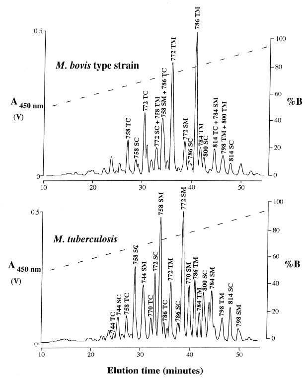
As with M. tuberculosis Erdman and H37Ra, the chloroform extract of the culture filtrate of the M. bovis type strain contained a large family of exochelins. More than 20 peaks exhibiting a high A450 eluted from the C18 reverse-phase HPLC column (Fig. 1). Based on the similarity of their elution pattern to that of exochelins from M. tuberculosis (Fig. 1) (7), we tentatively identified 18 of these peaks as ferri-exochelins and subsequently confirmed that they were exochelins by MS analysis (only the major peaks were analyzed). The patterns of elution were very similar at 3, 6, and 8 weeks of culture (data not shown). The yield was highest at 8 weeks (750 μg/liter).
FIG. 1.
Elution profile of an 8-week culture filtrate from the M. bovis type strain (top) and of a 6-week culture filtrate from M. tuberculosis Erdman (bottom) on a C18 reverse-phase HPLC column. The chloroform extract of 1 liter of the M. bovis culture filtrate or 750 ml of the M. tuberculosis culture filtrate was loaded onto the column. Iron-binding molecules were monitored at 450 nm. The dashed line in each graph represents the concentration of buffer B. Each labeled peak was analyzed by MS and shown to contain an exochelin. Exochelin designations are given above each peak (see the legend to Fig. 3).
Most of the M. bovis exochelins were found in M. tuberculosis, but they differed in relative abundance. The two most abundant exochelin species of M. bovis, 772TM and 786TM (designated according to the nomenclature for exochelins described in the legend to Fig. 3), are not the most abundant in M. tuberculosis but are still among the top 10 in abundance. The third most abundant M. bovis exochelin species, 772TC, has not been identified in M. tuberculosis. Two other exochelin species of M. bovis—814TC and 800TM—have also not yet been found in M. tuberculosis.
FIG. 3.
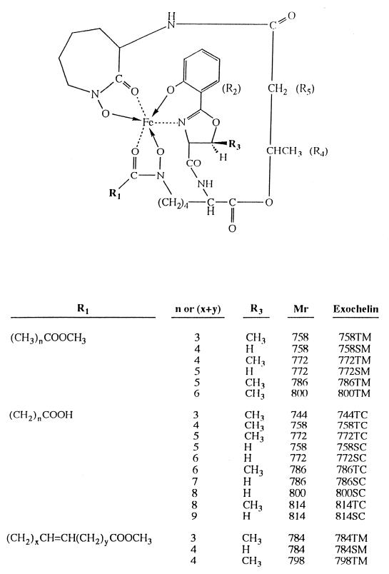
General structure of exochelins of the M. bovis type strain. The exochelins of M. bovis differ from each other at R1 and R3. R1 is either saturated or singly unsaturated and terminates with either a methyl ester (COOCH3) or a carboxylic acid (COOH) moiety. R3 is either H or CH3. The exochelins of M. bovis and M. tuberculosis display the same variations at R1 and R3 and also are identical at R4 and R5. The exochelins of M. avium display the same variations at R1 as M. bovis and M. tuberculosis, but R3 is always CH3. In addition, M. avium differs from the other two species at R4, which is CHCH2CH3, and R5, which is CHCH3. The exochelins are named according to (i) their mass in daltons in the iron-loaded form, (ii) whether R3 is H (serine) (S) or CH3 (threonine) (T), and (iii) whether R1 terminates in a methyl ester (M) or carboxylate (C) moiety.
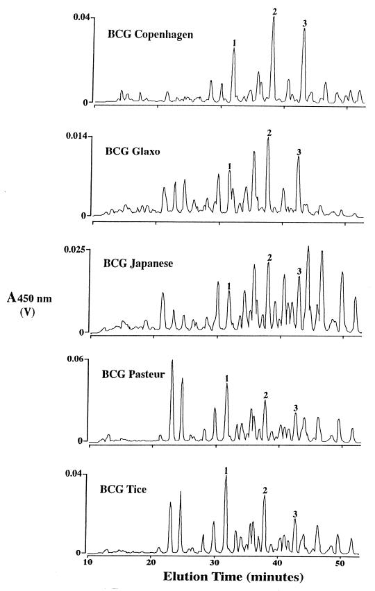
All five substrains of M. bovis BCG produced the same set of exochelins (Fig. 2). The overall yields of exochelins from the BCG strains were comparatively low—75 μg/liter for BCG Glaxo, 140 μg/liter for BCG Copenhagen, 150 μg/liter for BCG Japanese, 345 μg/liter for BCG Pasteur, and 390 μg/liter for BCG Tice versus 750 μg/liter for the M. bovis type strain and 2 to 4 mg/liter for M. tuberculosis. Furthermore, there was great variation in the relative quantities of individual exochelins. However, exochelins 772TM, 800SC, and 814SC were the major species of all BCG strains.
FIG. 2.
Elution profiles of 8-week culture filtrates from M. bovis BCG Copenhagen, Glaxo, Japanese, Pasteur, and Tice on a C18 reverse-phase HPLC column. The chloroform extract of 250 ml of each culture filtrate was loaded onto the column. Iron-binding molecules were monitored at 450 nm. Peaks 1, 2, and 3 correspond to exochelins 772TM, 800SC, and 814SC, respectively.
Based on the elution times after HPLC separation and the MS mass and fragmentation data on individual exochelins, the M. bovis, M. bovis BCG, and M. tuberculosis Erdman and H37Ra exochelins appear to be structurally identical. As in the case of M. tuberculosis, the core structures of the exochelins of M. bovis and M. bovis BCG are identical to those of the mycobactins of the same species and contain 3 amino acid moieties—2 N-hydroxylysines and either a serine or a threonine, depending on the absence or the presence of a methyl group at R3 (Fig. 3). This core contains the functional groups that bind the iron atom. As with M. tuberculosis and M. avium, the difference between the exochelins and mycobactins of M. bovis resides exclusively in the R1 side group. The R1 side chains of the M. bovis exochelins are either saturated or unsaturated and terminate with either a methyl ester or a carboxylic acid, as previously described for M. tuberculosis (7) and M. avium (20).
Conclusions.
This study demonstrates that the pathogenic mycobacteria M. tuberculosis and M. bovis produce the same set of iron-binding exochelins. Moreover, BCG, an attenuated strain of M. bovis, produces the same set of exochelins as the pathogenic mycobacteria M. tuberculosis and M. bovis. There are only two differences between the exochelins of M. tuberculosis and M. bovis among the strains studied. One difference is in the overall amount of exochelins produced: greatest in M. tuberculosis, intermediate in M. bovis, and smallest in M. bovis BCG. The second difference is in the relative amounts of specific exochelin species produced, as each mycobacterial species and strain has its own unique pattern of exochelin production.
Acknowledgments
This work was supported by grant AI35275 from the National Institutes of Health and by grant R96-LA1302/SF1301 from the California Universitywide AIDS Research Program. J. Gobin was supported by a fellowship from The Will Rogers Memorial Fund, and D. K. Wong was supported by a fellowship from The American Foundation for Pharmaceutical Education. We are indebted to PerSeptive Biosystems for the generous loan of the Voyager instrument to UCSF and to the UCSF Mass Spectrometry facility, which is partially supported by the National Center for Research Resources (RR 01614).
REFERENCES
- 1.Barclay R, Ratledge C. Mycobactins and exochelins of Mycobacterium tuberculosis, M. bovis, M. africanum and other related species. J Gen Microbiol. 1988;134:771–776. doi: 10.1099/00221287-134-3-771. [DOI] [PubMed] [Google Scholar]
- 2.Bouvet E, Casalino E, Mendoza-Sassi G, Lariven S, Valle E, Pernet M, Gottot S, Vachon F. A nosocomial outbreak of multidrug-resistant Mycobacterium bovis among HIV-infected patients. A case-control study. AIDS. 1993;7:1453–1460. doi: 10.1097/00002030-199311000-00008. [DOI] [PubMed] [Google Scholar]
- 3.Casanova J L, Blanche S, Emile J-F, Jouanguy E, Lamhamedi S, Altare F, Stéphan J-L, Bernaudin F, Bordigoni P, Turck D, Lachaux A, Pocidalo M-A, Le Deist F, Gaillard J-L, Griscelli C, Fischer A. Idiopathic disseminated bacillus Calmette-Guérin infection: a French national retrospective study. Pediatrics. 1996;98:774–778. [PubMed] [Google Scholar]
- 4.Colditz G A, Brewer T F, Berkey C S, Wilson M E, Burdick E, Fineberg H V, Mosteller F. Efficacy of BCG vaccine in the prevention of tuberculosis. Meta-analysis of the published literature. JAMA. 1994;271:698–702. [PubMed] [Google Scholar]
- 5.Danker W M, Waecker N J, Essey M A, Moser K, Thompson M, Davis C. Mycobacterium bovis infections in San Diego: a clinicoepidemiologic study of 73 patients and a historical review of a forgotten pathogen. Medicine. 1993;72:11–37. [PubMed] [Google Scholar]
- 6.Gobin J, Horwitz M A. Exochelins of Mycobacterium tuberculosis remove iron from human iron-binding proteins and donate iron to mycobactins in the M. tuberculosis cell wall. J Exp Med. 1996;183:1527–1532. doi: 10.1084/jem.183.4.1527. [DOI] [PMC free article] [PubMed] [Google Scholar]
- 7.Gobin J, Moore C H, Reeve J R, Jr, Wong D K, Gibson B W, Horwitz M A. Iron acquisition by Mycobacterium tuberculosis: isolation and characterization of a family of iron-binding exochelins. Proc Natl Acad Sci USA. 1995;92:5189–5193. doi: 10.1073/pnas.92.11.5189. [DOI] [PMC free article] [PubMed] [Google Scholar]
- 8.Lotte A, Waz-Höcket O, Poisson N, Dumitrescu N, Verron M, Couvet M. BCG complications. Adv Tuberc Res. 1984;21:107–245. [PubMed] [Google Scholar]
- 9.Macham L P, Ratledge C. A new group of water soluble iron-binding compounds from mycobacteria: exochelins. J Gen Microbiol. 1975;89:379–382. doi: 10.1099/00221287-89-2-379. [DOI] [PubMed] [Google Scholar]
- 10.Macham L P, Ratledge C, Nocton J C. Extracellular iron acquisition by mycobacteria: role of the exochelins and evidence against the participation of mycobactin. Infect Immun. 1975;12:1242–1251. doi: 10.1128/iai.12.6.1242-1251.1975. [DOI] [PMC free article] [PubMed] [Google Scholar]
- 11.Medzihradszky K F, Maltby D A, et al. Protein sequence and structural studies employing matrix-assisted laser desorption ionization-high energy collision-induced dissociation. Int J Mass Spectrom Ion Processes. 1997;160:357–369. [Google Scholar]
- 12.Ratledge C. Metabolism of iron and other metals by mycobacteria. In: Kubica G P, Wayne L G, editors. The Mycobacteria: a sourcebook. New York, N.Y: Marcel Dekker; 1984. pp. 603–627. [Google Scholar]
- 13.Samper S, Martin C, Pinedo A, Rivero A, Blásquez J, Baquero F, van Soolingen D, van Embden J. Transmission between HIV-infected patients of multidrug-resistant tuberculosis caused by Mycobacterium bovis. AIDS. 1997;11:1237–1242. doi: 10.1097/00002030-199710000-00006. [DOI] [PubMed] [Google Scholar]
- 14.Sharman G J, Williams D H, Ewing D F, Ratledge C. Determination of the structure of exochelin MN, the extracellular siderophore from Mycobacterium neoaurum. Chem Biol. 1995;2:553–561. doi: 10.1016/1074-5521(95)90189-2. [DOI] [PubMed] [Google Scholar]
- 15.Sharman G J, Williams D H, Ewing D F, Ratledge C. Isolation, purification and structure of exochelin MS, the extracellular siderophore from Mycobacterium smegmatis. Biochem J. 1995;305:187–196. doi: 10.1042/bj3050187. [DOI] [PMC free article] [PubMed] [Google Scholar]
- 16.van Soolingen D, de Haas P E W, Haagsma J, Eger T, Hermans P W M, Ritacco V, Alito A, van Embden J D A. Use of various genetic markers in differentiation of Mycobacterium bovis strains from animals and humans and for studying epidemiology of bovine tuberculosis. J Clin Microbiol. 1994;32:2425–2433. doi: 10.1128/jcm.32.10.2425-2433.1994. [DOI] [PMC free article] [PubMed] [Google Scholar]
- 17.Vestal M L, Juhasz P, et al. Delayed extraction matrix-assisted laser desorption time-of-flight mass spectrometry. Rapid Commun Mass Spectrom. 1995;9:1044–1050. [Google Scholar]
- 18.Walls F C, Baldwin M A, et al. Experience with multichannel array detection in tandem mass spectrometric characterization of biopolymers at the picomole level. In: Burlingame A L, McCloskey J A, editors. Biological mass spectrometry. Amsterdam, The Netherlands: Elsevier; 1990. pp. 197–216. [Google Scholar]
- 19.Wayne L G. Mycobacterial speciation. In: Kubica G P, Wayne L G, editors. The Mycobacteria: a sourcebook. New York, N.Y: Marcel Dekker; 1984. pp. 25–65. [Google Scholar]
- 20.Wong D K, Gobin J, Horwitz M A, Gibson B W. Characterization of exochelins of Mycobacterium avium: evidence for saturated and unsaturated and for acid and ester forms. J Bacteriol. 1996;178:6394–6398. doi: 10.1128/jb.178.21.6394-6398.1996. [DOI] [PMC free article] [PubMed] [Google Scholar]
- 21.World Health Organization. Zoonotic tuberculosis (Mycobacterium bovis) memorandum from WHO meeting (with participation of FAO) Bull W H O. 1994;72:851–857. [PMC free article] [PubMed] [Google Scholar]


