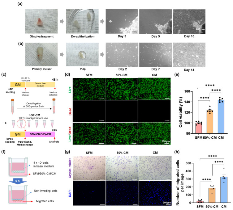
Figure 1.
Effect of human gingival fibroblast-derived conditioned medium (hGF-CM) on the proliferation and migration of DPSCs. (a,b) Primary culture of hGF and hDPSC. (a) hGF started to spread out from de-epithelized gingiva masses at Days 3−5 after plating. It was highly proliferated and subcultured 10−14 days after seeding. (b) Primary DPSC isolated from pulp was observed at 16−24 h after seeding and then clustered on days 4−6. It was subcultured on Day 14 after seeding. Both hGF and hDPSC have a typical fibroblast-like appearance. (c–e) The effect of hGF-CM on proliferation. (c) Schematic images of the preparation for hGF-CM and the proliferation test on DPSCs to investigate the effect of hGF-CM. (d) Representative live/dead images of DPSCs 6 h after treatment with either hGF-CM, 50% hGF-CM, or SFM. (e) Quantified graphs of the viability of DPSCs measured by the CCK-8 assay. n = 6. (f–h) The effect of hGF-CM on migration. (f) Schematic image of the migration assay. A total of 4 × 10⁴ cells in 100 µL basal medium were seeded in the upper well, and the lower compartment contained 350 µL hGF-CM, 50% hGF-CM, or SFM. (g) Representative images of migrated DPSCs to the lower side of the membrane stained with crystal violet and DAPI after treatment for 6 h. (h) Cells were counted on the lower side of the membrane with DAPI images in 6 random fields. All data are represented as the mean ± SEM. One-way analysis of variance followed (ANOVA) by Tukey’s multiple comparisons test was used to detect statistically significant differences between groups. Represents **** p < 0.0001, scale bars represent 200 µm. SFM: Serum free medium, GM: Growth medium, CM: Conditioned medium, 50% CM: Conditioned medium diluted by 50% with serum free medium.

