

Abstract
Background and purpose:
Prolonging the drug release can be a suitable approach to overcome the challenges related to topical ophthalmic administration of drugs especially the ones prescribed for chronic ailments. The sustained delivery of the drug would reduce the required frequency of administration which could extremely improve patient compliance and feeling of well-being. This study aimed to develop nanofibrous inserts for sustained ophthalmic delivery of timolol maleate (TIM) for the treatment of glaucoma.
Experimental approach:
Polycaprolactone-based nanofibers containing TIM were prepared using pure polycaprolactone or a blend of it with cellulose acetate or Eudragit RL100 polymers by the electrospinning method. Following the preparation, polymeric inserts were evaluated for morphological and physicochemical properties. The in vitro drug release was assessed and the in vivo efficacy of a selected insert in decreasing the intraocular pressure (IOP) was also evaluated in the equine eyes.
Findings / Results:
Prepared nanofibers indicated diameter ranged between 122-174 nm. The formulations showed suitable physicochemical properties and stability for ophthalmic administration. In vitro release study showed prolonged release of drug during more than 3 days. In vivo evaluation revealed that the prepared insert is non-irritant and non-toxic to the equine eyes while having suitable efficacy in decreasing the IOP during 6 days.
Conclusions and implication:
Prepared TIM inserts indicated a higher efficacy than commercial TIM eye drop in lowering IOP during a prolonged period. Thus, these formulations can be considered suitable for enhancing patient compliance by reducing the frequency of administration in the treatment of glaucoma.
Keywords: Electrospinning, Equine, Glaucoma, Nanofibers, Ophthalmic drug delivery, Timolol maleate
INTRODUCTION
Glaucoma is an eye disorder characterized by the degeneration of retinal cells, which eventually leads to complications such as loss of visual acuity, impaired vision, and irreversible blindness (1). This disorder is classified as a serious pathological condition since it is the second leading cause of blindness among eye diseases and possessed a high prevalence involving 70 million cases worldwide, recently.
The abovementioned urged scientists to develop novel drug delivery systems to accelerate the therapeutic efficacy of currently available therapeutic agents (2). Increased intraocular pressure (IOP) is one of the major risk factors responsible for glaucoma; hence, IOP-lowering drugs like β-blockers are the first-line therapy in this regard. These drugs act mainly by regulating the inflow and outflow of the aqueous humor (3). Despite being efficacious, so far, β-blockers have been formulated majorly as eye drops which come across with a limited intraocular bioavailability and absorption of these drugs. Accordingly, novel delivery formulations such as sustained-release ocular inserts that could provide targeted delivery to the site of action have been introduced in different studies. Multiple studies have suggested beneficial properties for targeted drug delivery (4).
In conformity with what was mentioned, most of the conventional topical therapeutic agents that are commonly prescribed for the treatment of glaucoma, possess a short contact time with the eye; hence, requiring repetitive administration during the day which leads to reduced compliance of patient and feeling of malaise (5). Moreover, some of these topical preparations cause discomfort to the eye; for example, suspension eye drops would cause irritation and the feeling of an external agent in the eye and ointments could cause blurry vision. Another drawback of conventional eye drops is the requirement for preservatives that can be harmful to the eye, occasionally. Additionally, due to the special physiology and anatomy of the eye, besides the eliminating function of the tear film and blinking reflex, a small amount (1-5%) of the topically administrated drugs can remain on the cornea for more than a few minutes or permeate to deeper layers of the eye (6). Also, part of the prescribed drug is inactivated as a result of binding to the proteins present in the tear film (7).
Consequently, the abovementioned obstacles persuade the researchers to design novel formulations including vesicular, nano-based, and polymer-based formulations which can facilitate drug delivery and improve bioavailability by causing longer retention of drugs on the cornea (8). These novel systems provide a more efficient and targeted drug delivery by various approaches such as sustaining the drug release, increasing the permeation through the cornea, and improving retention or contact of the drug with the cornea owing to mucoadhesive property and negative surface charge (9).
Accordingly, colloidal and polymeric carriers are suitable alternatives to conventional delivery systems for enhancing the ophthalmic delivery of drugs (10). To conquer the discussed issues and achieve an effective therapeutic response while reducing side effects, nanofibers have been designed and developed as ophthalmic inserts. These inserts could prolong drug release by increasing the retention time and having a more gradual elimination from the conjunctival sac. The unique properties of these systems that make them different from conventional delivery systems include the high surface-to-volume ratio, sustained-release behavior, and mucoadhesive property (11,12). Of note, despite eye drops these systems have the advantage of being preservative-free. These characteristics make nanofibers one of the most desired candidates for targeted drug delivery.
Timolol maleate (TIM) is an IOP-lowering β-blocker, commonly prescribed for the treatment of open-angle glaucoma. Since the systemic administration of this drug is related to multiple side effects, topical administration is preferred (10). The main mechanism behind the IOP lowering effect of TIM is still unknown but it is supposed to be related to the beta-adrenergic blocking effect. Due to the amphiphilic nature (13) and water-solubility of 2.74 mg/mL (14), this drug is mostly eliminated from the surface of the eye right after administration. Therefore, formulating it as prolonged-release nanofiber has the advantage of more targeted delivery, reduced frequency of administration, and accordingly increased patient compliance (15).
A literature review revealed that there were a few studies focused on the development of TIM-loaded nanofibers and many of them exclude in vivo evaluation (16,17); hence, the therapeutic effects of these inserts were not well-discussed. Accordingly, the present study is one of the first that design TIM-loaded nanofibers and evaluate their in vivo therapeutic efficacy. In this study, the nanofibrous insert of TIM was developed and optimized to control IOP for a longer duration compared to the eye drop formulation, which is typically administrated twice daily. The nanofibers were prepared using polycaprolactone (PCL) blended with cellulose acetate (CA) and Eudragit RL100 (EUD) polymers by the electrospinning method. The physicochemical properties, in vitro release, and in vivo efficacy in lowering the equine IOP were evaluated for the developed inserts.
MATERIALS AND METHODS
TIM, CA (acetyl content 39.8%, MW = 30,000 g/mol), and PCL (MW = 80,000 g/mol) were purchased from Sigma-Aldrich (Steinheim, Germany). EUD was procured from Evonik Degussa (Darmstadt, Germany). Acetone, dichloromethane (DCM), dimethylformamide (DMF), tryptic soy broth (TSB), fluid thioglycollate media (FTM), sabouraud dextrose broth (SDB), sodium dihydrogen phosphate dodecahydrate were purchased from Merck (Darmstadt, Germany). All materials were of analytical grade.
Preparation of electrospinning solutions
PCL solution at a concentration of 10% (w/v) was dissolved in DCM:DMF (9:1) solvent mixture by continuous stirring at room temperature. TIM was added to the solution at 10 and 20% w/w of PCL to the solution and stirred at a 1000 rpm rate for 1 h. Finally, a clear solution was obtained for the preparation of pure PCL nanofibers entitled PCL-1 (containing 10% w/w of TIM) and PCL-2 (containing 20% w/w of TIM).
To prepare PCL-CA and PCL-EUD nanofibers, solutions of PCL, CA, and EUD at 10% (w/v) concentration were prepared separately using a 9:1 mixture of DCM:DMF, and DCM:acetone (3:7), respectively for dissolving PCL and CA and methanol for EUD. TIM was added at 10% w/w of polymers to each solution under continuous stirring with 1000 rpm for 1 h to obtain completely dissolved transparent solutions for the electrospinning procedure.
Electrospinning procedure
To prepare PCL-CA and PCL-EUD nanofibers with a mixed structure double-jet electrospinning with nozzles in the frontal position was utilized. In separated double-nozzle electrospinning procedures 20 mL of PCL/TIM solution were filled in an injectable reservoir while the other nozzle was filled with 20 mL of either EUD/TIM or CA/TIM solutions. The solutions were injected, concurrently toward an aluminum-covered rotary collector (diameter10 cm) with a 1 mL/h rate, while a 15 kV voltage was applied by a high-voltage DC power supplier between the injector and rotating collector. The injector-collector distance was modified to 20 cm and the temperature maintains at 25 °C throughout the process. After evaporation of solvents overnight the deposited nanofibrous mats obtained from each electrospinning process were collected. To prepare the pure PCL nanofibers the single-jet electrospinning of PCL/TIM solutions with drug concentration at 10% and 20% w/w of the polymer at a completely similar condition to the mix nanofibers was adopted.
Morphology characterization
The morphology and structure of prepared formulations were observed utilizing scanning electron microscopy (SEM) to evaluate the uniformity of nanofibers which is required for the preparation of drug-loaded ophthalmic inserts and the reproducibility of the obtained results. The samples were examined by EM-6200 (KYKY, China) microscope, under a high vacuum after being coated with gold (18). The process took place at 20 kV accelerating voltage. Finally, the size distribution of the prepared fibers in each sample was estimated using the ImageJ software by measuring the diameter at twenty different points of the obtained SEM photograph. An average was taken for the mean diameter of each examined formulation.
Fourier-transform infrared spectroscopy
To ensure that no major interaction took place between the polymers and pharmacologically active parts of the drug, the Fourier-transform infrared spectroscopy (FTIR) evaluation was utilized. The drug, polymers, and all prepared nanofibers were observed after being pressed with potassium bromide to obtain disks for examination (19). The spectrophotometer (Prestige-21, Shimadzu, Japan) was used to record the FTIR spectra of the samples between the 4000-400 cm-1 range, and the recorded data were analyzed for any significant shift, change, and elimination of characteristic peaks of the drug in the nanofibers.
Physicochemical characterization of nanofibers
Uniformity
The uniformity of prepared nanofibers was examined by measuring thickness and drug content at three different points across the nanofibrous mats. The thickness was estimated using a digital micrometer (Syntek, Zhejiang, China) with an accuracy of 0.001 mm. The nanofibers were put between the spindle and anvil of the device and the spindle moved toward the sample until it reached the surface of the nanofiber but did not press into lower thickness. The mean thickness (n = 3) was reported.
Drug loading and encapsulation efficiency
To estimate the drug loading and encapsulation efficiency, 25 mg pieces of nanofibers were dissolved in proper solvents and the amount of drug was calculated by an ultraviolet-visible (UV-Vis) spectrophotometer at 295 nm as the wavelength of maximum absorbance, after proper dilution. The values were calculated using the following equations:

Flexibility
The flexibility of nanofibers was judged by the obtained folding endurance and tensile strength. The resistance of a sample against being torn after repeated folding is known as folding endurance. Samples with equal dimensions (3×3 cm2) were folded manually until being torn. The procedure was performed in triplicated and an average was reported.
The tensile test is partially similar to folding endurance with the difference of being performed automatically and measuring the maximum stretching stress a sample can resist before tearing. Utilizing an STM-5 testing machine (Santam, Iran), the 20×30 mm2 samples with an almost 0.1 mm thickness, were pulled at a 5 mm/min rate by the mobile gauge (20). Three samples were examined for each formulation and the mean ± SD was reported.
Swelling degree
The swelling degree is usually an indicator of the release behavior. This parameter is defined as the ability of the formulation for water absorption multiple times its weight. The pre-weighed samples were immersed in phosphate buffer solution (PBS) with a pH of 7.4 for 24 h then were re-weighed and the swelling was calculated using the standard formula in which the W0 stands for initial weight and the Wf stands for the final weight (equation 3).

Stability
The stability of nanofibrous inserts which is essential for optimization was evaluated by calculating the moisture uptake and loss during humid and dry conditions. A desiccator containing calcium chloride or aluminum chloride was used as a stimulator of dry and humid conditions, respectively. The samples were placed in desiccators for 72 h and the moisture loss and uptake were measured using equation 4; W0 stands for initial weight and the Wf stands for final weight.

In vitro drug release
The TIM in vitro release from nanofibers was evaluated. A specific amount of each formulation was placed in a dialysis bag tied at both ends along with 0.5 mL of PBS (pH 7.4) and then immersed in 49.5 mL PBS as the receptor compartment at 37 °C under shaking conditions (21). Samples were collected at different time intervals and the receptor medium was fully replaced with fresh PBS after each sampling to remain in sink condition. The absorption of samples was determined in the UV-Vis analysis at 295 nm as the wavelength of maximum absorption. The amount of released TIM was estimated using the regression equation obtained by the plotted calibration curve.
Release mechanism
To predict the release mechanism, the data obtained in the first 24 h of the release study were fitted in different kinetical models and the model with the highest correlation was chosen as the release kinetics. Zero-order, first-order, Higuchi, and Korsmeyer-Peppas kinetical models were evaluated for each formulation.
Ex vivo mucoadhesion time
The mucoadhesive property of formulations was examined according to the method adopted by Tofighia et al. (22). A self-assembled device was utilized to perform the test. A disintegration apparatus was filled with 900 mL PBS (pH 7.4). Freshly excised sheep cornea with an almost 3 cm diameter was provided, then rinsed with PBS and attached to a glass slide utilizing a two-sided glue tape. This assembled slide was vertically fixed on the baskets of the disintegration apparatus. Pieces of inserts (1×1 cm2) were hydrated with PBS at one side and then contacted to the cornea and the apparatus was set to sweep upward and downward at a constant rate at 37 °C. Eventually, the time required for the entire detachment of inserts from the cornea was reported as the mucoadhesion time (n = 3).
Sterility test
To ensure the sterility of formulations before in vivo study, the sterility test was performed. All formulations were prepared under aseptic conditions to remain sterile. Also, the formulations were exposed to UV radiation in order to eliminate any surface contamination by microorganisms prior to the test. As per the USP guidelines, samples were incubated in the FTM, TSB, and SDB to observe the growth and contamination of anaerobic bacteria, aerobic bacteria, and fungi. Positive and negative controls were prepared for accurate comparison. The negative control included sterile culture media without immersion of any inserts or inoculation of any microorganisms. The positive controls were developed by inoculation of Bacillus subtilis (ATCC: 21332) in FTM, Escherichia coli (ATCC: 25922) in TSB, and Candida albicans (PFCC: 62194) in SDB. The culture media were observed at 7-, 14-, 21-, and 28-day intervals.
In vivo evaluation and irritancy test in equine eyes
Fourteen glaucomatous horses of different Iranian breeds with high levels of IOP were selected, in which a total of 22 eyes had IOP higher than the normal range. A single-dose administration of optimized nanofibrous patch with the best performance through the in vitro study (PCL-CA 10%) containing 2 mg of TIM was used in the 11 cases with high-pressure eyes while 11 eyes received TIM 0.5% eye drop twice on the first day. Changes in IOP were evaluated using an Air Puff tonometer (Keeler Instruments Inc, Broomall, Pennsylvania). The IOP was measured for a 6-day duration and the equine eyes were controlled daily for any sign of irritancy, damage, and erythema.
All the experiments were approved by the Institutional Animal Ethics Committee (Ethics No. IR.KUMS.REC.1399.744), Kermanshah University of Medical Sciences, Kermanshah, I.R. Iran.
Statistical analysis
To statistically compare the results of physicochemical characterizations, one-way ANOVA followed by Tukey's post hoc test was performed while the results of animal study were statistically analyzed by Kruskal-Wallis one-way ANOVA followed by Mann-Whitney U test. All statistical examinations were performed by SPSS software (version 25.00). P values ≤ 0.05 were considered statistically significant.
RESULTS
Morphology characterization
As it is represented in Fig. 1, SEM images showed that the plain PCL-1 and PCL-2 formulations had a uniform structure with a mean diameter of 110 ± 41 nm and 136 ± 32 nm. A slightly increased diameter in PCL-2 compared to PCL-1 was observed. PCL-CA nanofibers indicated a mean diameter of 176 ± 42 nm which was expected based on the reported values for plain PCL and CA nanofibers. In a similar study in 2019, PCL nanofibers were reported to have a mean diameter of more than 350 nm (23). Also, CA nanofibers were reported to have a mean diameter of 180 nm in a similar study (24). PCL-EUD nanofiber showed a mean diameter of 104 ± 20 nm.
Fig. 1.

The scanning electron microscopy images of (A) PCL-1, (B) PCL-2, (C) PCL-EUD, and (D) PCL-CA nanofibers at magnification 40000× and the histograms of diameter distribution. PCL, Polycaprolactone; CA, cellulose acetate; EUD, Eudragit RL100.
FTIR
The FTIR spectra obtained for the drug, polymers, and nanofibers are indicated in Fig. 2. The characteristic peaks of polymers, and the drug appear in the spectra obtained for all nanofibers. Peaks at around 2941 cm-1and 2866 cm-1, respectively, indicate asymmetric and symmetrical stretching bands of the CH2 in PCL and TIM appearing at the spectra of all formulations. Peaks appearing in 1622 cm-1and 1720 cm-1are attributed to the C=N and C=O stretching of TIM, respectively. The peaks at around 1720 cm-1 appeared at the spectra of PCL-CA and PCL-EUD is also assigned to C=O vibration of PCL, CA, and EUD polymers. There are peaks at 1246 cm-1 attributed to the vibration of C-O-C in PCL, CA, and EUD polymers. PCL-CA and PCL-EUD also represent peaks at 1381 cm-1 related to symmetrical C-CH3 vibrations. The peak at 960 cm-1 is assigned to the N-S band of the heterocyclic ring in TIM.
Fig. 2.
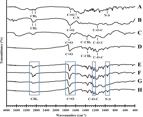
The FTIR spectra of (A) timolol maleate, (B) PCL, (C) CA, (D) EUD, (E) PCL-1, (F) PCL-2, (G) PCL-CA, and (H) PCL-EUD. PCL, Polycaprolactone; CA, cellulose acetate; EUD, Eudragit RL100.
Physicochemical characterization
Table 1 represents the measured physicochemical properties of formulations. All the nanofibers possessed suitable and uniform thicknesses of almost 0.100-0.400 mm to fit in the conjunctival sac. The drug content was also uniform all across the nanofibrous mat.
Table 1.
The physicochemical characteristic obtained for different formulations.
| Parameters |
|||||||||
|---|---|---|---|---|---|---|---|---|---|
| Formulations | Folding endurance (times) | Thickness(mm) | Swelling(%) | Moisture loss(%) | Moisture uptake(%) | Tensile strength (MPa) | Elongation at break (%) | Drug loading(%) | Encapsulation efficacy(%) |
| PCL-1 | 379 ± 2 | 0.352 ± 0.003 | 150.3 ± 6.3 | 0.67 ± 0.02 | 0.52 ± 0.01 | 5.04 ± 0.35 | 34.0 ± 0.7 | 9.49 ± 0.14 | 95.21 ± 1.24 |
| PCL-2 | 347 ± 2 | 0.408 ± 0.002 | 154.9 ± 2.3 | 0.90 ± 0.02 | 0.64 ± 0.06 | 2.61 ± 0.10 | 14.6 ± 0.3 | 18.74 ± 0.37 | 93.86 ± 1.67 |
| PCL-CA | 209 ± 3 | 0.087 ± 0.002 | 188.2 ± 7.4 | 1.16 ± 0.02 | 1.05 ± 0.04 | 1.78 ± 0.05 | 4.7 ± 0.1 | 9.65 ± 0.15 | 96.93 ± 1.40 |
| PCL-EUD | 186 ± 4 | 0.201 ± 0.003 | 183.3 ± 4.1 | 1.24 ± 0.05 | 0.94 ± 0.02 | 0.46 ± 0.05 | 9.8 ± 0.2 | 9.59 ± 0.10 | 96.21 ± 0.92 |
PCL, Polycaprolactone; CA, cellulose acetate; EUD, Eudragit RL100.
The formulations showed encapsulation efficiency of 93-96%. The drug loading was measured to be extremely close to the expected values as electrospinning is an appropriate method with a high yield. The data obtained for drug loading and encapsulation efficiency is classified in Table 1.
The highest and lowest folding endurance of nanofibers were related to PCL-1 and PCL-EUD nanofibers, respectively. All formulations showed >180 times folding endurance. This suitable folding endurance indicated that nanofibers possessed sufficient flexibility to be used as an ocular insert.
Tensile testing is a characteristic examination indicating the strength and flexibility of a formulation. An ocular insert is required to be strong enough to preserve its integrity while being flexible enough to be non-irritant to the eye. As Fig. 3 is representing, the stress-strain curve of all formulations followed the manner of an elastic structure. The strongest formulation was PCL-1 comprised of pure PCL while the weakest one was the PCL-EUD. The PCL-1 and PCL-2 formulations showed more elasticity compared to PCL-CA with an elongation percentage of more than 20% against almost 5% elongation of PCL-CA.
Fig. 3.

Stress-strain curve of the developed timolol maleate-loaded nanofibers. PCL, Polycaprolactone; CA, cellulose acetate; EUD, Eudragit RL100.
The swelling degree of the prepared formulations was decreased in the following order: PCL-CA > PCL-EUD > PCL-2 > PCL-1 and the formulations indicated suitable stability with less than 2% moisture loss or uptake through humid and dry conditions.
In vitro drug release assay
In vitro release test indicated that nanofibers possessed a two-step release profile in which the TIM release initially in a burst-release manner which is related to the release of the surface-loaded TIM of nanofibers during the initial 12 h followed by a sustained-release phase through more than 3 days (Fig. 4). At the time of 12 h PCL-1, PCL-2, PCL-CA, and PCL-EUD released 85.96 ± 1.78%, 85.80 ± 0.03%, 81.98 ± 0.10%, and 81.78 ± 0.18% of their drug content, respectively. At the end of 72 h, the PCL-1 and PCL-2 released 90.87 ± 1.61% and 91.99 ± 0.21%; meanwhile 93.64 ± 0.10% and 85.87 ± 0.47% release was observed for PCL-CA and PCL-EUD, respectively. Therefore, PCL-CA with the highest amount of released drug was chosen for further studies.
Fig. 4.

The in vitro release profile of timolol maleate from different nanofibers (PCL-1, PCL-2, PCL-CA, PCL-EUD) in PBS (pH 7.4) at 37 °C for 80 h with the magnification of the first 12 h. PCL, Polycaprolactone; CA, cellulose acetate; EUD, Eudragit RL100.
Release mechanism
Table 2 represents the regression coefficients obtained from fitting the 24-h release data in different kinetical models. All formulations followed the Korsmeyer-Peppas kinetical model. Accordingly, the drug release from the formulations is governed mainly by the diffusion mechanism. In fact, the release mechanism began with the diffusion of the surface-loaded drug into the PBS and was followed by the penetration of the medium into the porous structure of nanofibers. Eventually, the loaded drug in the nanofibers dissolved in the penetrated medium and was released gradually. This mechanism could also be related to the hydrophobic nature of PCL. In a similar study, a PCL-based nanofibrous insert that was developed for the delivery of artemisinin obeyed Korsmeyer-Peppas kinetics of drug release (25).
Table 2.
The regression coefficient of formulations obtained by fitting the formulations in different kinetical models.
| Formulation | Zero-Order | First-Order | Higuchi | Korsmeyer-Peppas |
|---|---|---|---|---|
| PCL-1 | 0.5722 | 0.7135 | 0.7222 | 0.8123 |
| PCL-2 | 0.6887 | 0.8409 | 0.8254 | 0.8990 |
| PCL-CA | 0.6031 | 0.7904 | 0.7505 | 0.8199 |
| PCL-EUD | 0.6646 | 0.8600 | 0.8016 | 0.8829 |
PCL, Polycaprolactone; CA, cellulose acetate; EUD, Eudragit RL100.
Also, the diffusion exponent (n) was measured to be 0.42, 0.48, 0.70, and 0.84 for PCL-1, PCL-CA, PCL-EUD, and PCL-2. Hence, the PCL-1 and PCL-CA with n-values in the vicinity of 0.5, released their drug content majorly through Fickian diffusion while PCL-EUD and PCL-2 with n-values between 0.5-1.0 released their drug through an anomalous transport. Anomalous transport is the characteristic of systems that in addition to diffusion, other mechanisms are involved in release (26).
Ex vivo mucoadhesion time
The PCL-1, PCL-2, PCL-EUD, and PCL-CA indicated 35 ± 7, 32 ± 9, 128 ± 12, and 133 ± 21 s mucoadhesion time. The PCL-2 showed the least mucoadhesive property while PCL-CA possessed the highest mucoadhesive property.
Sterility test
The formulation is required to be sterile prior to administration in the animal eyes. None of the formulations showed any turbidity or sign of microorganism growth during the 28 days of sterility test owing to preserved aseptic condition during the preparation of inserts. This result indicated the sterility of formulations.
In vivo evaluation and irritancy test in equine eyes
As PCL-EUD did not show promising tensile strength and in vitro release, it was ruled out of in vivo study. Among PCL-1, PCL-2, and PCL-CA formulations that all indicated suitable physicochemical properties with acceptable strength, flexibility, and in vitro release, the optimized formulation was chosen based on the highest mucoadhesive property for in vivo evaluation to avoid inserts falling out of the conjunctival sac. According to previous studies, PCL-based nanofibers showed a lower level of mucoadhesive property compared to nanofibers comprised of hydrophilic polymers (16). Hence, PCL-CA nanofiber with a higher mucoadhesive property was selected for in vivo evaluation.
Fourteen glaucomatous horses with a total of 22 high-level IOP eyes were examined, considering that the normal IOP of equine eyes is between 17-28 mm Hg. All horses had one or two eyes affected by IOP higher than the normal range, which was checked with an air puff-Keeler tonometer. Ophthalmic TIM-loaded insert of PCL-CA that was chosen as optimized nanofiber, based on the results of in vitro release study was placed in 11 affected equine eyes. Fig. 5 indicates the procedure of IOP measurement during in vivo study and the IOP changes from baseline. Less than one day after administration of the nanofiber, the IOP of most cases lowered to the normal range. The insert formulation had a maximum IOP lowering effect during the 5th day of administration with almost 5 mmHg changes in IOP. Based on the reported data the conventional marketed eye drops of TIM indicated a rapid release of the drug led to a maximum effect 4 h after administration reaching up to 9 mmHg changes in IOP (27).
Fig. 5.

(A) The procedure of IOP measurement during in vivo study; (B) comparison of mean changes in the equine IOP after administration of ophthalmic insert (n = 11) and conventional eye drop of TIM (n = 11). TIM, Timolol maleate; IOP, intraocular pressure.
Accordingly, the insert formulation prepared in the present study could lower the IOP up to almost 20% of baseline for an extended duration of 6 days.
DISCUSSION
Glaucoma is a serious pathological condition of the eye that can lead to impaired vision, and irreversible blindness if not managed appropriately. There is a significant relationship between a rise in IOP and the occurrence of glaucoma, hence, most therapeutic agents aimed to reduce the abnormally increased IOP. Conventional β-blocker eye drops are typically prescribed for patients with glaucoma-affected eyes, which faced obstacles of limited intraocular bioavailability and absorption along with a requirement for frequent administration. Accordingly, the present study aimed to develop nanofibrous inserts for sustained ophthalmic delivery of TIM for the treatment of glaucoma.
Different formulations were developed using pure PCL or a blend of it with CA or EUD by the electrospinning process. Although PCL is a polymer with multiple beneficial properties such as suitable mechanical strength and flexibility, its hydrophobic nature may limit the drug release from the nanofibrous structure. The main rationale behind blending PCL with CA and EUD was to implant hydrophilic pockets in the nanofibrous structures to improve the release behavior of the drug. Moreover, using pure CA or EUD was expected to fabricate nanofibers with lower strength and flexibility which were not appropriate to be used for ocular aims. The obtained inserts were uniform in texture, high strength, and flexible enough to be separated from the collector, effortlessly. Another main reason for the addition of CA and EUD was to increase the mucoadhesive properties of nanofibers to avoid inserts falling out of the conjunctival sac. The morphology characterization by SEM analysis indicated an almost uniform nanofibrous structure for all prepared formulations with a mean diameter in a range of 104-176 nm. The slightly increased diameter in PCL-2 compared to PCL-1 was because of the higher drug loading. This result was in accordance with the reports of a previous study (28). While the addition of CA to the PCL nanofibers led to an increased diameter, the addition of EUD to the formulation did not cause a significant change in the mean diameter. The nano-sized diameter of fibers would result in a higher surface-to-volume ratio which could enhance the drug delivery.
The FTIR spectroscopy was performed to investigate drug-excipient compatibility. Polymer-drug interactions usually result in the removal of characteristic peaks or the appearance of additional peaks in the spectrum. With a glance at spectra obtained for each formulation (Fig. 2), it is obvious that characteristic peaks of the drug have not been removed or have not undergone a significant shift, which confirms the lack of any interaction between the pharmacologically active groups of the drug and the polymers.
There is an optimal range for the thickness of ocular inserts in which the inserts are thick enough to preserve their integrity in the eye and thin enough to allow patient acceptance and not be irritant to the eye (29). This optimal thickness is presumed to be almost 0.4 mm. The formulations indicated uniform thickness in the mentioned range which could ensure the mechanical stability and non-irritancy of the formulation.
The nanofibers showed suitable encapsulation efficiency of more than 93%. The formulations did not show any statistically significant difference in encapsulation efficiency. The drug loading was measured to be extremely close to the expected values as electrospinning is an appropriate method with a high yield. The PCL-1, PCL-CA, and PCL-EUD that were intended to achieve 10% loading values indicated more than 9% loading, and PCL-2 was intended to achieve 20% loading, demonstrating almost 18% of drug loading.
The visual appearance, weight, strength, and flexibility of nanofibers remained intact during these 12 months of storage at room temperature. Also, no significant changes in the drug loading were observed meanwhile. This could ensure the long-term stability of nanofibers.
All formulations showed good flexibility based on the reported desirable value for folding endurance of nanofibers which is reported to be above 40 times (30). This suitable folding endurance ensured sufficient flexibility of formulations. Similar results were reported in a previous study that developed TIM-loaded PCL nanofibers with 415 times folding endurance (16). As it is obvious, the addition of both CA and EUD significantly decreased the folding endurance of PCL-CA and PCL-EUD compared to PCL-1 and PCL-2 formulations due to the much lower folding endurance of CA and EUD plain nanofibers which was also reported by previous studies compared to PCL nanofibers (31,32).
The stress-strain curve of all formulations followed the manner of an elastic structure according to the result of tensile testing. Increasing the drug content in PCL-2 led to a significantly decreased tensile strength compared to PCL-1. It was previously reported that decreasing the drug content would cause a rise in the strength of a nanofibrous structure (33). Also, the addition of CA and EUD resulted in a significantly decreased strength due to the low inherent strength of these polymers; but EUD caused a more significant change in the strength of formulation. In a previous study, a tensile strength of 1.3 MPa was acquired for CA nanofiber (34). Although PCL-EUD showed suitable elongation of 10%, the lack of desired strength made it less favorable among the formulation.
The reason for the observed swelling pattern for nanofibers (Table 1) could be that the more hydrophilic nature and small diameter of CA and EUD nanofibers led to a higher surface-to-volume ratio and more space to absorb water. PCL-1 and PCL-2 nanofibers indicated an almost similar degree of swelling (P = 0.760) which showed that increasing the drug loading would not alter the swelling capacity of formulations, significantly; but due to their relatively hydrophobic nature, they absorbed a significantly lower amount of water compared to PCL-CA and PCL-EUD (P < 0.05) (16). Of note, there was not a statistically significant difference between the swelling degree of PCL-CA and PCL-EUD (P = 0.701).
In vitro release test indicated a two-step release profile for all formulations. PCL-CA, PCL-1, and PCL-2 formulations released almost a similar percentage of their drug content during 72 h of the study. PCL-EUD indicated a significantly lower amount of released drug compared to other formulations (P < 0.05). While the release mechanisms of PCL-CA and PCL-EUD nanofibers were expected to be more affected by erosion, the release mechanism of PCL-1 and PCL-2 formulations was expected to be more affected by the diffusion phenomenon due to the hydrophobic nature of the PCL compared to CA and EUD. However, since all nanofibers contained PCL, the dominant mechanism of release was diffusion in all cases. It was reported in a previous study that the drug release from PCL-PEG nanofibers followed a similar behavior (35). In a 2017 study on TIM-loaded nanofibers, in vitro release for up to 24 h, with an initial burst phase was reported (36). PCL-based nanofibrous ophthalmic inserts showed a 55-day release of itraconazole (37).
The formulations showed 30-130 s mucoadhesion time to the sheep cornea. PCL-1 and PCL-2 showed an almost similar mucoadhesion time. The PCL-CA and PCL-EUD showed almost 4-times higher mucoadhesion time compared to pure PCL formulations (P < 0.05). This significantly higher mucoadhesive property is due to the addition of EUD and CA with more hydrophilic nature to hydrophobic PCL. Additionally, there was no statistically significant difference between the mucoadhesive property of PCL-CA and PCL-EUD. It should be noted that mucoadhesion time is not representative of the residence time as the condition of this test is exaggerated compared to the normal environment of the eye and conjunctival sac. The PCL-CA showed 6 days of residence in the equine eye.
In vivo evaluation of PCL-CA ophthalmic insert in glaucomatous equine eyes and comparison of it with twice-daily administration of TIM 0.5% eye drop. The IOP-lowering efficacy of formulations on each day (during 6 days) was compared by the Mann-Whitney U test. While statistical analysis showed a higher IOP-lowering effect for eye drop formulation compared to insert during the first day of administration (P < 0.05), the insert formulation showed a significantly higher efficacy from day 2 to day 6 of administration indicating a promising prolonged therapeutic effect of formulation (P < 0.05). Owing to the presence of CA in the PCL-CA formulation that underwent in vivo evaluation, the formulation indicated a suitable mucoadhesive property to remain in the cul-de-sac for a prolonged time of 6 days. Hence, nanofibrous inserts with single-dose administration are preferred compared to the eye drop that requires twice doses daily or other procedures, such as surgery which are invasive and cause irreversible complications. In a similar study, a novel nano-based formulation of encapsulated dendrimer containing the anti-glaucoma drug acetazolamide was developed. While the plain drug could make a 5 mmHg change in the IOP of rabbits, the dendrimer formulation lowers the IOP by 7 mmHg (38). In another study, timolol-dorzolamide nanofibers showed the potential to reduce and normalize the IOP of rabbits for 72 h. The nanofibers lower the IOP by 2-12 mmHg changes which indicated a maximum effect on the first day of administration by 12 mmHg changes followed by 5-6 mmHg changes during the second and third day of administration. Of note, using a combination of two drugs led to a higher reduction in IOP (16). Urtti et al. reported that silicone inserts of TIM could lower the IOP by 10 mmHg during 24 h (39). Also, during the daily examinations, the safety and non-irritancy of the insert were confirmed due to lack of any tissue damage or complications like redness, inflammation, abnormal discharge, etc.
CONCLUSIONS
In the present study, PCL-based nanofibrous inserts of timolol maleate were prepared using the pure solution of PCL and the mixture of it with CA or EUD polymers by the electrospinning technique. All formulations demonstrated appropriate physicochemical properties to be used as ophthalmic inserts. The nanofibers possessed suitable flexibility and strength, stability, and uniform structures. During the in vitro release study, it was observed that all formulations followed Korsmeyer Peppas-like kinetics with two-phase release behavior containing a burst release of drug through the first 12 h followed by a gradual and sustained phase during more than 3 days. The formulation containing PCL and CA showed a higher percentage of released drug and suitable mucoadhesive property, hence subjected to in vivo evaluation. The in vivo evaluation of ophthalmic timolol-loaded insert in the equine glaucomatous eyes showed that this formulation has efficacy to lower the IOP by almost 5 mmHg during 6 days after administration which is more favorable compared to marketed eye drop with rapid release during 4 h. No significant sign of irritancy and any tissue damage was observed after administration of the formulation. The obtained results suggested that the prepared formulations had the potential to sustained-release TIM with therapeutic concentrations for an extended duration of time. Thus, these formulations can be considered suitable for administration in glaucoma. The prolonged release of the drug from these inserts can make them more acceptable for patient administration compared to frequently administrated eye drops.
Conflict of interest statement
The authors declared no conflict of interest in this study.
Authors’ contributions
S. Mirzaeei, F. Daneshgar, and L. Rezaei contributed to the study concept and design, supervised the study, and drafted the manuscript; F. Bahrami Faryadras acquired the data; S. Mehrandish and A. Karami analyzed and interpreted the data; S. Mirzaeei and S. Mehrandish revised the manuscript critically for important intellectual content. The final version of the article was approved by all authors.
Acknowledgments
The authors would like to acknowledge the Research Council of Kermanshah University of Medical Sciences for financial support of this work (Grant No. 990761). Also, faithfully thank Rahesh Daru Novin knowledge-based company for cooperation in providing materials and equipment. The authors would like to thank the horse stable “Olympic Village” (Kermanshah, Iran), for providing the permission to carry out this work at their stables and for good-natured support throughout.
REFERENCES
- 1.Gooch N, Molokhia SA, Condie R, Burr RM, Archer B, Ambati BK, et al. Ocular drug delivery for glaucoma management. Pharmaceutics. 2012;4(1):197–211. doi: 10.3390/pharmaceutics4010197. doi: 10.3390/pharmaceutics4010197. [DOI] [PMC free article] [PubMed] [Google Scholar]
- 2.Zhang N, Wang J, Li Y, Jiang B. Prevalence of primary open angle glaucoma in the last 20 years: a meta-analysis and systematic review. Sci Rep. 2021;11(1):13762, 1–12. doi: 10.1038/s41598-021-92971-w. doi: 10.1038/s41598-021-92971-w. [DOI] [PMC free article] [PubMed] [Google Scholar]
- 3.Ghate D, Edelhauser HF. Barriers to glaucoma drug delivery. J Glaucoma. 2008;17(2):147–156. doi: 10.1097/IJG.0b013e31814b990d. doi: 10.1097/IJG.0b013e31814b990d. [DOI] [PubMed] [Google Scholar]
- 4.Singh RB, Ichhpujani P, Thakur S, Jindal S. Promising therapeutic drug delivery systems for glaucoma: a comprehensive review. Ther Adv Ophthalmol. 2020;12 doi: 10.1177/2515841420905740. 2515841420905740,1-17. doi: 10.1177/2515841420905740. [DOI] [PMC free article] [PubMed] [Google Scholar]
- 5.King-Smith PE, Reuter KS, Braun RJ, Nichols JJ, Nichols KK. Tear film breakup and structure studied by simultaneous video recording of fluorescence and tear film lipid layer images. Invest Ophthalmol Vis Sci. 2013;54(7):4900–4909. doi: 10.1167/iovs.13-11878. doi: 10.1167/iovs.13-11878. [DOI] [PMC free article] [PubMed] [Google Scholar]
- 6.Shen J, Lu GW, Hughes P. Targeted ocular drug delivery with pharmacokinetic/pharmacodynamic considerations. Pharm Res. 2018;35(11):217, 1–20. doi: 10.1007/s11095-018-2498-y. doi: 10.1007/s11095-018-2498-y. [DOI] [PubMed] [Google Scholar]
- 7.Tangri P, Khurana S. Basics of ocular drug delivery systems. Int J Res Pharm Biomed Sci. 2011;2(4):1541–1552. [Google Scholar]
- 8.Mehrandish S, Mirzaeei S. Design of novel nanoemulsion formulations for topical ocular delivery of itraconazole: development, characterization and in vitro bioassay. Adv Pharm Bull. 2021;12(1):93–101. doi: 10.34172/apb.2022.009. doi: 10.34172/apb.2022.009. [DOI] [PMC free article] [PubMed] [Google Scholar]
- 9.Mehrandish S, Mirzaei S. A review on ocular novel drug delivery systems of antifungal drugs: functional evaluation and comparison of conventional and novel dosage forms. Adv Pharm Bull. 2020;11(1):28–38. doi: 10.34172/apb.2021.003. doi: 10.34172/apb.2021.003. [DOI] [PMC free article] [PubMed] [Google Scholar]
- 10.Rawas-Qalaji M, Williams C-A. Advances in ocular drug delivery. Curr Eye Res. 2012;37(5):345–356. doi: 10.3109/02713683.2011.652286. doi: 10.3109/02713683.2011.652286. [DOI] [PubMed] [Google Scholar]
- 11.Khalil IA, Ali IH, El-Sherbiny IM. Noninvasive biodegradable nanoparticles-in-nanofibers single-dose ocular insert: in vitro, ex vivo and in vivo evaluation. Nanomedicine (Lond) 2019;14(1):33–55. doi: 10.2217/nnm-2018-0297. doi: 10.2217/nnm-2018-0297. [DOI] [PubMed] [Google Scholar]
- 12.Singla J, Bajaj T, Goyal AK, Rath G. Development of nanofibrous ocular insert for retinal delivery of fluocinolone acetonide. Curr Eye Res. 2019;44(5):541–550. doi: 10.1080/02713683.2018.1563196. doi: 10.1080/02713683.2018.1563196. [DOI] [PubMed] [Google Scholar]
- 13.Mackeben S, Müller-Goymann CC. Solubilization of timolol maleate in reversed micellar systems: measurement of particle size using SAXS and PCS. Int J Pharm. 2000;196(2):207–210. doi: 10.1016/s0378-5173(99)00423-8. doi: 10.1016/s0378-5173(99)00423-8. [DOI] [PubMed] [Google Scholar]
- 14.Natu MV, de Sousa HC, Gil MH. Effects of drug solubility, state and loading on controlled release in bicomponent electrospun fibers. Int J Pharm. 2010;397(1-2):50–58. doi: 10.1016/j.ijpharm.2010.06.045. doi: 10.1016/j.ijpharm.2010.06.045. [DOI] [PubMed] [Google Scholar]
- 15.Nashatizadeh MM, Lyons KE, Pahwa R. A review of ropinirole prolonged release in Parkinson's disease. Clin Interv Aging. 2009;4:179–186. doi: 10.2147/cia.s3358. doi: 10.2147/cia.s3358. [DOI] [PMC free article] [PubMed] [Google Scholar]
- 16.Gagandeep, Garg T, Malik B, Rath G, Goyal AK. Development and characterization of nano-fiber patch for the treatment of glaucoma. Eur J Pharm Sci. 2014;53:10–16. doi: 10.1016/j.ejps.2013.11.016. doi: 10.1016/j.ejps.2013.11.016. [DOI] [PubMed] [Google Scholar]
- 17.Mehta P, Al-Kinani AA, Arshad MS, Chang M-W, Alany RG, Ahmad Z. Development and characterisation of electrospun timolol maleate-loaded polymeric contact lens coatings containing various permeation enhancers. Int J Pharm. 2017;532(1):408–420. doi: 10.1016/j.ijpharm.2017.09.029. doi: 10.1016/j.ijpharm.2017.09.029. [DOI] [PubMed] [Google Scholar]
- 18.Nandhini S, Ilango K. Development and characterization of a nano-drug delivery system containing vasaka phospholipid complex to improve bioavailability using quality by design approach. Res Pharm Sci. 2021;16(1):103–117. doi: 10.4103/1735-5362.305193. doi: 10.4103/1735-5362.305193. [DOI] [PMC free article] [PubMed] [Google Scholar]
- 19.Yasin H, Al-Taani B, Salem MS. Preparation and characterization of ethylcellulose microspheres for sustained-release of pregabalin. Res Pharm Sci. 2021;16(1):1–15. doi: 10.4103/1735-5362.305184. doi: 10.4103/1735-5362.305184. [DOI] [PMC free article] [PubMed] [Google Scholar]
- 20.Bahri-Najafi R, Tavakoli N, Senemar M, Peikanpour M. Preparation and pharmaceutical evaluation of glibenclamide slow release mucoadhesive buccal film. Res Pharm Sci. 2014;9(3):213–223. PMID: 25657792. [PMC free article] [PubMed] [Google Scholar]
- 21.Rezazadeh M, Safaran R, Minaiyan M, Mostafavi A. Preparation and characterization of Eudragit L 100-55/chitosan enteric nanoparticles containing omeprazole using general factorial design: in vitro/in vivo study. Res Pharm Sci. 2021;16(4):358–369. doi: 10.4103/1735-5362.319574. doi: 10.4103/1735-5362.319574. [DOI] [PMC free article] [PubMed] [Google Scholar]
- 22.Tofighia P, Soltani S, Montazam SH, Montazam SA, Jelvehgari M. Formulation of tolmetin ocuserts as carriers for ocular drug delivery system. Iran J Pharm Res. 2017;16(2):432–441. [PMC free article] [PubMed] [Google Scholar]
- 23.Beachley V, Wen X. Effect of electrospinning parameters on the nanofiber diameter and length. Mater Sci Eng C Mater Biol Appl. 2009;29(3):663–668. doi: 10.1016/j.msec.2008.10.037. doi: 10.1016/j.msec.2008.10.037. [DOI] [PMC free article] [PubMed] [Google Scholar]
- 24.Jatoi AW, Kim IS, Ogasawara H, Ni Q-Q. Characterizations and application of CA/ZnO/AgNP composite nanofibers for sustained antibacterial properties. Mater Sci Eng C Mater Biol Appl. 2019;105(110077):1–8. doi: 10.1016/j.msec.2019.110077. DOI: 10.1016/j.msec.2019.110077. [DOI] [PubMed] [Google Scholar]
- 25.Huo P, Han X, Zhang W, Zhang J, Kumar P, Liu B. Electrospun nanofibers of polycaprolactone/collagen as a sustained-release drug delivery system for artemisinin. Pharmaceutics. 2021;13(8):1228, 1–14. doi: 10.3390/pharmaceutics13081228. doi: 10.3390/pharmaceutics13081228. [DOI] [PMC free article] [PubMed] [Google Scholar]
- 26.Fosca M, Rau JV, Uskoković V. Factors influencing the drug release from calcium phosphate cements. Bioact Mater. 2022;7:341–363. doi: 10.1016/j.bioactmat.2021.05.032. doi: 10.1016/j.bioactmat.2021.05.032. [DOI] [PMC free article] [PubMed] [Google Scholar]
- 27.Adelli GR, Bhagav P, Taskar P, Hingorani T, Pettaway S, Gul W, et al. Development of a Δ9-tetrahydrocannabinol amino acid-dicarboxylate prodrug with improved ocular bioavailability. Invest Ophthalmol Vis Sci. 2017;58(4):2167–2179. doi: 10.1167/iovs.16-20757. doi: 10.1167/iovs.16-20757. [DOI] [PMC free article] [PubMed] [Google Scholar]
- 28.Karuppuswamy P, Venugopal JR, Navaneethan B, Laiva AL, Ramakrishna S. Polycaprolactone nanofibers for the controlled release of tetracycline hydrochloride. Mater Lett. 2015;141:180–186. doi: 10.1016/j.matlet.2014.11.044. [Google Scholar]
- 29.Aburahma MH, Mahmoud AA. Biodegradable ocular inserts for sustained delivery of brimonidine tartarate: preparation and in vitro/in vivo evaluation. AAPS Pharm Sci Tech. 2011;12(4):1335–1347. doi: 10.1208/s12249-011-9701-3. doi: 10.1208/s12249-011-9701-3. [DOI] [PMC free article] [PubMed] [Google Scholar]
- 30.Kamble RN, Mehtre RV, Mehta PP, Nangare P, Patil SS. Albendazole electrospun nanofiber films: in-vitro and ex-vivo assessment. BioNanoScience. 2019;9(3):625–636. doi: 10.1007/s12668-019-00627-x. [Google Scholar]
- 31.AnjiReddy K, Karpagam S. Hyperbranched cellulose polyester of oral thin film and nanofiber for rapid release of donepezil; preparation and in vivo evaluation. Int J Biol Macromol. 2019;124:871–887. doi: 10.1016/j.ijbiomac.2018.11.224. doi: 10.1016/j.ijbiomac.2018.11.224. [DOI] [PubMed] [Google Scholar]
- 32.Ali A, Kumar N, Ahad A, Aqil M, Sultana Y. Enhanced delivery of diclofenac diethylamine loaded Eudragit RL 100® transdermal system against inflammation. J Polym Eng. 2015;35(7):699–708. doi: 10.1515/polyeng-2014-0352. [Google Scholar]
- 33.Chou S-F, Woodrow KA. Relationships between mechanical properties and drug release from electrospun fibers of PCL and PLGA blends. J Mech Behav Biomed Mater. 2017;65:724–733. doi: 10.1016/j.jmbbm.2016.09.004. doi: 10.1016/j.jmbbm.2016.09.004. [DOI] [PMC free article] [PubMed] [Google Scholar]
- 34.Tang C, Chen P, Liu H. Cocontinuous cellulose acetate/polyurethane composite nanofiber fabricated through electrospinning. Polym Eng Sci. 2008;48(7):1296–1303. doi: 10.1002/pen.21090. [Google Scholar]
- 35.Ravikumar R, Ganesh M, Senthil V, Ramesh YV, Jakki SL, Choi EY. Tetrahydro curcumin loaded PCL-PEG electrospun transdermal nanofiber patch: preparation, characterization, and in vitro diffusion evaluations. J Drug Deliv Sci Technol. 2018;44:342–348. doi: 10.1016/j.jddst.2018.01.016. [Google Scholar]
- 36.Karavasili C, Komnenou A, Katsamenis OL, Charalampidou G, Kofidou E, Andreadis D, et al. Self-assembling peptide nanofiber hydrogels for controlled ocular delivery of timolol maleate. ACS Biomater Sci Eng. 2017;3(12):3386–3394. doi: 10.1021/acsbiomaterials.7b00706. doi: 10.1021/acsbiomaterials.7b00706. [DOI] [PubMed] [Google Scholar]
- 37.Mehrandish S, Mohammadi G, Mirzaeei S. Preparation and functional evaluation of electrospun polymeric nanofibers as a new system for sustained topical ocular delivery of itraconazole. Pharm Dev Technol. 2022;27(1):25–39. doi: 10.1080/10837450.2021.2018609. doi: 10.1080/10837450.2021.2018609. [DOI] [PubMed] [Google Scholar]
- 38.Mishra V, Jain N. Acetazolamide encapsulated dendritic nano-architectures for effective glaucoma management in rabbits. Int J Pharm. 2014;461(1-2):380–390. doi: 10.1016/j.ijpharm.2013.11.043. doi: 10.1016/j.ijpharm.2013.11.043. [DOI] [PubMed] [Google Scholar]
- 39.Urtti A, Rouhiainen H, Kaila T, Saano V. Controlled ocular timolol delivery: systemic absorption and intraocular pressure effects in humans. Pharm Res. 1994;11(9):1278–1282. doi: 10.1023/a:1018938310628. DOI: 10.1023/a:1018938310628. [DOI] [PubMed] [Google Scholar]