Abstract
In the recent years, the increasing applications of brain–computer interface (BCI) in rehabilitation programs have enhanced the chances of functional recovery for patients with neurological disorders. We presented and validated a BCI system with a lower-limb robot for short-term training of patients with spinal cord injury (SCI). The cores of this system included: (1) electroencephalogram (EEG) features related to motor intention reported through experiments and used to drive the robot; (2) a decision tree to determine the training mode provided for patients with different degrees of injuries. Seven SCI patients (one American Spinal Injury Association Impairment Scale (AIS) A, three AIS B, and three AIS C) participated in the short-term training with this system. All patients could learn to use the system rapidly and maintained a high intensity during the training program. The strength of the lower limb key muscles of the patients was improved. Four AIS A/B patients were elevated to AIS C. The cumulative results indicate that clinical application of the BCI system with lower-limb robot is feasible and safe, and has potentially positive effects on SCI patients.
Supplementary Information
The online version contains supplementary material available at 10.1007/s11571-022-09801-6.
Keywords: Brain-computer interface (BCI), Electroencephalogram (EEG), Spinal cord injury (SCI), Rehabilitation robot
Introduction
As a direct communication and control pathway between brain and computer, brain–computer interface (BCI) system has been applied in the field of neurological rehabilitation. Much advancement has been made in the field in recent years. (Shindo et al. 2011; Ramos-Murguialday et al. 2013; Donati et al. 2016; Shokur et al. 2018; He et al. 2018; Tariq et al. 2018) However, patients with severe spinal cord injury (SCI) still have low recovery chances. (Shokur et al. 2018; Kirshblum et al. 2004) Previous studies have explored the rehabilitation therapy of SCI using BCI. The findings of Shokur, et al. indicated that long-term training with noninvasive BCI could induce functional recovery in patients with chronic SCI. Eight subjects with chronic motor complete injury (American Spinal Injury Association Impairment Scale (AIS) A/B) exhibited significant improvements in motor and sensory functions after 28 months of training. (Donati et al. 2016; Shokur et al. 2018) Ganzer, et al. conducted a 5-month training program for a complete SCI patient (AIS A) using invasive BCI, which also demonstrated improvements in motor and sensory functions. (Ganzer et al. 2020) Previous studies have suggested that BCI has positive rehabilitation effects on patients with motor complete SCI.
Our team has been engaged in a study that aims to restore mobility in SCI patients through short-term training with a BCI system. Current treatments for SCI involve unidirectional stimulation. For example, exercise therapy can produce bottom-up stimulation from the peripheral neurons to the SCI area (Takeoka 2020), and transcranial magnetic stimulation (TMS) can produce top-down stimulation of the SCI area (Hallett 2000; Goudman et al. 2019). However, BCI can generate both descending control signals and ascending sensory signals (bidirectional stimulation), thereby stimulating the SCI area effectively. The goal of our system is to stimulate the SCI area simultaneously by using the descending motion signal and the ascending sensory signal and to improve the effective and targeted stimulation in a single training session. Consequently, our system reduces the time required for the patients to learn and use it and augments the proportion of brain motor imagery along with the effective training revolutions. Experimental results have suggested that our system can induce neurological recovery in patients with SCI in short-term training.
The two cores of our system are the design of the motor intent decoder and the decision tree of the rehabilitation mode: (1) The design of the motor intention decoder: The descending motor control signal is emitted from the brain and is transmitted to the muscles of the limbs through the spinal cord, which causes muscle contraction and generates motor function. We hope that more targeted stimulation can be produced in the neurons related to motor signal transduction in the SCI region. Hence, it is necessary to ascertain the brain region and frequency domain characteristics related to motor function. In this regard, we adopted a new idea, that is, by comparing the spatial position and frequency domain characteristics of the electroencephalogram (EEG) between patients with motor dysfunction and healthy people, we tried to determine a group of signals that may be more theoretically conducive for the descending stimulation of the injured spinal motor neurons(Sheorajpanday et al. 2011, 2010; Finnigan and van Putten 2013; Hall et al. 2014; Lopez et al. 2019; Ramanathan et al. 2018). In the experiment, we found significant differences in several signals between the prefrontal lobes of the two groups. By designing the motor intent decoder and experimenting on healthy people, we showed that these signals can reflect the motor intention of the subjects to some extent. Despite the disrupted connection between the brain and the spinal cord, the brain can still generate appropriate motor commands (Donoghue et al. 2002; Collinger et al. 2013). Therefore, we moved this decoder in the motor intention recognition of the SCI rehabilitation robot. Motor intention can help patients recover through the combination of the BCI technology and the robot. It should be noted that the paradigm we adopt is different from the existing paradigm of motor imagery based on Event-related desynchronization (ERD)/Event-related synchronization (ERS). The motor imagery paradigm based on ERD/ERS has large individual differences, and requires a certain time of training, which limits its application in clinical rehabilitation (Donati et al. 2016; Edelman1* et al. 2019; Morash et al. 2008; Zuo et al. 2020). Moreover, in the study of rehabilitation medicine, the engagement and involvement of patients in the training process are the core requirements (Carr and Shepherd 1998). The EEG indicators we used can reflect the motor intention of patients to a certain extent, but also can reflect the degree of attention of patients (Gaume et al. 2019; Wang et al. 2018), which is more in line with the requirements of rehabilitation medicine. (2) The decision tree of the rehabilitation mode: To ensure that patients in different stages of SCI can produce enough bidirectional stimuli in the training process, we designed a decision tree mechanism that combines BCI control and machine force control. Meanwhile, multimodal feedbacks such as proprioception, EEG signals, and muscle strength were provided to the patients. Lower limb movement driven by the robot can produce upward sensory stimulation.
Materials and methods
System
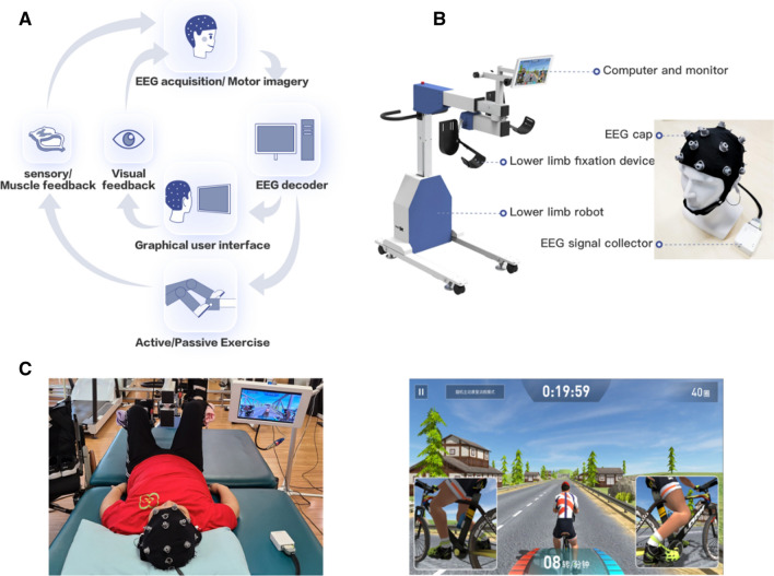
A BCI system with lower-limb robot was developed with an EEG acquisition and analysis device and a lower-limb robot. This system is illustrated in Fig. 1.
Fig. 1.
System introduction. A The flow of proposed system. While the system is operated, the patients are instructed to imagine themselves performing lower-limb movements. The EEG cap receives the raw EEG signals of the patient, which are then transmitted to a computer for analysis via Bluetooth. Then, the computer sends the control command to the lower-limb robot, and the robot conducts lower-limb training for the SCI patients. The robot has an angle sensor and sends the crank angle information to the display when running. The crank angle on the screen matches the actual crank angle. B The system includes an EEG cap with 8 electrodes, an EEG collector, a computer, a monitor, and a lower-limb driving robot (maximum torque: 12 N m; height: 0.9–1.2 m; crank length: 0.1 m; rotation speed: 0–100r/min). C Robot usage and game display interface. The patient is asked to lie down on a bed and their lower limbs are fixed to the robot, and the moving parts of the system drive the patient’s lower limb in a pedaling motion, giving muscle and sensory feedback to the patient. The monitor displays a riding game to simulate actual pedaling. The speed of the pedaling movement in the riding game is consistent with that of the patient. The game shows the remaining training time (top center panel), virtual riding distance (top right panel), speed (bottom center panel), muscle strength value (bottom panel, to the left of “speed”), and motor intentions (bottom panel, to the right of “speed”). The inset windows on the left and right display the cyclist’s legs, such that the patient can directly observe the changes in the movement speed of both the legs. These views provide real-time feedback to patients
The flow of the proposed system is depicted in Fig. 1A. In the experiment, the patient’s scalp EEG was collected through the brain cap, and the motor intention was decoded by the computer. The decoded information was used to determine to drive the robot. When it met with certain needs, the robot could drive the patient’s lower limb movement and provide feedback to the patient’s body sensation, while simultaneously giving visual feedback to the patient on the display screen.
The system structure is shown in Fig. 1B. The EEG signals were collected and analyzed in the EEG acquisition and analysis device using an EEG cap comprising of 2 ear clip electrodes (1 reference, 1 bias) and 8 wet (saline) electrodes. Reference electrode and bias electrode are clipped in left and right earlobes respectively. The 8 electrodes include FP1, FP2, F3, F4, C3, C4, FZ and CZ. The EEG collector uses the ADS1299 front-end analog chip (TI, Dallas, United States) as an analog-to-digital converter with a sampling rate of 250 Hz. The collected EEG signals are transmitted via Bluetooth to the lower-limb robot’s computer.
The lower-limb robot includes a computer and a monitor as well as a lower extremity drives actuator. The computer is mainly used to analyze and decode the EEG signals and send control instructions to the lower limb driving the actuator and display interface. The display can be used to adjust equipment parameters and display training games, EEG status, and muscle strength status to provide users with their performance feedback. The lower extremity drive consists of a servo motor system, a high precision force sensor, and support components. The servo motor can control the speed and position accurately based on the instructions, and the control accuracy can reach the level of 0.01%. The sampling frequency of our force sensor is 80 Hz, while the force measurement accuracy is 0.05 Nm. We installed a force sensor on the servo motor drive shaft, and, through the proportional coefficient of adjusting force and speed, as well as the smooth parameters of the servo motor acceleration and deceleration, accurate force control can be achieved. Robot and BCI jointly form a three-layer decision tree processing mechanism.
Robot usage and game display interface is depicted in Fig. 1C. During training, the patients wore EEG caps and imagined riding a bike; their lower limbs were driven by the robot. Meanwhile, the patients could receive constant feedback through the display.
The display showcases a riding game, and the game interface displays the speed of the device movement, the patient’s brain state, and the patient’s autonomous force and movement speed changes. The key portions of our system are motor intention analysis and the decision tree of the rehabilitation mode.
Motor intention analysis
The paradigm selection of motion intention considers three factors: the selection of indicators needs to have a certain mapping relationship with motor function; it should integrate the requirements of rehabilitation training in rehabilitation medicine; it can be generally applied to different patients in clinic, and the learning time is relatively short. In order to obtain the paradigm of motor intention recognition that meets the above requirements, we conducted two experiments in this part. In the first experiment, we compared the EEG characteristics between stroke patients with dyskinesia and healthy subjects in order to find indicators that were significantly different between the two groups. In the second experiment, through the combination of these indicators, we conducted the imagination experiment of motor intention of healthy subjects under cue stimulation in order to find the correlation between these indicators and motor intention.
Experiment 1: the experiment of extracting EEG characteristic parameters
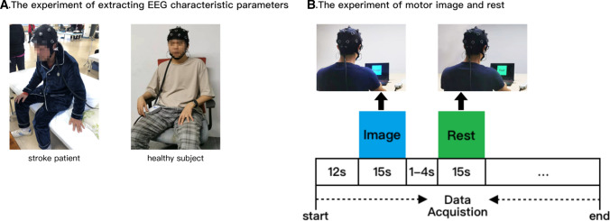
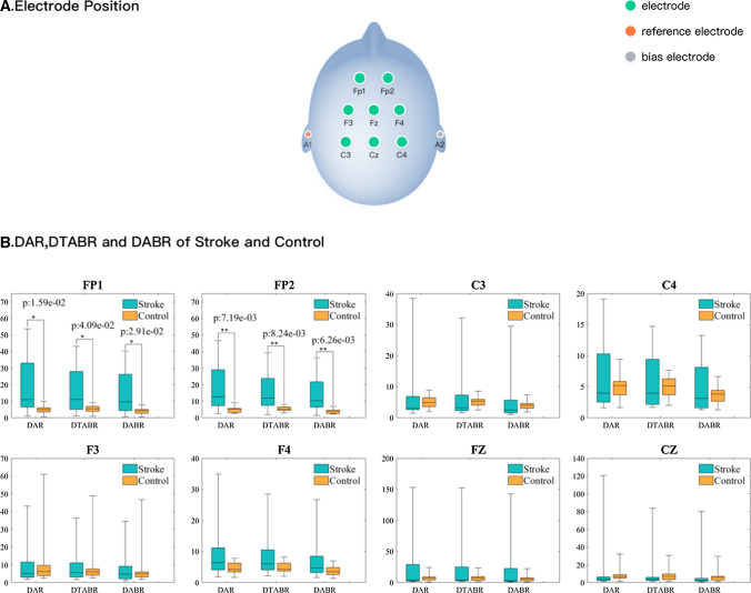
In the first experiment, due to the different conditions of stroke patients with dyskinesia, it is difficult to adopt a unified task-based experiment. In order to explore the differences in EEG characteristics between patients and healthy subjects in the same experimental condition, we conducted eye-closure resting experiments on patients and healthy subjects, which is shown in Fig. 2A. In this experiment, the above 8-channel EEG caps were used, and the positions of the 8 electrodes are shown in Fig. 3A. A total of 30 subjects (a stroke group of 15 stroke subjects, 11 males and 4 females, Mean ± SD of age: 37.5 ± 13.9; a control group of 15 healthy volunteers, 12 males and 3 females, Mean ± SD of age: 29.1 ± 4.5, p = 0.09, p > 0.05 compared with the stroke subjects; post-hoc Wilcoxon Signed Rank Test for the age) participated in the experiment.
Fig. 2.
Experiment 1 and Experiment 2. A The experiment of extracting EEG characteristic parameters. All subjects were asked to close their eyes for 5 min to collect the EEG data for analyses. The subjects should wear EEG caps correctly and ensure the quality of EEG signals. During the experiment, the subjects were instructed to stay relaxed without thinking of anything particular and without sleeping, and try to avoid unnecessary movements. All subjects were asked to refrain from using drugs or foods that additionally promote or suppress brain activity since 24 h prior to the experiment. The subjects could clearly understand the experimental process. B The experiment of motor image and rest. Before the experiment started, we explained the steps of the experiment and the matters needing attention in detail to each participant. The subjects were correctly wearing an EEG cap and were instructed to minimize unnecessary movements to ensure the quality of EEG signals. When the experiment started, the first is 12 s of preparation time. Then two stimuli (rest or image, lasting for 15 s respectively) were randomly presented. Participants were required to rest or imagine their legs’ movements according to the corresponding cue stimuli. The interval of cue stimulus is 1 ~ 4 s randomly. EEG data were collected throughout the experiment
Fig. 3.
Electrode positions and comparison of DAR, DTABR and DABR values between the stroke subjects and healthy subjects. A Electrode positions B The comparative results of DAR, DTABR and DABR on FP1, FP2, F3, F4, C3, C4, FZ and CZ channels between the stroke group and the control group, respectively (turquoise represents the experimental group and yellow represents the control group). The center of the color block is the mean value, and the upper and lower sides of the box are third-quartile and first-quartile values. The top and bottom whiskers indicate the maximum and minimum values. *p < 0.05, **p < 0.01 indicates significant differences in comparison by Wilcoxon Signed Rank Test. When significant difference is found, p value is placed on the sign of significant difference
After the above mentioned pretreatment steps, the obvious low-frequency artefact (caused by body or eye movements) and high-frequency noise in the data was filtered through 0.5–30 Hz 5th-order IIR band pass filter, after which the following methods were adopted for feature extraction. The power spectrum characteristics of EEG were calculated, and the spectrum energy ratio (delta/alpha power ratio (DAR), (delta + theta)/(alpha + beta) power ratio (DTABR), delta/(alpha + beta) power ratio(DABR)) and other EEG characteristics were extracted to compare the differences about several indicators between the stroke patients and healthy subjects.
Then, we compared the EEG signals between stroke patients and healthy subjects based on the above EEG characteristics. We found that the mean of DAR, DTABR and DABR in stroke subjects were higher than those in healthy subjects at FP1 and FP2. Then we compared two groups of subjects using the Wilcoxon Signed Rank Test. The results revealed that DAR, DTABR and DABR had significant differences between the two groups at FP1 and FP2 electrodes, as shown in Fig. 3B.
According to literature research, there is a strong mapping relationship between the indicators of DAR, DTABR, DABR and the function recovery of stroke patients (Sheorajpanday et al. 2011, 2010; Finnigan and van Putten 2013). Therefore, we extracted the characteristic parameters of DAR, DTABR and DABR, which were combined into a comprehensive indicator. That is the motor intention score, which we called Mscore.
We assumed motor intention as the instruction to drive the BCI and set the difficulty level from 1 to 11, according to the numerical difference between healthy people and patients. Taking the average difficulty level as level 6, the system becomes harder to drive as the difficulty level increases. According to each difficulty level, the patients’ motor intention is converted to 0–100 motor intention score by comparing with the threshold value. When the composite EEG score of the DAR, DTABR and DABR of the prefrontal cortex is higher than the threshold value, the robot drives the patient’s limbs for circular motion.
Experiment 2: the experiment of motor image and rest
In the second experiment, we performed an experiment on 5 healthy subjects, because the brain functions of patients with SCI were no significant difference with healthy subjects (Dobkin and Havton 2004; Carmena et al. 2003). The experimental process is shown in Fig. 2B.
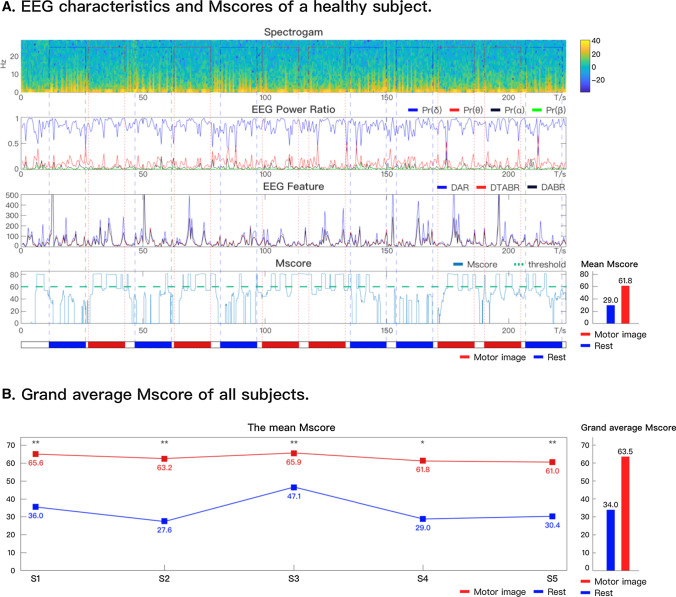
The EEG characteristics of FP1 were extracted for analysis, as shown in Fig. 4.
Fig. 4.
EEG characteristics and Mscore of FP1. A EEG characteristics and Mscores of a healthy subject. The subgraph with the title of Spectrogram represents the time–frequency EEG diagram for training of patients throughout the training process, with a color bar on the right; the subgraph with the title of EEG Power Ratio represent the power ratios of Delta, Theta, Alpha and Beta throughout the training process; the subgraph with the title of EEG Feature represent the changes of DAR, DTABR and DABR features in different states; the subgraph with the title of Mscore indicates the change of motor intention scores of patients in different states; the right part represents the specific representation of different colors of solid lines in the graph with the title of EEG Power Ratio, EEG Feature and Mscore; the bottom subgraph represents the state distribution throughout the training process, with red blocks representing motor imagery, blue blocks representing rest, and white blocks representing background throughout the training process, a total of 6 motor image and 6 rest were performed in each trials; segmentation between different states in subgraphs entitled Spectrogram, EEG Power Ratio, EEG Feature and Mscore by corresponding dashed lines. The average Mscore for rest and movement tasks is shown at the bottom right. B Grand average Mscore of all subjects. The mean Mscores represent the mean Mscores of each subject, and the grand average Mscore represents average Mscore of all subjects, with red blocks representing motor imagery, blue blocks representing rest. S* represent the subjects’ number. *p < 0.05, **p < 0.01 compared with the mean Mscore of rest; post-hoc Wilcoxon Signed Rank Test for the mean Mscore
From the Fig. 4A, a correlation can be noted between the subjects’ EEG score and motor intention. When the subjects imagined cycling with both the legs, the EEG score was higher. Similarly, when the subjects relaxed, the score decreased.
The EEG data were analyzed, and the Mscore value was obtained per second (when the Mscore was greater than 60 in one second, the subjects were considered to be in motor imagination). By comparing the Mscore and the cue value, the overall correct rate of the subjects was 72.2%, of which the correct rate of recognition under motion was 64.4% and that under rest was 80.0% (the rate of false trigger was 20.0%). The mean Mscore of motor image (61.8) is higher than that of rest (29.0). There were significant differences in the mean Mscores of motor image and rest (p = 0.0016, p < 0.01). The correlation coefficient between Mscore and cue value was 0.45, which was moderate correlation. Considering that all the subjects are using the system for the first time, the statistical analysis of this experiment shows that the system can better realize the recognition of motor intention for the first users. At the same time, in the process of rehabilitation training, patients need continuous motor imagery to help them better grasp the use of the system. Moreover, it should be noted that since our paradigm is used for continuous drive devices, it is more important to prevent false triggering. A lower false trigger rate can reduce the number of cases where the patient is distracted while the system continues to run. And 60% of the correct imagination rate gives the initial user a certain challenge. With visual, auditory and other feedback given by the system during actual clinical rehabilitation training, it can encourage patients to train with engagement and involvement. All the subjects’ mean Mscore and the grand average Mscore of subjects can be found in Fig. 4. All the subjects’ mean Mscores of motor image is higher than that of rest, and there were significant differences (S1: p = 1.7385e-07, S2: p = 1.7485e-04, S3: p = 0.0047, S4: p = 0.0016, S5: p = 2.3677e-08).
Decision tree of rehabilitation mode
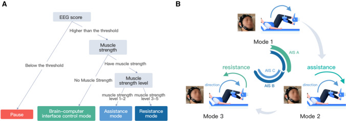
The decision tree of the rehabilitation mode consisted of 3 layers (Fig. 5). The 3 rehabilitation modes were switched through the decision tree.
Fig. 5.
The decision tree of rehabilitation mode. A The decision tree. The first layer (also the bottom layer) was used to monitor the active motion state for the BCI. When the patient’s EEG score does not reach the threshold through analysis and decoding, it prompts the patient; at the same time, when the patient’s interruption time exceeds a certain time, it stops the exercise training. On the second layer, when the patient has no muscle strength, the patient used the BCI control mode. The rotation speed depends on the patient’s Mscore. The third layer, patient’s muscle strength is greater than grade 1. Through real-time analysis of EEG signals, when Mscore is greater than the threshold, the system control robot will provide patients with the lowest speed of rehabilitation training. A torque sensor was installed at the motor output shaft to detect the patient’s active force and control the speed change by the force control algorithm to provide assistance and resistance. B Three modes were available for the patients. For AIS A patients, the training generally switched from mode 1 to mode 2; for AIS B patients, the training generally switched from mode 1 to mode 3; and for AIS C patients, the training generally switched from mode 2 to mode 3
The 3 modes of the system are as follows:
Mode 1: Brain control mode. When the patients had no muscle strength, the system controls the motor speed by detecting the Mscore.
Mode 2: Assistance mode. When the patient’s own muscle strength is insufficient to drive the limb movement and the robot helps the patients to produce movement, which is to amplify the signal of the sensor and improve the speed of the movement. In clinical rehabilitation training, the muscle strength of patients from 0 to 1 is irregular in the kick exercise, although the size of the force is uncertain. The phase and amplitude of the signal in the kick movement were analyzed. When the amplitude was oscillating, the systematic error was eliminated. Moreover, it is considered that when the patient begins to show muscle strength, the assistance mode is started.
Mode 3: Resistance mode. When the patient’s muscle strength can overcome the resistance of his/her limbs, the system increases the rotational resistance of the robot, making the patient feel resistant.
When the patient’s muscle strength increased to > 3 levels, a certain amount of resistance exercise was required. When the muscle strength of the patient continued to exceed the threshold, the system performed resistance mode. The threshold considered the muscle strength and the muscle tension of the patients.
Participant recruitment and training
Seven SCI patients (5 male, 2 female; 1 AIS A, 3 AIS B, and 3AIS C) were recruited for the training, and the age of these patients ranged from 20 to 58 years (mean = 43.0, SD = 13.2). The detailed patient information is shown in Table 1. All patients were naïve BCI subjects. Our experiments were performed at the Zhongshan Rehabilitation Branch of The First Affiliated Hospital of Nanjing Medical University in the Jiangsu province, China. The clinical protocol of the study was approved by the Ethics Committee of The First Affiliated Hospital of Nanjing Medical University; all patients signed informed consent before their participation. The purpose of the experiment is to evaluate the patient’s use of the system and find out the positive impact of our system on the possible functional improvement of SCI patients.
Table 1.
Information about the patients
| Subject | Sex | Age | AIS | Neurological level | Time since lesion (months) | Etiology | Training period (weeks) | Training trials |
|---|---|---|---|---|---|---|---|---|
| Patient 1 | M | 57 | A | T11 | 6 | Car accident | 6 | 61 |
| Patient 2 | M | 37 | B | C7 | 10 | High altitude fall | 7 | 69 |
| Patient 3 | M | 45 | B | T10 | 8 | High altitude fall | 4 | 41 |
| Patient 4 | M | 20 | C | L2 | 30 | High altitude fall | 3 | 28 |
| Patient 5 | F | 47 | C | T4 | 9 | Surgery | 7 | 73 |
| Patient 6 | M | 58 | B | C5 | 4 | Car accident | 4 | 20 |
| Patient 7 | F | 37 | C | T10 | 20 | Surgery | 4 | 25 |
Patients were required to meet the following criteria for inclusion: age ≥ 18 years and ≤ 70 years, regardless of their gender; SCI lesion AIS: A-C (without walking ability) on physical examination supplemented with MR/CT imaging; If an increase in the muscular tone of the lower limb was noted, the degree was not supposed to affect the wearable robot; the passive range of motion of the hip and knee were generally normal; patients could understand and participate actively in the training.
In the traditional rehabilitation treatment, AIS A/B SCI patients and chronic SCI patients are difficult to achieve muscle strength recovery and improvement. Especially, it is nearly impossible to achieve improvement in 3–7 weeks of the rehabilitation training. (Wirz et al. 2006; Marino et al. 2011) In the existing 2 studies of BCI in SCI rehabilitation, the control group was not set and the AB control or comparison with the general experience of existing rehabilitation treatment was used. (Shokur et al. 2018; Ganzer et al. 2020; Olivieri et al. 2009; Hejazi-Shirmard et al. 2020; Mehdi Rassafiani 2010) We believe that the spontaneous recovery of patients or the help of traditional rehabilitation treatment for muscle strength improvement can be excluded in these cases. Therefore, we compared the recovery of patients before receiving our BCI robot system training with that while receiving the BCI training, and the results were significantly different, which has also been applied in clinical trials of refractory diseases(also considering ethical limits) (Shokur et al. 2018; Ganzer et al. 2020; Donati et al. 2016). We thus believe that AB control can be used for these types of patients as a feasible option for BCI robot rehabilitation system to conduct experiments in practical clinics at the present stage; the results obtained can explain the potential intervention effect to a certain extent.
Among the patients, 4 had motor complete injury (AIS A/B) and 3 had motor incomplete injury (AIS C). We did not conduct any rehabilitation training for patients with AIS D, because such patients generally recover better (Harrop et al. 2011; Curt et al. 2008). The time after injury ranged from 4 to 30 months, indicating that all the patients had passed the early stage of recovery. Thus, spinal shock could be excluded from the study, and all the patients were basically in a stable period (Ditunno et al. 2004). The three motor incomplete SCI patients (AIS C) showed the longest duration post-injury, which was 9 months, 20 months, and 30 months, respectively. Patients with different injury levels ranged from cervical segments (C5) to the lumbar segments (L2).
Before the BCI training, all patients received routine rehabilitation therapy. The BCI training period for each patient was 3–7 weeks, with 20 min of training during each session. According to the degree of injury, the patients’ rehabilitation scheme was different. Due to the large muscle tension, patient 2 showed the “kicking” action during the training process. Therefore, after the first training, we selected mode 2 for him. The selected schemes of patients are shown in Fig. 6. The rehabilitation mode of patients’ subsequent training was automatically selected according to the decision tree.
Fig. 6.
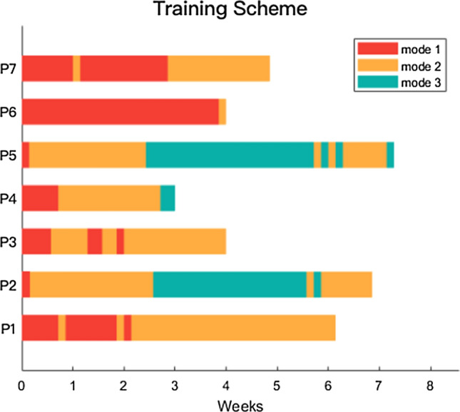
Patients’ rehabilitation training schemes. The training schemes of the patients, red represents brain-controlled mode, yellow represents brain-controlled with robot-assistance mode, and green represents brain-controlled with robot-resistance mode
Evaluation
We evaluated the patients based on 2 aspects: system usage and rehabilitation effect. System usage reflected the patients’ learning to use the system and the amount of effective training. It was evaluated by the continuous motor state switching time, the motor state percent, and the effective training revolutions. The first indicator reflected beginning time when the patient start to engage in the training process, rather than doing intermittent training. The second indicator focused on evaluating the stimulus intensity of descending motor signals, while the third indicator focused on evaluating the stimulus intensity of ascending sensory signals. The evaluation of the rehabilitation effect included the LEMS, recovery rates of the lower limb muscle strength, surface electromyogram (sEMG), sensation, and the activities of daily living (ADL).
Evaluation of system usage
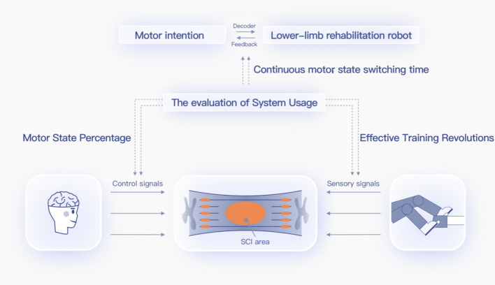
The evaluation of system usage mainly included continuous motor state switching time, motor state percent, and effective training revolutions. The relationship among system indicators, BCI system and SCI rehabilitation mechanism is depicted in Fig. 7.
Fig. 7.
The relationship between System Usage and SCI Rehabilitation. The system drives the rehabilitation robot to drive the patient’s limb movement by decoding the motor intention and provides feedback to the patients. The continuous motor state switching time of patients is an evaluation of the performance of the BCI paradigm. The descending control signals and the ascending sensory signals stimulate the spinal cord injury area. The motor state percent and the effective training revolutions can measure whether the descending control signal and the ascending sensory signal of the BCI can generate sufficient stimulation to the SCI area during training. The following portion is the schematic diagram of the system to stimulate the injured area of SCI patients. Motor state percent is used to evaluate the intensity of descending motor control signal in a single training. Effective training revolutions are employed to assess the intensity of ascending sensory signals. Continuous motor state switching time is used to evaluate the patient’s state of training using BCI system. The dash line represents the assessment. The solid lines represent signal correlation
Continuous motor state switching time
Continuous motor state switching time is the time the patients take to switch from a resting state to a concentrated motor intention state that can continuously drive the system after the start of a training session. The standard of concentrated motor intention state is the patient’s continuous effective imagination that lasts for > 10 s. It means that the patient is engaged in the training process, rather than doing intermittent training. As patients may experience different states before training begins, it is difficult to get into continuous engagement and involvement. The shorter the continuous motor state switching time, faster would be the patients entering the training state.
Motor state percent
The motor state percent was the proportion of the time when the patients’ motor intention is greater than the selected threshold. It indicates the time of effective motor function imagination in the training program. The motor state percent varies according to the patient’s understanding of the system and the modulations of their brain rhythms. The patients’ daily sleep, mental state, and other factors cause variance in their motor state percent. The higher the motor state percent of the patients, longer is the stimulation time of the descending motion signal to the injured area during the training process, which plays a key role in the bidirectional electrical stimulation of the brain–computer interface.
Effective training revolutions
Effective training revolutions refers to the number of times that the patients control the lower-limb robot to rotate during each training session. Sufficient training revolutions can ensure sufficient motor training of patients’ lower limbs, proving benefit to the patients. The higher the effective training revolutions, sufficient are the lower limb movement of patients and greater is the stimulation intensity of the ascending sensory signals to the SCI area. Evaluation of Rehabilitation Effect The International Standard for Neurological Classification of Spinal Cord Injury (ISNCSCI) developed jointly by the American Spinal Cord Injury Association and the International Spinal Cord Injury Society has been widely used as a standardized assessment tool (Kirshblum et al. 2004, Waters et al. 1994, Scivoletto, Morgantin and Molinari 2004, McKinley et al. 2004, Poynton et al. 1997).
As the main indicator of muscle strength recovery in the rehabilitation effect of patients in the International Standard AIS Evaluation System, the LEMS score is widely used in the evaluation of rehabilitation clinical trials. With the recovery of patients’ main muscle functions as the main observation index, it is consistent with the overall principle of rehabilitation medicine (Salmon et al. 2014; Poncumhak et al. 2013; Tsai et al. 2020; Jackson et al. 2008). In this experiment, 7 patients involved in the experiment demonstrated a short-term rapid increase in the muscle strength. Therefore, in this experiment, the LEMS score was used as the main evaluation index. The sensory score, surface electromyography (EMG), and other indicators were used as a reference. Therefore, an increase of 1 point in the LEMS score is of great significance for patients with motor complete SCI. In the evaluation process of patients, the experts of the hospital where the experiment was performed were highly cautious, and each patient’s weekly LEMS score was recorded in detail. The evaluations were achieved by a group, and the growths of LEMS scores were reviewed by doctors in addition to the therapist team. The evaluation data generated by the whole experiment were finally reviewed by the main experts of the hospital and recorded in patients’ files.
The LEMS was derived for evaluating the somatic function of the individuals with SCI according to ISNCSCI in patients. The scores of key lower extremity muscle groups were determined and the sum was calculated in order to assess the patient’s lower limb functions. The total LEMS was 50 points, representing the total score of 5 key muscles of the lower extremity (hip flexor, knee extensors, ankle dorsiflexors, long toe extensors, and ankle plantar flexors). The muscle strength of each key muscle is assessed by a freehand muscle strength test, and the muscle strength is evaluated on a 6 interval scale, with the score ranging from 0 to 5 (0 = complete paralysis, 1 = accessible or visible muscle contraction without motion of joints, 2 = active motion of the whole joint range of motion (ROM) under gravity condition, 3 = active motion against the full ROM under gravity condition, 4 = active motion of the full ROM under medium resistance of the special muscle position, and 5 = active motion of the full ROM under maximum resistance of the special muscle position). The maximum resistance was estimated based on the assumption that the patient’s function is normal (Kirshblum et al. 2011). The higher the score is, less is the motor function injury and better is the recovery (Marino and Graves 2004; Graves et al. 2006). Our pedaling training focuses on the improvement of hip flexor and knee extensor.
The patients were assessed based on activities of daily living (ADL) scores before and after the BCI training. ADL is an assessment scale used to assess patients’ ability of daily activities (Pentland* et al. 1999; Schonherr et al. 2005, Elizabeth, Yerxa and Locker 1990), it reflects people’s most basic ability in the family (or in a medical institution) and in the community, so it is the most basic and important content in rehabilitation medicine. It included ratings for eating, bathing, grooming, dressing, controlling defecation, controlling urination, going to the toilet, wheelchair transfer, walking and walking up and down stairs. 100 for good daily living, not dependent on others; A score of more than 60 and less than 100 indicates mild dysfunction, but they can take care of themselves in daily life. A score of 41–60 indicates moderate dysfunction and they need some help in daily life. A score of 21–40 indicates severe dysfunction and significant dependence on others in daily life; less than 20 is classified as completely disabled and completely dependent on others for daily life.
Results
Continuous motor state switching time
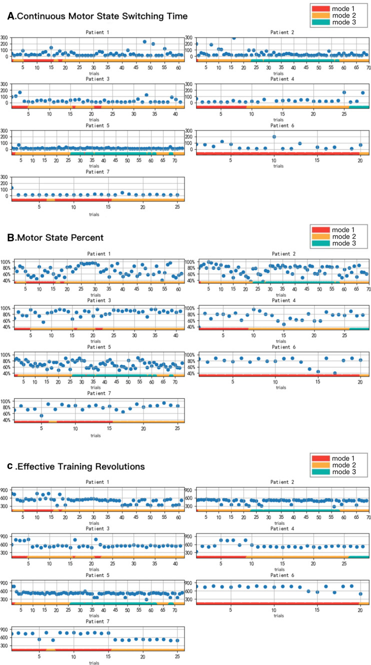
The continuous motor state switching time of 7 patients is displayed in Fig. 8A. The average continuous motor state switching time of all patients was 38.5 ± 80.1 s. The average continuous motor state switching time of patient 5 was the shortest (14.9 ± 8.6 s). Patients 5 and 7 kept a low continuous motor state switching time with little occasional fluctuations during the entire training process. The continuous motor state switching time of patients 2 and 3 decreased after 16 and 3 training sessions, respectively, and it was low during the subsequent training sessions. The continuous motor state switching time of patient 1 fluctuated significantly throughout the training, which may have been due to his poor compliance and poor rehabilitation engagement. The results revealed that the average motion state switching time of all patients only accounted for 3.2 ± 6.7% in the 20 min training session, and all patients could complete the motion state switching in a relatively short time.
Fig. 8.
The evaluation of system usage. Red represents brain-controlled mode, yellow represents brain-controlled in robot-assistance mode, and green represents brain-controlled with robot resistance mode. A The box plot of the change in the continuous motor state switching time of 7 patients with an increase in the training times, the blue dots represent the continuous motor state switching time; B The box plot of the changes in the motor state percent of 7 patients with an increase in the training times, the blue dots represent the motor state percent; C The box plot of the changes in the effective training revolutions of 7 patients with an increase in the training times, the blue dots represent the effective training revolutions
Motor State Percent
Figure 8B depicts the motor state percentages of the seven patients. The average motor state percentage of all the patients was 73.9 ± 14.4%. The motor state percentage of patient 3 was the highest (85.4 ± 9.8%), while that of patient 5 was the lowest (68.6 ± 10.7%). During the first training, except for patient 4 whose motor state percentage was about 40%, the values of the other patients were > 65%. However, during the second training, patient 4 reached 80% and maintained the high value. Even though there were some fluctuations in the training process, the patients were able to return to the normal training state after some adjustments. The results established that the patients were able to stay dedicated during the training and complete it with a high motor state percentage.
Effective training revolutions
Figure 8C illustrates the effective training revolutions of the seven patients. The mean effective training revolutions of the patients were 540.0 ± 109.6r and the mean speed was 27.0 ± 5.5r/min, which was close to the speed of normal rehabilitation training. The number of effective training revolutions in patient 6 was the highest, reaching 718.7 ± 89.4r. Patient 2 had the least number of effective training revolutions, which was 486.6 ± 67.9r. When the patient switched from the first mode to the second mode, the reduction in the effective training revolutions was obvious. The second mode required not only motor imagery but also joint action of the patients’ lower limb muscle strength. Since the patients had weak lower limbs, the number of revolutions was reduced. The results assert that the patients can complete the same amount of training revolutions as in existing rehabilitation training.
Evaluation of the rehabilitation effect
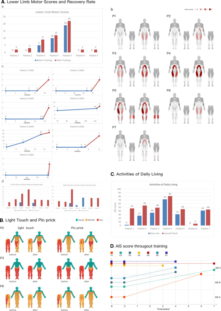
The patients were assessed using LEMS before and after BCI training. The mean LEMS of the patients was increased by 2.6 ± 0.8 points. The score of patient 1 (AIS A) increased by 2 points, with the left and right knee extensors increasing by 1 point each. Among the patients with AIS B, the score of patient 2 increased by 3 points, with the left hip flexors and knee extensors were increasing by 1 point each and the right knee extensors increasing by 1 point. The score of patient 3 increased by 4 points, with both sides of the hip flexor and knee extensors increasing by 1 point each. The score of patient 6 improved by 2 points, with the left and right knee extensors were increasing by 1 point each. Among the patients with AIS C, the score of patient 4 increased by 2 points, with the left hip flexors and knee extensors increasing by 1 point each. The score of patient 5 increased by 3 points, with the left hip flexors, left knee extensors, and left long toe extensors increasing by 1 point each. The schematic diagram of the patients’ LEMS scores and the change in muscle strength are displayed in Fig. 9A (a) and Fig. 9A (b), respectively. Comprehensive statistical results of LEMS are shown in Table 2.
Fig. 9.
The evaluation of the rehabilitation effect. A a Patients before BCI training (blue) and after BCI training (brown) LEMS scoring statistics. b The schematic diagram of the increase in the key muscle strength of patients. The darker the color, stronger is the muscle strength. c Patients before BCI training (blue) and after BCI training (brown) LEMS with changes in the injury time. d The left and the right hip flexors and knee extensors before BCI training (blue) and after BCI training (brown) recovery rate. B The changes in the light tough sensation and pink prick in patients 2, 5, and 6 before and after BCI training. Green represents normal sensation, yellow represents sensory disturbance, and red represents the disappearance of sensation. C The statistical chart of the ADL score of patients before BCI training (blue) and after training (brown). D Changes in the AIS scores in 7 patients before and after training
Table 2.
Mean ± SD score for Rehabilitation Effects
| Before | After | DIFF (After–Before) | |
|---|---|---|---|
| LEMS | 4.9 ± 7.3 | 7.4 ± 7.6 | 2.6 ± 0.8 |
| Left hip flexors and knee extensor recovery rates | 0.042 ± 0.072/week | 0.35 ± 0.19/week** | 0.31 ± 0.19/week |
| Right hip flexor and knee extensor recovery rates | 0.060 ± 0.10/week | 0.19 ± 0.18/week | 0.13 ± 0.23/week |
| Bilateral hip flexors and knee extensor recovery rates | 0.10 ± 0.16/week | 0.55 ± 0.26/week** | 0.44 ± 0.29/week |
| ADL | 29.7 ± 24.5 | 46.6 ± 22.4 | 16.9 ± 14.9 |
**p < 0.01 compared with the rate of before; post-hoc Wilcoxon Signed Rank Test for the recovery rates
Figure 9A (c) illustrates the relationship between the course of injury and the strength of the left and right hip flexors and the left and right knee extensors. The muscle strength increased in all the patients.
In this study, the recovery rate of the right and left hip flexors and knee extensors, which are the target muscles of the training, were also analyzed. We calculated the recovery rate as follows: we considered the difference in the hip flexor and knee extensor muscle strength score before and after the training and divided it by the number of training weeks.
On the left side of Fig. 9A (d), left hip flexors and knee extensor recovery rates of all the patients are illustrated. The average recovery rate of the patients before BCI rehabilitation training was 0.042 ± 0.072/week; only patient 3 and patient 5 displayed an increase in muscle strength, and the recovery rates were 0.16/week and 0.13/week respectively. After the BCI rehabilitation training, the average recovery rate of the sum of left hip flexors and knee extensors for all the patients was 0.35 ± 0.19/week, which was 8.5 times higher than that before BCI training. There were differences in the recovery rate of left hip flexors and knee extensor before and after training (p = 5.83e-04, p < 0.01). The average recovery rate for all the patients was > 0.19/week; among them, patient 4 had the highest recovery rate, reaching 0.70/week. On the right side of Fig. 9A (d), right hip flexor and knee extensor recovery rates of all the patients are illustrated. The average recovery rate before BCI training for all the patients was 0.060 ± 0.10/week; only patients 3, 4, and 5 showed an increase in muscle strength, and the recovery rates were 0.11/week, 0.047/week, and 0.27/week, respectively. During BCI rehabilitation training, the average recovery rate for all the patients was 0.19 ± 0.18/week, which was 3.2 times greater than that before BCI training. Patient 4 and patient 5 showed no increase in muscle strength, while the recovery rates of all other patients were > 0.15/week. Among them, patient 3 had the highest recovery rate, reaching 0.52/week. Patient 4 and patient 5 exhibited no increase in right hip flexor and knee extensor muscle strength. At the same time, there were differences in the recovery rate of bilateral hip flexors and knee extensor before and after training (p = 0.0023, p < 0.01). Comprehensive statistical results of left/right hip flexors and knee extensor recovery rates are shown in Table 2.
Sensory scores of patients 2, 3, and 6 before and after the training were evaluated. The improvements in the patients are presented in Fig. 9B. The sensation of the three patients improved to different degrees after the training: eight pin prick areas and eight light touch areas improved in patient 2; four areas of pin prick score and two areas of light touch score improved in patient 3; three areas of pin prick score and two points of light touch score improved in patient 6.
The ADL scores of all the patients improved, with an average improvement of 16.9 ± 14.9 points. The ADL score of patient 1 increased the most, that is, by 45 points. The score of patient 2 increased by 28 points, the scores of patients 3 and 5 increased by 14 points each, and the scores of patients 4, 6, and 7 increased by 9, 5, and 3 points, respectively. The ADL scoring results are portrayed in Fig. 9C. Comprehensive statistical results of ADL score are shown in Table 2.
The AIS scores of the seven patients before and after the training are shown in Fig. 9D. The grades of all the patients reached AIS C. While patient 1 was upgraded from AIS A to AIS C, patients 2, 3 and 6 were upgraded from AIS B to AIS C. The muscle strength of patients 4, 5, and 7 was also improved, but the AIS rating remained at AIS C.
The number of training sessions per week was 10 theoretically, but individual patients such as patients 6 and 7 were only maintained at 5 times/week. The reason was that the two patients had other complications, and the number of training sessions was maintained at the above level after doctors’ evaluation. From our actual clinical results, the time taken by these two patients to obtain significant muscle strength improvement was similar to that of the other five patients.
Discussion
According to previous studies, the rehabilitation effects of SCI are mainly related to injury location and injury level. Research has indicated that 75.2% of the complete SCI patients have no change in LEMS 1–6 months after the injury, while a mere 24.8% of the patients experience improvement. Besides, it has been shown that LEMS is increased by 1 point on average (Wirz et al. 2006). In another 1 year study, only 29.8% of the patients with AIS A showed improvements in the first year after injury, and 70.2% of the patients continued to have complete motor injuries in the lower limbs (Marino et al. 2011).
In the study of chronic AIS A/B by Shokur et al. (Shokur et al. 2018), the research process lasted for 28 months. The patients underwent several auxiliary training before BCI rehabilitation robot training. They use the paradigm of left and right arm motor imagery, but it was difficult for patients to master the use of BCI rehabilitation robots. The patients faced a long recovery time, and the lower limb muscle strength increased in 7–8 months. Our study enabled the patients to master the use of the system within a short time, and they displayed augmented muscle strength after 3–7 weeks of training. Unlike the study by Shokur et al. (Shokur et al. 2018), our patients were mainly AIS A/B within one year of the injury or chronic AIC C SCI patients; however, these patients are the ones requiring treatment in clinical rehabilitation. The short-term rehabilitation effect has a greater improvement in patients’ confidence in treatment. To a certain extent, it also reduces the economic burden of those who have already faced a tragedy. Generally speaking, the results indicate that clinical application of our system with lower-limb robot is easily for patients to use. And it shows potentially positive effects on SCI patients.
In our study, all AIS A and B patients were raised to the AIS C level, which is a significant improvement, indicating that the patients were elevated from complete to incomplete motor injury. Subsequently, the doctor’s treatment plan for the patients has also undergone fundamental changes. For patients with complete motor injury, the doctor only recommends training of the upper limb and increase in ADL ability and does not expect to restore the muscle strength of the lower limb. On the other hand, for patients with incomplete sports injury, the doctors suggest training to increase the strength of the patients’ limbs, hoping to restore lower limb muscle strength.
The patient with AIS A in our study was 57 years old, and the injury level was T11. He experienced an improvement in the lower limb muscle strength through a 6-week training program. Both left and right knee extensors increased by 1 level, and the LEMS score increased by 2 points. Unlike existing training, it is difficult to achieve rehabilitation effect through short-term training.
The rehabilitation effects were the most obvious for the three AIS B patients. For patients with AIS B, muscle strength recovery is very difficult. Such patients are also defined as motor complete injury. They only have residual sensation, and there is hardly any improvement in muscle strength after training for less than 1 year (Harrop et al. 2011). In existing treatment, the training of lower limb muscle strength is mostly abandoned. Of particular note, both patients 2 and 6 in our study had neck injuries with AIS B. According to previous studies, the higher the injury level, the lower is the possibility of recovery (Harrop et al. 2011). Currently, there are few studies on the improvement of lower limb muscle strength in patients with complete motor injuries of the neck. Patient 2 demonstrated no increase in muscle strength before the BCI training, and after 8 weeks of training, the left lower limb muscle strength increased by 2 points, while the right lower limb muscle strength improved by 1 point. Patient 6 showed no increase in muscle strength before the BCI training, and after 4 weeks of training, the left and right lower limb muscle strength increased by 1 point each. After 8 weeks of training, the left and right lower limb muscle strength of patient 3 increased by 2 points. Patients 2, 3, and 6 also reported an increase in sensation.
Once the SCI patients enter the chronic period, the rehabilitation effect is greatly compromised, and it is difficult to achieve obvious rehabilitation effects via short-term training (Kirshblum et al. 2004). However, three chronic patients with AIS C were also recruited and had rehabilitation effects. Among them, the left hip flexor of patient 4 improved from level 2 to level 3 and the left knee extensors improved from level 2 to level 3. After 3 weeks of BCI training, patient 4 was able to walk with braces. The patient’s course of disease had been as long as 2.5 years. Before using our system, the patient had been hospitalized for more than a year, and the possibility of spontaneous recovery was ruled out.
Patient 5 also had a course of disease that was longer than 9 months. After 8 weeks of training, the left hip flexor improved from level 1 to level 2, the left knee extensors improved from level 1 to level 2, and the left ankle plantar flexors improved from level 1 to level 2. After training, this patient was also able to walk with the aid of braces.
It should also be noted that seven patients had no adverse events during the use of our system. This proves to some extent that our system is safe.
The core of our system is to strengthen the bidirectional stimulation and provide rehabilitation training for the patients according to the decision tree. To enhance the bidirectional stimulation, we selected the EEG characteristics related to the motor intention of the patients in the training process as the main control indicator. This facilitated the guiding of the patients to give descending motor control signals and strengthen the intensity of the stimulation. Through the experimental study of patients with post-stroke dyskinesias and healthy people, we found that the obvious abnormality of DAR, DTABR and DABR in the prefrontal lobe of the patients may be related to the lack of motor function. The prefrontal cortex is associated with decision-making; hence, we assume that there is a certain correlation between the three prefrontal indicators (DAR, DTABR and DABR) and motor control. By experimenting with healthy people, we found that these indicators can reflect the motor intention of the subjects to some extent; therefore, they were used as the core parameters in the decoder design. We believe that the control of these indicators may strengthen the issuance of motor-related EEG rhythms in the state of motion, thereby strengthening the descending motor control signal. On the one hand, such a design can accurately apply descending control signal stimulation to the SCI area; on the other hand, repeated high-intensity training may also have a certain strengthening effect on specific brain areas. The accurate selection of indicators for brain decoders is in line with the future development trend of BCI technology. Although this study cannot provide conclusive evidence for the relationship between these indicators and motor control function, our experimental results suggest that the motor functions of SCI patients can be restored in the short term. We believe that this restoration may be related to our appropriate selection of motor-related EEG indicators. In addition, in rehabilitation training, emphasizing the patient’s engagement and involvement is the basic requirement of rehabilitation medicine for many years. In the paradigm that we use, we have EEG indicators for attention assessment (Beebe, Rose and Amin 2010; Gaume et al. 2019; Wang et al. 2018). While the patient is imagining the bicycle movement, it is also a process to urge the patient to concentrate on the training. Our system can detect attention during treatment, allowing patients to focus on the training process. Comparatively speaking, existing BCI paradigms, especially several paradigms of spontaneous EEG based on ERD/ERS require a long period of training (Donati et al. 2016, Edelman1* et al. 2019, Morash et al. 2008). Around 20% of subjects are unable to control the BCI system at all, despite undergoing training (Jeunet, Jahanpour and Lotte 2016, Zuo et al. 2020). In addition, the existing spontaneous EEG paradigm requires machine learning adjustment of the system based on individual training data, with great individual differences(Edelman1* et al. 2019; Morash et al. 2008; Jeunet et al. 2016). Systematic learning takes a long time, which is often unacceptable to hospitals and patients (Zuo et al. 2020). Although the BCI fused with P300 and MI can improve recognition accuracy and reduce training time to some extent, paradigms such as P300 based on external stimuli are prone to fatigue, which limits clinical applications (Zuo et al. 2020).
In previous studies, active motion was mainly realized by adding torque sensor or surface EMG sensor to the rehabilitation robot system (Huang et al. 2018, Meng et al. 2014, Benedetta Cesqui 2013, Manohar et al. 2017). The torque sensors and surface EMG sensors are placed at the end of the neural path. But it presents two problems. Problem 1: For patients with grade 0–1 muscle strength, the affected limb muscles cannot produce effective signals or the signals are very weak, which cannot be accurately captured by the above-mentioned sensors. Problem 2: In the later stages of rehabilitation, because of the long and boring training, patients tend to engage in patterned and regular exercise rather than active exercise involving the motor cortex of the brain. The involvement of the motor cortex is important for motor rehabilitation (Bonizzato et al. 2018, van den Brand et al. 2012). The BCI system can be considered as an effective sensor for the robot to detect the motor intention of the patient. Through the design of decision tree mechanism of the training mode, the active participation of the patients in different injury stages and the whole process of rehabilitation training can be ensured. In the patients’ oral presentations, the intensity of systematic training in this study was significantly higher than that of the current rehabilitation training methods, and the system design mechanism of whole-course active rehabilitation played a decisive role. High intensity active rehabilitation training can be reflected by two indicators: the motor state percentage and the effective training revolutions, which correspond to the distribution of descending control signal and ascending sensory signal during the training. At the same time, increasing the intensity of bidirectional stimulation was the core part in our study, which was proved in all seven patients. In the short term, these patients, who did not demonstrate improvements with the existing rehabilitation technology, showed obvious muscle strength enhancement, which was strongly associated with high intensity bidirectional stimulation. From this point of view, our study attempts to explain the mechanism of BCI in the rehabilitation of SCI.
Bidirectional stimulation plays a key role in nerve function remodeling. According to Hebbian theory (Hebb 1949), when the presynaptic and postsynaptic neurons are activated or discharged in a certain order, repeatedly and with a certain intensity, the transmission efficiency of the synapse is altered, and the synapse is remodeled. We believe that through bidirectional stimulation, the residual neurons in the injured area of the spinal cord can be stimulated to reshape so that the transmission efficiency of the neurons controlling the patient’s motor function can be increased and their functioning improved.
Central nervous system regeneration is a very challenging topic. In our experiment, patients with AIS A/B had short-term motor function under the condition of complete loss of motor function. We believe that there are some limitations in explaining the concept by using neural remodeling alone. Recent studies have shown that after SCI, many inhibitory factors are produced in the injured area, which inhibit the nerve regeneration pathway and lead to a decrease in the recovery rate of the nerve cells. Moreover, changes in the microenvironment after SCI can reduce the secretion of factors that promote nerve regeneration. Alterations in the microenvironment after SCI are the main reasons for the inhibition of nerve cell regeneration. Improving the microenvironment in the area of SCI, boosting the secretion of growth factors, and reducing the secretion of inhibitory factors may promote nerve regeneration (Rodríguez-Barrera et al. 2017; Chen et al. 2020). In 1981, David et al. used autologous sciatic nerve to bridge the 35 mm medulla oblongata and the spinal cord and found that neuronal connective axons grew nearly 30 mm along the transplanted sciatic nerve. This study confirmed that the central nervous system cells could be regenerated under certain conditions (Samuel David 1981). Since then, researchers have explored various methods to improve the secretion of promoters and inhibitors in the microenvironment of the SCI and promote the regeneration of the central nervous system cells to treat the SCI. These methods include the use of neurotrophic factors (Bradbury et al. 1999; Hodgetts and Harvey 2017), stem cells (Huang et al. 2003; Lima et al. 2010), electrical stimulation (Batty et al. 2020; Beaumont et al. 2014; Griffin et al. 2009; Huang et al. 2013), and exercise therapy.
In experiments involving rats, a combination of electrical/magnetic stimulation therapy and exercise therapy produced significantly better results than monotherapy (Petrosyan et al. 2015; Hou et al. 2014). BCI has been used in the rehabilitation of SCI and combines the characteristics of transcranial magnetic/electrical stimulation, and exercise therapy. According to the research, synapse conductance signals need not combine with the target cells completely to transmit the signals. Existing research has proven that the axons of the synapses in the absence of point-to-point connections exhibit different degrees of improvements in nerve functions, that is, after SCI of synaptic connections inaccuracy can also make body restore some functions. (Hamid and Hayek 2008) Therefore, the design of a BCI system to generate high intensity and simultaneous bidirectional stimulation may promote the growth of spinal nerve cells to a certain extent, which may have potentially positive effect in the functional recovery of SCI patients.
Supplementary Information
Below is the link to the electronic supplementary material.
Author contributions
ZC, YL, SH, XW have contributed equally to this work. ZC, SZ, YL, XW, XF, FL, SH, WC, JL designed experiments. XW, XW, YZ, FC, collected the data. ZC, XW, XF, FL, analyzed the data. ZC, SZ, YL, XW, XF, FL, XW, WC, HQ wrote the manuscript. ZC, SZ, XF, FL, XW, PY, WC, created the system ZC, XF, FL designed the user interface.
Funding
Key Project of Jiangsu Provincial Department of Science and Technology (BE2017007-5, recipient: YL). Wuxi Taihu Talent Project. NO. WXTTP2020008.
Data availability
All data generated or analysed during this study are included in this published article (and its supplementary information files).
Declarations
Conflict of interest
The authors declares that they have no conflict of interest.
Footnotes
Publisher's Note
Springer Nature remains neutral with regard to jurisdictional claims in published maps and institutional affiliations.
Contributor Information
Shiqiang Zhu, Email: sqzhu@zju.edu.cn.
Jianan Li, Email: lijianan@carm.org.cn.
Weidong Chen, Email: chenwd@zju.edu.cn.
References
- Batty NJ, Torres-Espín A, Vavrek R, Raposo P, Fouad K. Single-session cortical electrical stimulation enhances the efficacy of rehabilitative motor training after spinal cord injury in rats. Exp Neurol. 2020;324:113136. doi: 10.1016/j.expneurol.2019.113136. [DOI] [PubMed] [Google Scholar]
- Beaumont E, Guevara E, Dubeau S, Lesage F, Nagai M, Popovic M. Functional electrical stimulation post-spinal cord injury improves locomotion and increases afferent input into the central nervous system in rats. J Spinal Cord Med. 2014;37:93–100. doi: 10.1179/2045772313Y.0000000117. [DOI] [PMC free article] [PubMed] [Google Scholar]
- Beebe DW, Rose D, Amin R. Attention, learning, and arousal of experimentally sleep-restricted adolescents in a simulated classroom. Journal of Adolesc Health. 2010;47:523–525. doi: 10.1016/j.jadohealth.2010.03.005. [DOI] [PMC free article] [PubMed] [Google Scholar]
- Benedetta Cesqui PT, Micera S, Krebs HI. EMG-based pattern recognition approach in post stroke robot-aided rehabilitation: a feasibility study. J Neuroeng Rehabil. 2013;10(1):1–5. doi: 10.1186/1743-0003-10-75. [DOI] [PMC free article] [PubMed] [Google Scholar]
- Bonizzato M, Pidpruzhnykova G, DiGiovanna J, Shkorbatova P, Pavlova N, Micera S, Courtine G. Brain-controlled modulation of spinal circuits improves recovery from spinal cord injury. Nat Commun. 2018;9:3015. doi: 10.1038/s41467-018-05282-6. [DOI] [PMC free article] [PubMed] [Google Scholar]
- Bradbury EJ, Khemani S, Von R, Priestley JV, McMahon SB. NT-3 promotes growth of lesioned adult rat sensory axons ascending in the dorsal columns of the spinal cord. Eur J Neurosci. 1999;11:3873–3883. doi: 10.1046/j.1460-9568.1999.00809.x. [DOI] [PubMed] [Google Scholar]
- Carmena JM, Lebedev MA, Crist RE, O’Doherty JE, Santucci DM, Dimitrov DF, Patil PG, Henriquez CS, Nicolelis MA. Learning to control a brain-machine interface for reaching and grasping by primates. PLoS Biol. 2003;1:E42. doi: 10.1371/journal.pbio.0000042. [DOI] [PMC free article] [PubMed] [Google Scholar]
- Carr JH, Shepherd RB (1998) Neurological rehabilitation: optimizing motor performance
- Chen Y-Q, Wang S-N, Shi Y-J, Chen J, Ding S-Q, Tang J, Shen L, Wang R, Ding H, Hu J-G. CRID3, a blocker of apoptosis associated speck like protein containing a card, ameliorates murine spinal cord injury by improving local immune microenvironment. J Neuroinflammation. 2020;17:1–18. doi: 10.1186/s12974-020-01937-8. [DOI] [PMC free article] [PubMed] [Google Scholar]
- Collinger JL, Foldes S, Bruns TM, Wodlinger B, Gaunt R, Weber DJ. Neuroprosthetic technology for individuals with spinal cord injury. J Spinal Cord Med. 2013;36:258–272. doi: 10.1179/2045772313Y.0000000128. [DOI] [PMC free article] [PubMed] [Google Scholar]
- Curt A, Van Hedel HJ, Klaus D, Dietz V, E.-S. S. Group (2008) Recovery from a spinal cord injury: significance of compensation, neural plasticity, and repair. J Neurotrauma 25(6):677–685 [DOI] [PubMed]
- Ditunno JF, Little JW, Tessler A, Burns AS. Spinal shock revisited: a four-phase model. Spinal Cord. 2004;42:383–395. doi: 10.1038/sj.sc.3101603. [DOI] [PubMed] [Google Scholar]
- Dobkin BH, Havton LA. Basic advances and new avenues in therapy of spinal cord injury. Annu Rev Med. 2004;55:255–282. doi: 10.1146/annurev.med.55.091902.104338. [DOI] [PMC free article] [PubMed] [Google Scholar]
- Donati AR, Shokur S, Morya E, Campos DS, Moioli RC, Gitti CM, Augusto PB, Tripodi S, Pires CG, Pereira GA, Brasil FL, Gallo S, Lin AA, Takigami AK, Aratanha MA, Joshi S, Bleuler H, Cheng G, Rudolph A, Nicolelis MA. Long-term training with a brain-machine interface-based gait protocol induces partial neurological recovery in paraplegic patients. Sci Rep. 2016;6:30383. doi: 10.1038/srep30383. [DOI] [PMC free article] [PubMed] [Google Scholar]
- Donoghue JP, Mijail D, Serruya GN, Hatsopoulos LP, Matthew FR. Brain-machine interface: Instant neural control of a movement signal. Nature. 2002;416:14. doi: 10.1038/416141a. [DOI] [PubMed] [Google Scholar]
- Edelman1* BJ, Meng2† J, Suma2† D, Zurn1 C, Nagarajan3 E, Baxter1‡ BS, Cline1§ CC, He2 B (2019) Noninvasive neuroimaging enhances continuous neural tracking for robotic device control. Sci Robot 4:eaaw6844 [DOI] [PMC free article] [PubMed]
- Elizabeth J, Yerxa, Locker SB (1990) Quality of time use by adults with spinal cord injllries. Am J Occup Ther 44(4) [DOI] [PubMed]
- Finnigan S, van Putten MJ. EEG in ischaemic stroke: quantitative EEG can uniquely inform (sub-)acute prognoses and clinical management. Clin Neurophysiol. 2013;124:10–19. doi: 10.1016/j.clinph.2012.07.003. [DOI] [PubMed] [Google Scholar]
- Ganzer PD, Colachis SC, Schwemmer MA, Friedenberg DA, Dunlap CF, Swiftney CE, Jacobowitz AF, Weber DJ, Bockbrader MA, Sharma G. Restoring the sense of touch using a sensorimotor demultiplexing neural interface. Cell. 2020;181(4):763–773. doi: 10.1016/j.cell.2020.03.054. [DOI] [PubMed] [Google Scholar]
- Gaume A, Dreyfus G, Vialatte FB. A cognitive brain-computer interface monitoring sustained attentional variations during a continuous task. Cogn Neurodyn. 2019;13:257–269. doi: 10.1007/s11571-019-09521-4. [DOI] [PMC free article] [PubMed] [Google Scholar]
- Goudman L, Brouns R, De Groote S, De Jaeger M, Huysmans E, Forget P, Moens M. Association between spinal cord stimulation and top-down nociceptive inhibition in people with failed back surgery syndrome: a cohort study. Phys Ther. 2019;99:915–923. doi: 10.1093/ptj/pzz051. [DOI] [PubMed] [Google Scholar]
- Graves DE, Frankiewicz RG, Donovan WH. Construct validity and dimensional structure of the ASIA motor scale. J Spinal Cord Med. 2006;29:39–45. doi: 10.1080/10790268.2006.11753855. [DOI] [PMC free article] [PubMed] [Google Scholar]
- Griffin L, Decker MJ, Hwang JY, Wang B, Kitchen K, Ding Z, Ivy JL. Functional electrical stimulation cycling improves body composition, metabolic and neural factors in persons with spinal cord injury. J Electromyogr Kinesiol. 2009;19:614–622. doi: 10.1016/j.jelekin.2008.03.002. [DOI] [PubMed] [Google Scholar]
- Hall TM, de Carvalho F, Jackson A. A common structure underlies low-frequency cortical dynamics in movement, sleep, and sedation. Neuron. 2014;83:1185–1199. doi: 10.1016/j.neuron.2014.07.022. [DOI] [PMC free article] [PubMed] [Google Scholar]
- Hallett M. Transcranial magnetic stimulation and the human brain. Nature. 2000;406:147–150. doi: 10.1038/35018000. [DOI] [PubMed] [Google Scholar]
- Hamid S, Hayek R. Role of electrical stimulation for rehabilitation and regeneration after spinal cord injury: an overview. Eur Spine J. 2008;17:1256–1269. doi: 10.1007/s00586-008-0729-3. [DOI] [PMC free article] [PubMed] [Google Scholar]
- Harrop JS, Naroji S, Maltenfort MG, Ratliff JK, Tjoumakaris SI, Frank B, Anderson DG, Albert T, Vaccaro AR. Neurologic improvement after thoracic, thoracolumbar, and lumbar spinal cord (conus medullaris) injuries. Spine (phila Pa 1976) 2011;36:21–25. doi: 10.1097/BRS.0b013e3181fd6b36. [DOI] [PubMed] [Google Scholar]
- He Y, Eguren D, Azorin JM, Grossman RG, Luu TP, Contreras-Vidal JL. Brain-machine interfaces for controlling lower-limb powered robotic systems. J Neural Eng. 2018;15:021004. doi: 10.1088/1741-2552/aaa8c0. [DOI] [PubMed] [Google Scholar]
- Hebb DO. The organization of behavior. New York: Wiley & Sons; 1949. [Google Scholar]
- Hejazi-Shirmard M, Taghizadeh G, Azad A, Lajevardi L, Rassafiani M. Sensory retraining improves light touch threshold of the paretic hand in chronic stroke survivors: a single-subject A-B design. Somatosens Mot Res. 2020;37:74–83. doi: 10.1080/08990220.2020.1736021. [DOI] [PubMed] [Google Scholar]
- Hodgetts SI, Harvey AR. Neurotrophic factors used to treat spinal cord injury. Vitam Horm. 2017;104:405–457. doi: 10.1016/bs.vh.2016.11.007. [DOI] [PubMed] [Google Scholar]
- Hou J, Nelson R, Nissim N, Parmer R, Thompson FJ, Bose P. Effect of combined treadmill training and magnetic stimulation on spasticity and gait impairments after cervical spinal cord injury. J Neurotrauma. 2014;31:1088–1106. doi: 10.1089/neu.2013.3096. [DOI] [PubMed] [Google Scholar]
- Huang H, Chen L, Wang H, Xiu B, Li B, Wang R, Zhang J, Zhang F, Gu Z, Li Y. Influence of patients’ age on functional recovery after transplantation of olfactory ensheathing cells into injured spinal cord injury. Chin Med J. 2003;116:1488–1491. [PubMed] [Google Scholar]
- Huang J, Zhang Y, Lu L, Hu X, Luo Z. Electrical stimulation accelerates nerve regeneration and functional recovery in delayed peripheral nerve injury in rats. Eur J Neurosci. 2013;38:3691–3701. doi: 10.1111/ejn.12370. [DOI] [PubMed] [Google Scholar]
- Huang Y, Lai WP, Qian Q, Hu X, Tam EWC, Zheng Y. Translation of robot-assisted rehabilitation to clinical service: a comparison of the rehabilitation effectiveness of EMG-driven robot hand assisted upper limb training in practical clinical service and in clinical trial with laboratory configuration for chronic stroke. Biomed Eng Online. 2018;17:91. doi: 10.1186/s12938-018-0516-2. [DOI] [PMC free article] [PubMed] [Google Scholar]
- Jackson A, Carnel C, Ditunno J, Read MS, Boninger M, Schmeler M, Williams S, Donovan W. Outcome measures for gait and ambulation in the spinal cord injury population. J Spinal Cord Med. 2008;31:487–499. doi: 10.1080/10790268.2008.11753644. [DOI] [PMC free article] [PubMed] [Google Scholar]
- Jeunet C, Jahanpour E, Lotte F. Why standard brain-computer interface (BCI) training protocols should be changed: an experimental study. J Neural Eng. 2016;13:036024. doi: 10.1088/1741-2560/13/3/036024. [DOI] [PubMed] [Google Scholar]
- Kirshblum S, Millis S, McKinley W, Tulsky D. Late neurologic recovery after traumatic spinal cord injury. Arch Phys Med Rehabil. 2004;85:1811–1817. doi: 10.1016/j.apmr.2004.03.015. [DOI] [PubMed] [Google Scholar]
- Kirshblum SC, Waring W, Biering-Sorensen F, Burns SP, Johansen M, Schmidt-Read M, Donovan W, Graves D, Jha A, Jones L, Mulcahey MJ, Krassioukov A. International standards for neurological classification of spinal cord injury (Revised 2011) J Spinal Cord Med. 2011;34:547–554. doi: 10.1179/204577211X13207446293695. [DOI] [PMC free article] [PubMed] [Google Scholar]
- Lima C, Escada P, Pratas-Vital J, Branco C, Arcangeli CA, Lazzeri G, Santana Maia CA, Capucho C, Hasse-Ferreira A, Peduzzi JD. Olfactory mucosal autografts and rehabilitation for chronic traumatic spinal cord injury. Neurorehabilit Neural Repair. 2010;24:10–22. doi: 10.1177/1545968309347685. [DOI] [PubMed] [Google Scholar]
- Lopez ME, Pusil S, Pereda E, Maestu F, Barcelo F. Dynamic low frequency EEG phase synchronization patterns during proactive control of task switching. Neuroimage. 2019;186:70–82. doi: 10.1016/j.neuroimage.2018.10.068. [DOI] [PubMed] [Google Scholar]
- Manohar A, Foffani G, Ganzer PD, Bethea JR, Moxon KA. Cortex-dependent recovery of unassisted hindlimb locomotion after complete spinal cord injury in adult rats. Elife. 2017;6:e23532. doi: 10.7554/eLife.23532. [DOI] [PMC free article] [PubMed] [Google Scholar]
- Marino RJ, Graves DE. Metric properties of the ASIA motor score: subscales improve correlation with functional activities. Arch Phys Med Rehabil. 2004;85:1804–1810. doi: 10.1016/j.apmr.2004.04.026. [DOI] [PubMed] [Google Scholar]
- Marino RJ, Burns S, Graves DE, Leiby BE, Kirshblum S, Lammertse DP. Upper- and lower-extremity motor recovery after traumatic cervical spinal cord injury: an update from the national spinal cord injury database. Arch Phys Med Rehabil. 2011;92:369–375. doi: 10.1016/j.apmr.2010.09.027. [DOI] [PubMed] [Google Scholar]
- McKinley W, Meade MA, Kirshblum S, Barnard B. Outcomes of early surgical management versus late or no surgical intervention after acute spinal cord injury. Arch Phys Med Rehabil. 2004;85:1818–1825. doi: 10.1016/j.apmr.2004.04.032. [DOI] [PubMed] [Google Scholar]
- Mehdi Rassafiani RS. Single case experimental design: an overview. Int J Ther Rehabil. 2010;17(6):285–289. [Google Scholar]
- Meng W, Liu Q, Zhou Z, Ai Q. Active interaction control applied to a lower limb rehabilitation robot by using EMG recognition and impedance model. Ind Robot Int J. 2014;41:465–479. [Google Scholar]
- Morash V, Bai O, Furlani S, Lin P, Hallett M. Classifying EEG signals preceding right hand, left hand, tongue, and right foot movements and motor imageries. Clin Neurophysiol. 2008;119:2570–2578. doi: 10.1016/j.clinph.2008.08.013. [DOI] [PMC free article] [PubMed] [Google Scholar]
- Olivieri A, Locatelli F, Zecca M, Sanna A, Cimminiello M, Raimondi R, Gini G, Mordini N, Balduzzi A, Leoni P, Gabrielli A, Bacigalupo A. Imatinib for refractory chronic graft-versus-host disease with fibrotic features. Blood. 2009;114:709–718. doi: 10.1182/blood-2009-02-204156. [DOI] [PubMed] [Google Scholar]
- Pentland W, Harvey AS, Smith T, Walker J. The impact of spinal cord injury on men’s time use. Spinal Cord. 1999;37:786–792. doi: 10.1038/sj.sc.3100908. [DOI] [PubMed] [Google Scholar]
- Petrosyan HA, Alessi V, Hunanyan AS, Sisto SA, Arvanian VL. Spinal electro-magnetic stimulation combined with transgene delivery of neurotrophin NT-3 and exercise: novel combination therapy for spinal contusion injury. J Neurophysiol. 2015;114:2923–2940. doi: 10.1152/jn.00480.2015. [DOI] [PMC free article] [PubMed] [Google Scholar]
- Poncumhak P, Saengsuwan J, Kamruecha W, Amatachaya S. Reliability and validity of three functional tests in ambulatory patients with spinal cord injury. Spinal Cord. 2013;51:214–217. doi: 10.1038/sc.2012.126. [DOI] [PubMed] [Google Scholar]
- Poynton AR, O’Farrell DA, Shannon F, Murray P, McManus F, Walsh MG. Sparing of sensation to pin prick predicts recovery of a motor segment after injury to the spinal cord. J Bone Jt Surg. 1997;79:952–954. doi: 10.1302/0301-620x.79b6.7939. [DOI] [PubMed] [Google Scholar]
- Ramanathan DS, Guo L, Gulati T, Davidson G, Hishinuma AK, Won SJ, Knight RT, Chang EF, Swanson RA, Ganguly K. Low-frequency cortical activity is a neuromodulatory target that tracks recovery after stroke. Nat Med. 2018;24:1257–1267. doi: 10.1038/s41591-018-0058-y. [DOI] [PMC free article] [PubMed] [Google Scholar]
- Ramos-Murguialday A, Broetz D, Rea M, Laer L, Yilmaz O, Brasil FL, Liberati G, Curado MR, Garcia-Cossio E, Vyziotis A, Cho W, Agostini M, Soares E, Soekadar S, Caria A, Cohen LG, Birbaumer N. Brain-machine interface in chronic stroke rehabilitation: a controlled study. Ann Neurol. 2013;74:100–108. doi: 10.1002/ana.23879. [DOI] [PMC free article] [PubMed] [Google Scholar]
- Rodríguez-Barrera R, Flores-Romero A, Fernández-Presas AM, García-Vences E, Silva-García R, Konigsberg M, Blancas-Espinoza L, Buzoianu-Anguiano V, Soria-Zavala K, Suárez-Meade P. Immunization with neural derived peptides plus scar removal induces a permissive microenvironment, and improves locomotor recovery after chronic spinal cord injury. BMC Neurosci. 2017;1:1–13. doi: 10.1186/s12868-016-0331-2. [DOI] [PMC free article] [PubMed] [Google Scholar]
- Salmon E, Carrico C, Nichols L, Reddy L, Salles S, Sawaki L (2014) Transcranial direct current stimulation to enhance motor function in spinal cord injury: Pilot data. In: 2014 IEEE 16th international conference on e-Health networking, applications and services (Healthcom), pp 1–6. IEEE
- Samuel David AJA. Axonal elongation into peripheral nervous system bridges after central nervous system injury in adult rats. Science. 1981;214:931 . doi: 10.1126/science.6171034. [DOI] [PubMed] [Google Scholar]
- Schonherr MC, Groothoff JW, Mulder GA, Eisma WH. Participation and satisfaction after spinal cord injury: results of a vocational and leisure outcome study. Spinal Cord. 2005;43:241–248. doi: 10.1038/sj.sc.3101683. [DOI] [PubMed] [Google Scholar]
- Scivoletto G, Morganti B, Molinari M. Sex-related differences of rehabilitation outcomes of spinal cord lesion patients. Clin Rehabil. 2004;18:709–713. doi: 10.1191/0269215504cr749oa. [DOI] [PubMed] [Google Scholar]
- Sheorajpanday RV, Nagels G, Weeren AJ, De Surgeloose D, De Deyn PP. Additional value of quantitative EEG in acute anterior circulation syndrome of presumed ischemic origin. Clin Neurophysiol. 2010;121:1719–1725. doi: 10.1016/j.clinph.2009.10.037. [DOI] [PubMed] [Google Scholar]
- Sheorajpanday RV, Nagels G, Weeren AJ, van Putten MJ, De Deyn PP. Quantitative EEG in ischemic stroke: correlation with functional status after 6 months. Clin Neurophysiol. 2011;122:874–883. doi: 10.1016/j.clinph.2010.07.028. [DOI] [PubMed] [Google Scholar]
- Shindo K, Kawashima K, Ushiba J, Ota N, Ito M, Ota T, Kimura A, Liu M. Effects of neurofeedback training with an electroencephalogram-based brain-computer interface for hand paralysis in patients with chronic stroke: a preliminary case series study. J Rehabil Med. 2011;43:951–957. doi: 10.2340/16501977-0859. [DOI] [PubMed] [Google Scholar]
- Shokur S, Donati ARC, Campos DSF, Gitti C, Bao G, Fischer D, Almeida S, Braga VAS, Augusto P, Petty C, Alho EJL, Lebedev M, Song AW, Nicolelis MAL. Training with brain-machine interfaces, visuo-tactile feedback and assisted locomotion improves sensorimotor, visceral, and psychological signs in chronic paraplegic patients. PLoS ONE. 2018;13:e0206464. doi: 10.1371/journal.pone.0206464. [DOI] [PMC free article] [PubMed] [Google Scholar]
- Takeoka A. Proprioception: bottom-up directive for motor recovery after spinal cord injury. Neurosci Res. 2020;154:1–8. doi: 10.1016/j.neures.2019.07.005. [DOI] [PubMed] [Google Scholar]
- Tariq M, Trivailo PM, Simic M. EEG-based BCI control schemes for lower-limb assistive-robots. Front Hum Neurosci. 2018;12:312. doi: 10.3389/fnhum.2018.00312. [DOI] [PMC free article] [PubMed] [Google Scholar]
- Tsai CY, Delgado AD, Weinrauch WJ, Manente N, Levy I, Escalon MX, Bryce TN, Spungen AM. Exoskeletal-assisted walking during acute inpatient rehabilitation leads to motor and functional improvement in persons with spinal cord injury: a pilot study. Arch Phys Med Rehabil. 2020;101:607–612. doi: 10.1016/j.apmr.2019.11.010. [DOI] [PubMed] [Google Scholar]
- van den Brand R, Heutschi J, Barraud Q, DiGiovanna J, Bartholdi K, Huerlimann M, Friedli L, Vollenweider I, Moraud EM, Duis S, Dominici N, Micera S, Musienko P, Courtine G. Restoring voluntary control of locomotion after paralyzing spinal cord injury. Science. 2012;336:1182–1185. doi: 10.1126/science.1217416. [DOI] [PubMed] [Google Scholar]
- Wang H, Dragomir A, Abbasi NI, Li J, Thakor NV, Bezerianos A. A novel real-time driving fatigue detection system based on wireless dry EEG. Cogn Neurodyn. 2018;12:365–376. doi: 10.1007/s11571-018-9481-5. [DOI] [PMC free article] [PubMed] [Google Scholar]
- Waters RL, Adkins RH, Yakura JS, Sie I. Motor and sensory recovery following incomplete tetraplegia. Arch Phys Med Rehabil. 1994;75:67–72. doi: 10.1016/0003-9993(94)90034-5. [DOI] [PubMed] [Google Scholar]
- Wirz M, van Hedel HJ, Rupp R, Curt A, Dietz V. Muscle force and gait performance: relationships after spinal cord injury. Arch Phys Med Rehabil. 2006;87:1218–1222. doi: 10.1016/j.apmr.2006.05.024. [DOI] [PubMed] [Google Scholar]
- Zuo C, Jin J, Yin E, Saab R, Miao Y, Wang X, Hu D, Cichocki A. Novel hybrid brain-computer interface system based on motor imagery and P300. Cogn Neurodyn. 2020;14:253–265. doi: 10.1007/s11571-019-09560-x. [DOI] [PMC free article] [PubMed] [Google Scholar]
Associated Data
This section collects any data citations, data availability statements, or supplementary materials included in this article.
Supplementary Materials
Data Availability Statement
All data generated or analysed during this study are included in this published article (and its supplementary information files).


