

Abstract
Background
Large territory middle cerebral artery (MCA) ischaemic strokes account for around 10% of all ischaemic strokes and have a particularly devastating prognosis when associated with malignant oedema. Progressive cerebral oedema starts developing in the first 24 to 48 hours of stroke ictus with an associated rise in intracranial pressure. The rise in intracranial pressure may eventually overwhelm compensatory mechanisms leading to a cascading secondary damage to surrounding unaffected parenchyma. This downward spiral can rapidly progress to death or severe neurological disability. Early decompressive craniectomy to relieve intracranial pressure and associated tissue shift can help ameliorate this secondary damage and improve outcomes. Evidence has been accumulating of the benefit of early surgical decompression in stroke patients. Earlier studies have excluded people above the age of 60 due to associated poor outcomes; however, newer trials have included this patient subgroup. This review follows a Cochrane Review published in 2012.
Objectives
To assess the effectiveness of surgical decompression in people with malignant oedema after ischaemic stroke with regard to reduction in mortality and improved functional outcome. We also aimed to examine the adverse effects of surgical decompression in this patient cohort.
Search methods
We searched the Cochrane Stroke Group Trials Register, the Cochrane Central Register of Controlled Trials (CENTRAL; 2022, Issue 7 of 12), MEDLINE Ovid, Embase Ovid, Web of Science Core Collection, Scopus databases, ClinicalTrials.gov, and the WHO ICTRP to July 2022. We also reviewed the reference lists of relevant articles.
Selection criteria
We included randomised controlled trials (RCTs) comparing decompressive craniectomy with medical management to best medical management alone for people with malignant cerebral oedema after MCA ischaemic stroke.
Data collection and analysis
Two review authors independently screened the search results, assessed study eligibility, performed risk of bias assessment, and extracted the data. The primary outcomes were death and death or severe disability (modified Rankin Scale (mRS) > 4) at 6 to 12 months follow‐up. Other outcomes included death or moderate disability (mRS > 3), severe disability (mRS = 5), and adverse events. We assessed the certainty of the evidence using the GRADE approach, categorising it as high, moderate, low, or very low.
Main results
We included nine RCTs with a total of 513 participants included in the final analysis. Three studies included patients younger than 60 years of age; two trials accepted patients up to 80 years of age; and one trial only included patients 60 years or older. The majority of included trials (six) mandated a time from stroke ictus to treatment of < 48 hours, whilst in two of them this was < 96 hours.
Surgical decompression was associated with a reduction in death (odds ratio (OR) 0.18, 95% confidence interval (CI) 0.12 to 0.27, 9 trials, 513 participants, P < 0.001; high‐certainty evidence); death or severe disability (mRS > 4, OR 0.22, 95% CI 0.15 to 0.32, 9 trials, 513 participants, P < 0.001; high‐certainty evidence); and death or moderate disability (mRS > 3, OR 0.34, 95% CI 0.22 to 0.52, 9 trials, 513 participants, P < 0.001; moderate‐certainty evidence). Subgroup analysis did not reveal any significant effect on treatment outcomes when analysing age (< 60 years versus ≥ 60 years); time from stroke ictus to intervention (< 48 hours versus ≥ 48 hours); or dysphasia. There was a significant subgroup effect of time at follow‐up (6 versus 12 months, P = 0.02) on death as well as death or severe disability (mRS > 4); however, the validity of this finding was affected by fewer participant numbers in the six‐month follow‐up subgroup. There was no consistent reporting of per‐participant adverse event rates in any of the included studies, which prevented further analysis.
Authors' conclusions
Surgical decompression improves outcomes in the management of malignant oedema after acute ischaemic stroke, including a considerable reduction in death or severe disability (mRS > 4) and a reduction in death or moderate disability (mRS > 3). Whilst there is evidence that this positive treatment effect is present in patients > 60 years old, it is important to take into account that these patients have a poorer prospect of functional survival independent of this treatment effect. In interpreting these results it must also be considered that the data demonstrating benefit are drawn from a unique patient subset with profound neurological deficit, reduced level of consciousness, and no pre‐morbid disability or severe comorbidity.
Keywords: Humans; Middle Aged; Brain Edema; Brain Edema/etiology; Brain Edema/surgery; Decompression, Surgical; Decompression, Surgical/adverse effects; Edema; Infarction, Middle Cerebral Artery; Infarction, Middle Cerebral Artery/complications; Infarction, Middle Cerebral Artery/surgery; Ischemic Stroke; Stroke
Plain language summary
Surgical decompression for people with severe brain swelling after stroke
Review question
What is the effect of surgical decompression on death or disability in people who have developed brain swelling after a stroke?
Background
Most strokes are caused by blockage of a blood vessel to the brain (ischaemic stroke), which is a major cause of death and disability worldwide. This blockage prevents the oxygen‐carrying blood from supplying the brain, and part of the brain being supplied by this vessel begins to die (infarct). Over the following 24 to 48 hours, the damaged brain begins to swell. Sometimes the swelling can be very dramatic, causing a rise in the pressure inside the skull which can lead to the surrounding brain being affected and rapidly progressing to death.
Surgical decompression can help relieve the pressure by creating a large enough hole in the skull and tissue layers around the affected brain (decompressive craniectomy). Recent studies suggest that early use of this treatment after a large stroke can prevent death or disability in survivors. Early evidence only studied the use of this technique in younger patients; however, more recent studies have begun to address its use in older patients. We wanted to find out whether the use of surgical decompression is better or worse than standard medical management alone in people who have had a large stroke.
Study characteristics
In July 2022, we searched the literature for randomised controlled trials (a type of study where participants are randomly assigned to one of two or more treatment groups) that compared outcomes for stroke patients who were treated with early surgical decompression compared to those who were treated without surgery. We found nine trials with a total of 526 participants, of which 13 participants were not included in the final analysis because they were either lost to follow‐up or did not follow the trial instructions. We therefore considered 248 participants who received early surgical decompression and 265 participants who received medical treatment alone after their stroke. The trials generally selected people with severe strokes with significant impairments who did not have any previous severe illnesses or disabilities. Two trials recruited patients up to 80 years of age, and one trial only included patients above 60 years old. Six trials treated patients within 48 hours of when their stroke was first noted.
Key results
Surgical decompression improved outcomes in people with large strokes when compared to medical treatment alone. The surgical decompression group had a significantly reduced chance of death and a significantly reduced rate of death or severe disability compared to the group receiving medical treatment alone. Using the more encompassing term 'moderate disability', we found there was also a reduced rate of death or moderate disability in the surgical group compared to the group receiving medical treatment alone. There was no difference between groups in the proportion of survivors with severe disability; however, there was a fair degree of uncertainty surrounding this result.
The harms of surgery, or any intervention including medical management, were not reported in a consistent manner across the included trials, therefore we could draw no meaningful conclusions on potential harms. When participants were categorised by age below or above 60 years, the results showed that older patients in general have a poorer prognosis than younger ones, although participants above the age of 60 also benefited from decompressive surgery.
Quality of the evidence
The overall quality of the evidence in this review was high, therefore we have a high degree of confidence in the main findings of this review.
Summary of findings
Summary of findings 1. Summary of findings table ‐ surgical decompression vs. medical treatment alone for malignant cerebral oedema after ischaemic stroke.
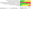
| Patient or population: malignant cerebral oedema after ischaemic stroke Setting: hospital Intervention: surgical decompression Comparison: medical treatment alone | ||||||
| Outcomes | Anticipated absolute effects* (95% CI) | Relative effect (95% CI) | № of participants (studies) | Certainty of the evidence (GRADE) | Comments | |
| Risk with medical treatment alone | Risk with surgical decompression | |||||
| Death at 6 to 12 months follow‐up | 683 per 1000 | 279 per 1000 (205 to 368) | OR 0.18 (0.12 to 0.27) | 513 (9 RCTs) | ⊕⊕⊕⊕ Higha | Surgical decompression results in large reduction in death at 6 to 12 months. |
| Death or severe disability (mRS > 4) at 6 to 12 months follow‐up | 740 per 1000 | 385 per 1000 (299 to 476) | OR 0.22 (0.15 to 0.32) | 513 (9 RCTs) | ⊕⊕⊕⊕ Higha,b | Surgical decompression results in a substantial reduction in death or severe disability (mRS > 4). |
| Death or moderate disability (mRS > 3) at 6 to 12 months follow‐up | 834 per 1000 | 631 per 1000 (525 to 723) | OR 0.34 (0.22 to 0.52) | 513 (9 RCTs) | ⊕⊕⊕⊝ Moderatea,b | Surgical decompression probably reduces death or moderate disability (mRS > 3); however, there is moderate inconsistency in the reported results across the included trials. |
| Severe disability (mRS = 5) at 6 to 12 months follow‐up | 179 per 1000 | 137 per 1000 (73 to 238) | OR 0.73 (0.36 to 1.44) | 262 (9 RCTs) | ⊕⊕⊝⊝ Lowa,c | The available evidence suggests that surgical decompression does not lead to a significant change in the proportion of survivors with severe disability (mRS = 5); however, this result is uncertain due to significant inconsistency in reported outcomes across the included trials and reduced participant numbers. |
| Adverse events | 0 per 1000 | 0 per 1000 (0 to 0) | Not estimable | (0 studies) | ‐ | There was no consistent reporting of adverse events across the included trials. |
| Hydrocephalus | 0 per 1000 | 0 per 1000 (0 to 0) | Not estimable | (0 studies) | ‐ | There was no consistent reporting of hydrocephalus across the included trials. |
| Reoperation | 0 per 1000 | 0 per 1000 (0 to 0) | Not estimable | (0 studies) | ‐ | There was no consistent reporting of reoperation across the included trials. |
| *The risk in the intervention group (and its 95% confidence interval) is based on the assumed risk in the comparison group and the relative effect of the intervention (and its 95% CI). CI: confidence interval; OR: odds ratio | ||||||
| GRADE Working Group grades of evidence High certainty: we are very confident that the true effect lies close to that of the estimate of the effect. Moderate certainty: we are moderately confident in the effect estimate: the true effect is likely to be close to the estimate of the effect, but there is a possibility that it is substantially different. Low certainty: our confidence in the effect estimate is limited: the true effect may be substantially different from the estimate of the effect. Very low certainty: we have very little confidence in the effect estimate: the true effect is likely to be substantially different from the estimate of effect. | ||||||
| See interactive version of this table: https://gdt.gradepro.org/presentations/#/isof/isof_question_revman_web_431153187149621800. | ||||||
a All included trials were unblinded; however, this did not substantially affect the outcomes of interest. b There was a moderate degree of inconsistency in the reported treatment effect between the included trials. c There was a significant degree of inconsistency in the reported outcomes across the included trials.
Background
Description of the condition
Ischaemic stroke is a leading cause of mortality and morbidity worldwide. Large territory middle cerebral artery (MCA) ischaemic strokes encompass approximately 10% of all ischaemic strokes and are particularly morbid, with a reported mortality of 80% when associated with malignant oedema (Berrouschot 1998; Das 2019; Hacke 1996). The large area involved may lead to the development of malignant MCA infarction characterised by significant mass effect with potentially avoidable secondary injury to surrounding brain tissue (Frank 1995; Shaw 1959). The initial ischaemic insult causes a rapid depletion of the major energy metabolite for neurons, adenosine triphosphate (ATP). Major ATP‐dependent membrane pumps begin to fail including the sodium‐potassium ATP‐dependent pump, leading to intracellular accumulation of sodium and calcium. The passive flow of water intracellularly follows the sodium influx, leading to cellular swelling called cytotoxic oedema. This process also results in damage to cerebral vasculature with impaired integrity of the blood‐brain barrier resulting in leakage of water into the extra‐cellular compartment (vasogenic oedema). Progressive cerebral oedema begins to develop during the first 24 to 48 hours after ischaemic insult and may then lead to a cascading series of secondary injuries within days of the initial infarction through raised intracranial pressure and transtentorial herniation (Das 2019). When the associated oedema is large enough to cause a decompensated rise in intracranial pressure, it is termed malignant oedema. Malignant oedema can lead to ischaemia in surrounding vascular territories as well as jeopardise the potentially salvageable brain tissue around the infarcted core. This is generally associated with deteriorating neurological function and worsening level of consciousness two to three days after the initial stroke.
Description of the intervention
Surgical decompression is widely utilised to alleviate raised intracranial pressure and prevent transtentorial herniation (Beez 2019). It is applied in a variety of neurosurgical settings including traumatic brain injury, intracerebral haemorrhage, and ischaemic stroke, and is generally employed as the last‐tier method when medical measures for controlling intracranial pressure (ICP) have failed. Whilst significant variation in the type of surgical decompression exist (including bifrontal craniectomy, hemicraniectomy, or sub‐occipital craniectomy), in the setting of MCA ischaemic stroke, the technical approach is approximately uniform; that is, given the unilateral hemispheric nature of the pathology, a unilateral fronto‐temporo‐parietal hemicranietomy is generally utilised. This typically involves a large, curved incision beginning close to the midline and traced around the skull to either behind or in front of the ear. The generated myocutaneous flap should cover most of the fronto‐temporo‐parietal region and is then elevated and retracted anteroinferiorly. Burr holes are then drilled in the skull and converted into a fronto‐temporo‐parietal craniectomy. The antero‐posterior diameter of the craniectomy should be at least 12 cm, with the recommended diameter in the setting of traumatic brain injury being 15 cm (Carney 2016). Expansile duroplasty is then performed to achieve greater decompression than simple bony decompression. Internal decompression, through the removal of infarcted tissue, is uncommonly utilised if maximal decompression is desired. Following the decompression, the bone flap is sent for storage, whilst the myocutaneous flap is closed. Surgical decompression is associated with attendant risks of surgery, including haemorrhage, infection, hydrocephalus, seizures, and longer‐term complications such as sunken‐flap syndrome (Beez 2019).
How the intervention might work
Decompressive hemicraniectomy allows swelling beyond the internal limits of the cranial vault and thus a lowering of intracranial pressure (Beez 2019). This reduces the resistance to blood flow and thus improves cerebral perfusion, preventing a potentially otherwise intractable rise in intracranial pressure, with associated global hypoperfusion and ischaemic damage. It can also salvage the brain regions affected by the initial ischaemic insult with suboptimal perfusion referred to as the ischaemic penumbra. Furthermore, a reduction in intracranial pressure may prevent potentially devastating transtentorial and sub‐falcine herniation. Through these two methods of improved cerebral perfusion and prevention of intracranial herniation, surgical decompression has thus been theorised to reduce mortality and improve functional outcomes in people suffering large territory MCA ischaemic strokes (Beez 2019; Vahedi 2007).
Why it is important to do this review
Ischaemic stroke causes a significant burden of mortality and morbidity to the community (Dasenbrock 2017; Rahme 2012). There is steadily accumulating evidence of the role of surgical decompression in reducing mortality in malignant oedema after stroke (Vahedi 2007). In addition, new studies have emerged that have examined the outcomes after decompressive craniectomy in a variety of patient cohorts, as well as examining the factors that may influence the decision to operate, such as the timing of surgery, patient age, and laterality of the stroke (Juttler 2007; Juttler 2014; Rahme 2012; Zhao 2012). By performing this review, we aimed to comprehensively evaluate the current evidence regarding these issues.
Objectives
To assess the effectiveness of surgical decompression in people with malignant oedema after ischaemic stroke with regard to reduction in mortality and improved functional outcome. We also aimed to examine the adverse effects of surgical decompression in this patient cohort.
Methods
Criteria for considering studies for this review
Types of studies
We included only randomised controlled trials (RCTs) that compared surgical decompression with best medical management to best medical management alone for people with malignant oedema after ischaemic stroke. We did not include non‐randomised studies.
Types of participants
Participants in the included studies had malignant cerebral oedema after MCA territory ischaemic stroke. This should be diagnosed by computed tomography (CT) or magnetic resonance imaging (MRI) brain scans, with the definition of malignant cerebral oedema left to the discretion of the authors of the individual studies. We included participants > 18 years of age with no upper age limit.
Types of interventions
We included trials with surgical decompression performed utilising a craniectomy. Surgical decompression should include bony decompression with fronto‐temporo‐parietal hemicraniectomy with expansile duroplasty. Additional internal decompression with removal of infarcted tissue, or associated haematoma, was not a requirement. Importantly, participants should be randomised shortly after ictus, with participants randomised to receive treatment within 96 hours of ictus. The additional limit to the timing of intervention is due to compelling evidence supportive of surgical intervention during this early period after stroke ictus.
We assessed the impact of surgery compared to no surgery, thus optimal medical management (mannitol/hypertonic saline, barbituates, hyperventilation, etc.) should be utilised in both groups. A difference in approach to the medical management of participants between the two arms is indicative of bias within the study.
Types of outcome measures
The presence of any one of the following outcome measures in a trial was an inclusion criterion of the review.
Primary outcomes
Death at 6 to 12 months follow‐up.
Death or severe disability defined as modified Rankin Scale (mRS) > 4 at 6 to 12 months.
Secondary outcomes
Death or moderate disability defined as mRS > 3 at 6 to 12 months.
Severe disability defined as mRS = 5 within surviving participants (mRS 0 to 5) at 6 to 12 months.
Adverse events: including the total number of reported adverse events, and specific stratifications of infection, hydrocephalus, and reoperation, where available.
Search methods for identification of studies
See the 'Specialised Register' information available at the Cochrane Stroke Group's website. We searched for trials in all languages and arranged for the translation of relevant articles where necessary.
Electronic searches
We searched the Cochrane Stroke Group Trials Register to 6 June 2022 and the following electronic databases:
Cochrane Central Register of Controlled Trials (CENTRAL; 2022, Issue 7) in the Cochrane Library (3 July 2022) (Appendix 1);
MEDLINE Ovid (from 1946; last searched 3 July 2022) (Appendix 2);
Embase Ovid (from 1974; last searched 3 July 2022) (Appendix 3);
Science Citation Index Expanded (SCI‐EXPANDED; from 1900), Conference Proceedings Citation Index ‐ Science (CPCI‐S; from 1900), and Emerging Sources Citation Index (2005 onwards) in the Web of Science Core Collection (Clarivate Analytics) (Appendix 4); and
Scopus abstract and citation database (Elsevier; from 1788) (Appendix 4).
We modified the subject strategies for databases modelled on the search strategy designed for MEDLINE by the Cochrane Stroke Group’s Information Specialist (Appendix 2; Appendix 4). We combined all search strategies deployed with subject strategy adaptations of the Highly Sensitive Search Strategy designed by Cochrane for identifying RCTs and controlled clinical trials, as referenced in Boxes 3.c and 3.d in the Technical Supplement to Chapter 4: Searching for and selecting studies in the Cochrane Handbook for Systematic Reviews of Interventions (Lefebvre 2022).
We did not include keywords related to cerebral oedema in our search strategy (Appendix 2), as they excluded potentially relevant references in preliminary test searches.
We searched the following ongoing trials registers:
US National Institutes of Health Ongoing Trials Register ClinicalTrials.gov (www.clinicaltrials.gov/) (Appendix 4);
World Health Organization International Clinical Trials Registry Platform (WHO ICTRP) (who.int/ictrp/en/) (Appendix 4).
Searching other resources
In an effort to identify further published, unpublished, and ongoing trials, we:
checked the bibliographies of included studies and any relevant systematic reviews identified for further references to relevant trials and searched Google Scholar (scholar.google.co.uk/) to forward track relevant references;
contacted original authors for clarification and further data if trial reports were unclear;
where necessary, contacted experts, trialists, and organisations in the field to obtain additional information on relevant trials; and
conducted searches in the British Library EThOS and ProQuest Dissertations & Theses Global (Appendix 4).
Data collection and analysis
Selection of studies
Two review authors (AD and MMu) independently screened the titles and abstracts of the references obtained as a result of the search and excluded irrelevant reports. We retrieved the full‐text articles for the remaining references, and two review authors (AD and MMu) independently screened the full‐text articles, identified studies for inclusion, and recorded reasons for exclusion of the ineligible studies. Any disagreements were resolved through discussion or consultation with a third review author (MMa). We collated multiple reports of the same study so that each study, rather than each reference, was the unit of interest in the review. We recorded the selection process and completed a PRISMA flow diagram.
Data extraction and management
Two review authors (AD and MMa) independently extracted data from the included studies onto data extraction tables. We extracted the following data:
study methods: study design, method of randomisation, study country, number of centres, time between ictus to randomisation;
participants: sample sizes, age, sex, the proportion of participants with dominant versus non‐dominant stroke, laterality of stroke, the proportion of participants receiving intended treatment, the proportion of participants with cross‐over treatment, method of diagnosis of malignant oedema;
intervention: time from ictus to intervention/surgery, time from randomisation to surgery, surgical technique, size of craniectomy, 'best medical management' strategies, operator (consultant/trainee);
outcomes: number of participants in each comparator group with the following outcomes: death, mRS > 4, mRS > 3, severe disability as defined by trial authors; we collected these outcome measures at six months, 12 months, and final follow‐up. Adverse events: hydrocephalus, reoperation, infection, other adverse events.
Any disagreements were resolved through discussion or by consulting a third review author (MS) where required. For dichotomous data, we extracted the number of participants experiencing the event and the total number of participants in each arm of the trial. For continuous data, we extracted mean values and standard deviations for participants experiencing an event, along with the total number in each trial arm. In the event that trials reported only effect estimates such as odds ratio or risk ratio without the number of participants or events, we extracted these data for inclusion in our review using the methods outlined in Chapter 6 of the Cochrane Handbook for Systematic Reviews of Interventions (Higgins 2022).
Assessment of risk of bias in included studies
Two review authors (AD and MMu) independently assessed the risk of bias for each study using the criteria outlined in Chapter 8 of the Cochrane Handbook for Systematic Reviews of Interventions (Higgins 2011). Any disagreements were resolved through discussion or by consulting a third review author (MS). We assessed risk of bias according to the following domains.
Random sequence generation
Allocation concealment
Blinding of participants and personnel
Blinding of outcome assessment
Incomplete outcome data
Selective outcome reporting
Other bias
We graded the risk of bias for each domain as low, high, or unclear and provided information from the study report together with a justification for our judgement in the risk of bias tables.
Measures of treatment effect
We used a fixed‐effect model for statistical analyses of all outcome measures, unless we identified a significant degree of heterogeneity (I2 > 60%), in which case we used a random‐effects model. We defined 'severe disability' measured by mRS using an mRS score of 5, and 'moderate disability' using an mRS score of 4 to 5, and conducted these analyses separately.
As all outcome measures, including death, functional outcomes, and adverse events, were dichotomised, we calculated the effect sizes using odds ratio (ORs) and relative risk reduction (RRR) with corresponding 95% confidence intervals (CIs). We conducted all analyses on an intention‐to‐treat basis.
Unit of analysis issues
We assessed the level of randomisation in each included study and expected that all included studies would be randomised on an individual basis, with the unit of analysis being individual participants with malignant oedema after ischaemic stroke. In the event we found non‐standard study designs, including cluster‐randomised studies, as well as other analysis issues, we applied the recommendations in Chapter 23 of the Cochrane Handbook for Systematic Reviews of Interventions (Higgins 2022).
Furthermore, if we found studies with multiple outcome measures within the same period, we included the longest follow‐up within each specified period, that is at 12 months. We analysed the data for the longest follow‐up period provided, as well as performing an analysis separating the effects of different periods, that is six months and 12 months (Higgins 2022).
Dealing with missing data
In the event that data points were missing from the included studies, we imputed the missing data with replacement values using the methods described in Section 10.12.2 of the Cochrane Handbook for Systematic Reviews of Interventions (Higgins 2022). When replacement values were entered, we performed a sensitivity analysis to determine the effect of the replacement values, including an assessment with the best‐ and worst‐case scenarios for the replacement data. Furthermore, we attempted to contact study authors to obtain missing data, where possible.
Assessment of heterogeneity
We used the I2 statistic to measure heterogeneity amongst the trials in each analysis. We interpreted the I2 statistic value according to Chapter 10 of the Cochrane Handbook for Systematic Reviews of Interventions (Higgins 2022), as follows:
0% to 40%: might not be important;
30% to 60%: moderate heterogeneity;
50% to 90%: substantial heterogeneity;
75% to 100%: considerable heterogeneity.
In the case of heterogeneity in an outcome measure, we sought to identify the underlying cause by examining the studies and subgroup characteristics.
Assessment of reporting biases
We assessed reporting bias using a qualitative analysis of the included studies as outlined in Chapter 13 of the Cochrane Handbook for Systematic Reviews of Interventions (Higgins 2022). We investigated protocols of the included studies, including a search for missing data within included trials and trials without published results. If we identified more than 10 studies for inclusion, we used a funnel plot, and employed Egger's test to assess the role of reporting and associated small‐study bias.
Data synthesis
Where we considered studies to be sufficiently similar, we conducted a meta‐analysis by pooling the appropriate data using RevMan Web (RevMan Web 2022). We used a random‐effects model to analyse pooled outcome data from the included studies if there was a significant degree of heterogeneity in the included studies (i.e. I2 > 60%), otherwise we performed a fixed‐effect model analysis. We conducted statistical analysis according to the recommendations outlined in Chapter 10 of the Cochrane Handbook for Systematic Reviews of Interventions (Higgins 2022). We utilised the Mantel‐Haenszel method for fixed‐effect model meta‐analysis of dichotomous outcomes and DerSimonian and Laird inverse variance for a random‐effects model analysis. If we were unable to perform a meta‐analysis, due to either wide variance between the studies or lack of studies (< 2), we described the studies narratively.
Subgroup analysis and investigation of heterogeneity
Where there was a sufficient number of studies, we performed a subgroup analysis of both primary and secondary outcome measures using the following factors.
Time from ictus to surgery (defined as the time between the onset of stroke symptoms and surgical intervention).
Age (> 60 years of age versus ≤ 60 years of age).
Laterality of the stroke (dominant versus non‐dominant side stroke; this was based on the presence or absence of dysphasia, respectively).
Dysphasia (the presence versus absence of dysphasia; this may be a more accurate predictor of dominant versus non‐dominant side stroke).
Short‐ versus long‐term follow‐up (final follow‐up at six months versus final follow‐up greater than six months; we planned to carry out this subgroup analysis for both primary and secondary outcome measures, excluding adverse events).
We utilised the formal test for 'subgroup analysis' provided in RevMan Web (RevMan Web 2022).
Sensitivity analysis
We conducted sensitivity analyses for primary outcome measures by excluding the following studies.
Studies with a high risk of bias (see Assessment of risk of bias in included studies).
Studies where the mean time from ictus to surgery was greater than 48 hours.
Summary of findings and assessment of the certainty of the evidence
We created a summary of findings table using the following outcomes: death, death or severe disability defined as mRS > 4, death or moderate disability defined as mRS > 3, severe disability (mRS = 5), adverse events, hydrocephalus, and reoperation.
We used the five GRADE considerations (study limitations, consistency of effect, imprecision, indirectness, and publication bias) to assess the certainty of a body of evidence as it relates to the studies that contribute data to the meta‐analyses for the prespecified outcomes (Atkins 2004). We used the methods and recommendations described in Chapter 14 of the Cochrane Handbook for Systematic Reviews of Interventions and the GRADE Handbook (Higgins 2022; Schünemann 2013), employing GRADEpro GDT software (GRADEpro GDT). We justified all decisions to downgrade the certainty of the evidence using footnotes, and made comments to aid the reader's understanding of the review where needed.
Results
Description of studies
See Characteristics of included studies; Characteristics of excluded studies.
Results of the search
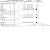
Our searches yielded the following results (Figure 1): CENTRAL (739 records); MEDLINE (1341 records); Embase (5649 records); Scopus (1038 records); Web of Science (792 records); ClinicalTrials.gov (119 records); WHO ICTRP (250 records).
1.

Study flow diagram.
We screened a total of 8691 references, of which we identified 34 separate RCTs for full‐text analysis (DECIMAL; Demitur; DESTINY; Destiny II; HAMLET; HeADDFIRST; Slezins 2012; Zhao 2012). Of these, eight studies were completed, published, and met the criteria for inclusion (DECIMAL; DESTINY; Destiny II; HAMLET; HeADDFIRST; HeMMI; Slezins 2012; Zhao 2012). One study was found to be withdrawn from publication. However, we contacted the study authors who confirmed that the study had been completed as outlined and who provided the complete study results (Demitur). This study was eligible for inclusion in the review, for a total of nine included studies.
Included studies
Summary details for the studies are provided in Characteristics of included studies. We included nine studies with a total of 526 randomised participants, of which 13 participants were not included in the final analysis either because of loss to follow‐up or major protocol violations. We therefore included 248 participants in the surgical arm and 265 participants in the medical treatment‐only arm at final analysis.
All included studies followed similar surgical decompression techniques, and the standardised medical treatment protocol was also largely homogenous across the studies. The only notable deviation in surgical protocol was by Slezins 2012, which implanted an intracranial pressure (ICP) monitor for participants randomised to surgery who did not display midline shift on neuroimaging. They proceeded with surgical decompression only if ICPs were elevated on the parenchymal ICP monitor. Slezins 2012 randomised a total of 28 participants, of whom four participants assigned to the surgical cohort did not undergo decompressive craniectomy and were excluded from the final analysis. In three cases, they received surgical decompression over 100 hours after stroke ictus, which was a time frame breach for the study design, and in one case a participant showed no signs of raised ICP on parenchymal ICP monitor and was therefore excluded from surgical intervention.
HeMMI randomised 29 participants out of 156 that were screened. However, five participants were excluded from the final analysis as they were lost to follow‐up (three in the surgical group versus two in the medical treatment‐only arm). Moreover, three participants in the medical treatment‐only arm underwent surgical intervention due to clinical deterioration, whilst one participant in the surgical arm did not undergo surgical decompression due to myocardial infarction.
HeADDFIRST randomised a total of 25 participants, of which one of the 15 participants in the surgical arm withdrew due to the family withdrawing consent. This was the smallest cohort of the included studies. Demitur had the largest cohort, with a total of 151 randomised participants.
Furthermore, three of the included trials stopped recruitment prematurely (DECIMAL; DESTINY; HAMLET). In two trials, a pooled analysis of data from the three trials was considered, whilst in the case of HAMLET, an interim analysis of the data determined it was unlikely to achieve a statistically significant difference with the planned sample size.
There were variations in the radiological criteria used for defining a malignant MCA infarction across the included studies. However, in general they all required an infarction of at least the majority of the MCA territory. This was defined as infarction affecting two‐thirds or more of the MCA territory in five studies (Demitur; DESTINY; Destiny II; HAMLET; Zhao 2012). DESTINY and Destiny II required associated involvement of the ipsilateral basal ganglia territory. DECIMAL required at least 50% involvement of the MCA territory with an infarct volume of at least 145 cm3 on diffusion‐weighted imaging (DWI). HeMMI and Slezins 2012 similarly required infarction size of ≥ 50% of MCA territory on neuroimaging. HeADDFIRST defined the neuroimaging criteria as infarction area being ≥ 50% of MCA territory if CT was < 5 hours from stroke onset, or complete MCA territory involvement if it was > 5 hours and within 48 hours from stroke ictus. Furthermore, participants were required to have a minimum amount of midline shift on repeat CT before randomisation.
The clinical criteria used to assess stroke severity for eligibility was the National Institutes of Health Stroke Scale (NIHSS) score in seven of the studies (DECIMAL; Demitur; DESTINY; Destiny II; HAMLET; HeADDFIRST; Slezins 2012), whilst the other two studies primarily relied upon the Glasgow Coma Score (GCS) (HeMMI; Zhao 2012). The minimum cut‐off NIHSS scores for inclusion varied across all studies, although this was generally within a narrow range selecting for moderate‐severe strokes. The lowest minimum NIHSS score across the included studies was 14 for non‐dominant strokes in Destiny II, whilst the highest was 21 for dominant strokes in DESTINY. Five studies used differential inclusion criteria for dominant and non‐dominant strokes to account for the confounding speech deficit (Demitur; DESTINY; Destiny II; HAMLET; HeMMI). Furthermore, eight studies mandated a reduced level of consciousness in their inclusion criteria (DECIMAL; Demitur; DESTINY; Destiny II; HAMLET; HeADDFIRST; HeMMI; Zhao 2012).
All of the included studies excluded patients with severe comorbidities and limited life expectancy. The pre‐morbid mRS was utilised in all studies, with the majority excluding patients with a pre‐morbid mRS > 1 (DECIMAL; Demitur; DESTINY; Destiny II; HAMLET; Slezins 2012), and three studies setting the cut‐off at mRS > 2 (HeADDFIRST; HeMMI; Zhao 2012). Six studies excluded patients with absence of pupillary reaction (Demitur; DESTINY; Destiny II; HAMLET; Slezins 2012; Zhao 2012).
Three studies included participants 60 years of age or younger (DECIMAL; DESTINY; HAMLET), whilst HeMMI had a maximum inclusion age of 65, and HeADDFIRST had a maximum age of 75. Zhao 2012 and Demitur enrolled participants up to the age of 80. Destiny II was the only trial to exclusively enrol elderly patients, including participants 61 years of age or older. This trial had the highest mean age of participants, being 69.9 years (standard deviation (SD) ± 4.4) in the surgical cohort and 70.4 years (SD ± 4.8) in the medical treatment‐only cohort. The lowest mean age of participants was in DECIMAL, which had a mean age of 43.5 years (SD ± 9.7) for the surgical cohort and 43.3 years (SD ± 7.1) for the medical treatment‐only cohort. All of the other studies had a mean age of participants within this range. The majority of studies were well‐matched for age between the two randomised treatment cohorts; however, Slezins 2012 had the most significant variation in age, with a mean age of 57.2 years in the surgical cohort compared to 65 years in the medical treatment‐only cohort (P = 0.02).
The majority of studies randomised participants to treatment within a maximum of 48 hours after stroke ictus (DECIMAL; Demitur; DESTINY; Destiny II; Slezins 2012; Zhao 2012). Of these studies, DECIMAL had the shortest time from ictus to intervention, including participants after a maximum of 24 hours from stroke ictus; the average time to surgical intervention in their cohort was 20.5 hours. On the other hand, HAMLET included participants presenting up to 96 hours after stroke onset, and required treatment within three hours of randomisation. The median time to randomisation was 41 hours in the surgical cohort and 45 hours in the control cohort. Similarly, HeADDFIRST allowed the inclusion of participants with stroke symptoms up to 96 hours after onset, whilst the median time from stroke onset to randomisation was 53 hours. HeMMI randomised participants for treatment up to 72 hours after stroke ictus; however, of these only two participants in the surgical arm underwent surgery after 48 hours, and the overall time to randomisation was 9.0 (± 10.6) hours.
Outcome measures reported in all the studies included both death and mRS data at final follow‐up. The reported mRS outcomes could be dichotomised at both 0 to 3 versus 4 to 6 and 0 to 4 versus 5 to 6 in all of the included studies. Adverse events were only systematically reported in two studies, and in an inconsistent manner that was not comparable (DECIMAL; Destiny II), thus a meta‐analysis of adverse events was not performed. The follow‐up period for outcome measures was between six and 12 months in all studies. Primary outcome measures were reported at both six and 12 months in five studies (DECIMAL; Demitur; DESTINY; Destiny II; Zhao 2012), at six months only in two studies (HeADDFIRST; HeMMI), and at 12 months only in two studies (HAMLET; Slezins 2012).
Excluded studies
There were no excluded studies.
Risk of bias in included studies
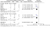
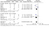
The results of the risk of bias assessment are presented in Figure 2 and Figure 3.
2.

Risk of bias summary: review authors' judgements about each risk of bias item for each included study.
3.
Risk of bias graph: review authors' judgements about each risk of bias item presented as percentages across all included studies.
Allocation
We judged all of the included studies to be at either low or unclear risk of bias for this domain. We assessed five studies as at unclear risk of bias for random sequence generation as insufficient information was provided to determine if the allocation sequence was genuinely randomised. We assessed six studies as at unclear risk of bias for allocation concealment because there was no description of the method of concealment.
Blinding
We acknowledge that it is not possible to blind the participant or clinician for this intervention, therefore we judged all of the included studies as at high risk of bias for blinding of participants and personnel. We assessed five studies as at low risk of bias for blinding of outcome assessment. The remaining four studies did not blind assessors and were judged to be at high risk of detection bias.
Incomplete outcome data
We judged seven studies as at low risk of attrition bias. We assessed one study as having a high risk of bias due to the proportion of missing data (HeMMI). We judged one study as at unclear risk of bias as the description of the patient flow from inclusion to primary outcome follow‐up was insufficient to assess the degree of missing data (Slezins 2012).
Selective reporting
We judged two studies as at low risk of reporting bias. We assessed the remaining seven studies as at unclear risk of bias as insufficient information was provided to ensure that prespecified outcomes had been reported in the prespecified way. Four trials had no protocol available and were registered on clinical trial registries after recruitment had commenced; two trials had no protocol available and were registered on clinical trial registries after the trial was completed; and one trial had no protocol and was not registered on a clinical trial registry (HeADDFIRST).
Other potential sources of bias
We did not identify any other source of bias in one trial. We judged six trials to be of unclear risk of other bias: five trials were terminated early, and in one trial there was no sample size calculation or primary outcome endpoint. We judged two trials to have additional biases sufficient to deem them at high risk of bias for this domain. One trial was a pilot study with small numbers and inequity between the two treatment arms (HeADDFIRST). The other trial was withdrawn from publication by the authors (Demitur).
Effects of interventions
See: Table 1
See Table 1.
Death
Nine trials evaluated the effect of surgical decompression compared to medical treatment only on the outcome of death at 6 to 12 months follow‐up, with a total of 526 participants, of whom 513 were included in the final analysis. There was a statistically significant reduction in death at 6 to 12 months follow‐up with surgical decompression (odds ratio (OR) 0.18, 95% confidence interval (CI) 0.12 to 0.27, P < 0.001, Analysis 1.1; high‐certainty evidence). There was a low degree of statistical heterogeneity (I2 = 1%).
1.1. Analysis.

Comparison 1: Comparison: surgical decompression versus medical treatment alone for malignant cerebral oedema after ischaemic stroke, Outcome 1: Outcome 1: death at 6 to 12 months
Death or severe disability defined as mRS > 4
Nine trials evaluated the effect of surgical decompression compared to medical treatment only on death or severe disability (mRS > 4). There was a statistically significant reduction in this outcome by surgery (OR 0.22, 95% CI 0.15 to 0.32, 513 participants, P < 0.001, Analysis 1.2; high‐certainty evidence). There was a moderate degree of heterogeneity in the reported outcomes (I2 = 44%).
1.2. Analysis.

Comparison 1: Comparison: surgical decompression versus medical treatment alone for malignant cerebral oedema after ischaemic stroke, Outcome 2: Outcome 2: death or severe disability defined as mRS > 4 at 6 to 12 months
Death or moderate disability defined as mRS > 3
There was also a significant reduction in death or moderate disability (mRS > 3) in the surgical cohort compared to medical treatment alone (OR 0.34, 95% CI 0.22 to 0.52, 9 RCTs, 513 participants, P < 0.001, Analysis 1.3; moderate‐certainty evidence). Similarly, there was a moderate degree of heterogeneity in the treatment effect (I2 = 54%).
1.3. Analysis.

Comparison 1: Comparison: surgical decompression versus medical treatment alone for malignant cerebral oedema after ischaemic stroke, Outcome 3: Outcome 3: death or moderate disability defined as mRS > 3 at 6 to 12 months
Severe disability (mRS = 5)
There was no significant difference in the proportion of surviving participants with severe disability (mRS = 5) in the surgical cohort compared to medical treatment alone, although there was a trend for a lower rate in the surgical arm (OR 0.73, 95% CI 0.36 to 1.44, P = 0.36, I2 = 41%, Analysis 1.4; low‐certainty evidence).
1.4. Analysis.

Comparison 1: Comparison: surgical decompression versus medical treatment alone for malignant cerebral oedema after ischaemic stroke, Outcome 4: Outcome 4: severe disability (mRS = 5) at 6 to 12 months
Adverse events
There was no consistent reporting of adverse events across the included studies. Studies either did not report on adverse events (Demitur; DESTINY; HAMLET; HeADDFIRST; Slezins 2012; Zhao 2012), or reported them in an inconsistent manner (DECIMAL; HeMMI). Destiny II was the only trial to consistently report adverse events; however, this was defined as "serious adverse events". They reported 88 adverse events in the hemicraniectomy group (49 participants) compared to 84 adverse events in the control cohort (63 participants). There was no per‐participant analysis, thus we did not include the results in our meta‐analysis.
Hydrocephalus
There was no uniform reporting of results for the specific outcome of hydrocephalus in any of the included studies, thus no final meta‐analysis was performed.
Reoperation
There was no uniform reporting of results for the specific outcome of reoperation in any of the included studies, thus no final meta‐analysis was performed.
Subgroup analysis
Time from stroke ictus, < versus ≥ 48 hours
We performed a subgroup analysis evaluating the effect of time from stroke ictus to intervention with a cut‐off at 48 hours. No significant subgroup effect was detected on death at 6 to 12 months follow‐up, although there was a non‐significant trend of greater reduction in death in the < 48 hours subgroup (P = 0.08, Analysis 2.1). However, there was uneven distribution of the covariate, with fewer trials and participants in the ≥ 48 hours subgroup (1 trial, 25 participants) compared to seven trials and 440 participants in the < 48 hours subgroup.
2.1. Analysis.

Comparison 2: Subgroup analysis for time from stroke ictus, Outcome 1: Outcome 1: death at 6 to 12 months
The subgroup analysis evaluating the effect of time from stroke ictus on death or severe disability (mRS > 4) similarly found no significant subgroup effect of time from stroke ictus (P = 0.11, Analysis 2.2). There were substantially fewer trials and participants in the ≥ 48 hours subgroup (1 trial, 25 participants) compared to seven trials and 440 participants in the < 48 hours subgroup.
2.2. Analysis.

Comparison 2: Subgroup analysis for time from stroke ictus, Outcome 2: Outcome 2: mRS > 4 at end of follow‐up
Age < versus ≥ 60 years
We performed a subgroup analysis evaluating the effect of age with a cut‐off of 60 years. Five studies had a cohort of 215 participants aged < 60 years, compared to three studies with a cohort of 226 participants aged ≥ 60 years of age. There was no significant subgroup effect of age on the outcome of death (P = 0.56, Analysis 3.1) with no significant heterogeneity with the subgroups (I2 = 0%). There was also no significant subgroup effect of age on death or severe disability (mRS > 4) (P = 0.91, Analysis 3.2) or death or moderate disability (mRS > 3) (P = 0.46, Analysis 3.3).
3.1. Analysis.
Comparison 3: Subgroup analysis for age </≥ 60, Outcome 1: Outcome 1: death at 6 to 12 months
3.2. Analysis.
Comparison 3: Subgroup analysis for age </≥ 60, Outcome 2: Outcome 2: mRS > 4 at 6 to 12 months
3.3. Analysis.
Comparison 3: Subgroup analysis for age </≥ 60, Outcome 3: Outcome 3: mRS > 3 at 6 to 12 months
Dysphasia presence/absence
We performed a subgroup analysis comparing the effect of presence versus absence of dysphasia on death or severe disability (mRS > 4). No significant subgroup effect was detected (P = 0.42, Analysis 4.1). There was an even distribution of the covariate between the subgroups and little heterogeneity of the trials within the subgroups (4 trials, 147 participants, I2 = 0% in the dysphasia‐present subgroup versus 4 trials, 138 participants, I2 = 17% in the dysphasia‐absent subgroup).
4.1. Analysis.

Comparison 4: Subgroup analysis for dysphasia presence/absence, Outcome 1: Outcome 2: mRS > 4 at 6 to 12 months
Short (6‐month) versus long (12‐month) follow‐up
A subgroup analysis comparing 6‐ to 12‐month follow‐up trials found a statistically significant subgroup effect on the outcome of death (P = 0.02, Analysis 5.1). Trials reporting outcome measures at 12 months had a greater degree of reduction in death compared to trials reporting outcomes at six months. There was no significant heterogeneity between the trials of each subgroup. However, there were significantly fewer trials and participants in the 6‐month outcome subgroup (2 trials, 48 participants) compared to the 12‐month outcome subgroup (7 trials, 465 participants).
5.1. Analysis.

Comparison 5: Subgroup analysis for time of follow‐up, 6 months vs 12 months follow‐up, Outcome 1: Outcome 1: death
Similarly, we performed a subgroup analysis comparing 6‐ to 12‐month data for death or severe disability (mRS > 4) and death or moderate disability (mRS > 3). In both cases, there was a statistically significant subgroup effect (P = 0.02 and P = 0.02 respectively, Analysis 5.2; Analysis 5.3). For both outcome measures, there was an improved treatment effect in the studies reporting outcomes at 12 months compared to the trials reporting outcomes at six months. In both cases there was reduced heterogeneity in the results between trials of each subgroup. There were also significantly fewer trials and participants in the 6‐month outcome subgroup (2 trials, 48 participants) compared to the 12‐month outcome subgroup (7 trials, 465 participants).
5.2. Analysis.

Comparison 5: Subgroup analysis for time of follow‐up, 6 months vs 12 months follow‐up, Outcome 2: Outcome 2: mRS > 4
5.3. Analysis.

Comparison 5: Subgroup analysis for time of follow‐up, 6 months vs 12 months follow‐up, Outcome 3: Outcome 3: mRS > 3
Sensitivity analyses
We assessed no studies as having a high risk of bias. There were also no studies with fewer than 10 participants.
We performed a sensitivity analysis by excluding two studies in which > 10% of randomised participants were not included in the final analysis (HeMMI; Slezins 2012). We found no difference in the primary outcomes.
We performed a sensitivity analysis by excluding HeADDFIRST, the only study with an average time from stroke ictus to intervention of over 48 hours. We found no difference in the primary outcomes.
We performed a sensitivity analysis by excluding Demitur, the largest study in the meta‐analysis, which had also been withdrawn from publication. There was no change in the primary outcomes.
Discussion
The evidence in this new and updated systematic review reaffirms the main findings of the previous review published in 2012 (Cruz‐Flores 2012). However, it adds a further body of evidence that expands on the role of surgical decompression in the management of malignant cerebral oedema after ischaemic stroke. It provides evidence for reducing disability in patients and utilises a broader evidence base that is generalisable to older age groups.
Summary of main results
The currently available evidence demonstrates a substantial reduction in death with the use of surgical decompression for the management of malignant oedema after ischaemic stroke (OR 0.18, 95% CI 0.12 to 0.27). This has been a consistent finding across trials with little heterogeneity in treatment effect (I2 = 1%). Furthermore, the effect is quite substantial, with a relative risk reduction of 0.42 (95% CI 0.34 to 0.52). Surgical decompression similarly showed a strong and significant effect on reducing death or severe disability defined as mRS > 4 (OR 0.22, 95% CI 0.15 to 0.32). This was again a consistent finding across the included trials; however, the magnitude of treatment effect did differ across trials with moderate heterogeneity of treatment effects (I2 = 44%). This mirrors the finding of the previous review, which demonstrated a profound reduction in death or severe disability (Cruz‐Flores 2012). Our findings further corroborate this treatment effect across all age groups, including elderly patients above the age of 60, who were not included in the previous review.
Surgical decompression has also been associated with a reduction in death or moderate disability (mRS > 3) (OR 0.34, 95% CI 0.22 to 0.52). This is a substantial finding given that the previous review did not find a significant reduction. However, there was moderate heterogeneity in the treatment effect (I2 = 54%). The majority of studies individually did not find a reduction in death or moderate disability (mRS > 3) (DECIMAL; Demitur; DESTINY; Destiny II; Slezins 2012; Zhao 2012). This may indicate that previous trials have been underpowered to assess the validity of this effect. Furthermore, in at least three of the included studies, there was either no difference, or an observed worsening of death or moderate disability (HAMLET; HeADDFIRST; HeMMI). It is noteworthy that these were the only studies that allowed the inclusion of patients outside of 48 hours from stroke ictus. This indicates that earlier surgical decompression may be important for improved functional outcomes.
Earlier treatment is, in principle, a prophylactic approach to preventing the irreversible ischaemic damage that would otherwise be a cascading result of malignant oedema. It would therefore follow that earlier treatment may result in greater prevention of neurological injury, improved functional outcome and, consequently, more significant treatment effect. However, the subgroup analysis did not demonstrate a significant difference in outcomes when analysing the time from stroke ictus to intervention (< 48 hours versus ≥ 48 hours). Despite a trend towards better outcomes in the earlier‐treatment subgroup, there was no difference in either death (P = 0.08, I2 = 67.7%) or death or severe disability (P = 0.11, I2 = 61.0%). There were, however, significantly fewer participants in the ≥ 48 hours cohort (25 participants, 1 trial) compared to 440 participants and seven trials in the earlier‐treatment cohort (< 48 hours). As a result, we were not able to draw a conclusive answer, and further research may help clarify the true treatment effect of earlier decompression.
The utility of even earlier treatment cut‐off is also not understood, with no included trials mandating the inclusion of participants within 24 hours of stroke ictus. However, the median time for patient treatment in many of the included studies is within 24 hours of stroke ictus. A recent meta‐analysis of RCTs comparing surgical decompression to best medical management for hemispheric infarction demonstrated no difference between participants randomised at < 24 hours versus 24 to 48 hours from ictus (Reinink 2021). They did, however, show worse outcomes in participants randomised at > 48 hours. This suggests that there may be a maximal benefit at 48 hours, earlier than which may make little difference. Indeed the development of malignant oedema generally occurs after 48 hours of ischaemic insult, after which the rise in intracranial pressure would be destructive without treatment (Das 2019).
Moreover, surgical decompression has demonstrated a treatment benefit in older age groups that was comparable to younger patients. This is significant, given that the evidence has consistently revealed considerably poorer outcomes for older patients after a large MCA ischaemic stroke (Arac 2009). Indeed, they have been shown to have substantially worse mortality, as well as functional recovery. Consequently, the majority of earlier RCTs have excluded patients > 60 years of age from trials addressing the effectiveness of surgical decompression. Our review, however, demonstrated a treatment effect in participants ≥ 60 years of age, with a reduction in death (OR 0.17, 95% CI 0.10 to 0.31), death or severe disability (mRS > 4) (OR 0.18, 95% CI 0.10 to 0.33), and death or moderate disability (mRS > 3) (OR 0.23, 95% CI 0.11 to 0.50). The subgroup analysis did not find a difference in treatment effect when analysing age < 60 and ≥ 60 years as a factor. This demonstrates that surgical decompression is an effective treatment approach in patients with advanced age with comparable efficacy to its application in younger cohorts.
However, this finding must be interpreted with caution given that it does not change the underlying poorer functional outcomes in older patients. Whilst 67% of participants (144 of 215 participants) in the cohort < 60 years of age were classified as dead or disabled (mRS > 3) at final follow‐up, this was the case for comparatively 80% of participants in cohorts ≥ 60 years of age (180 of 226 participants). Similarly, in Destiny II, the only trial to include participants > 60 years of age, 93.8% (46 of 49 participants) of participants in the surgical cohort and 96.8% (61 of 63 participants) in the medical cohort were either dead or disabled (mRS > 3) at follow‐up. This indicates that elderly patients are likely to have a poorer functional outcome independent of surgical intervention. Consequently, whilst the application of surgical decompression for elderly patients is effective in improving outcomes, clinicians should consider the overall prognosis of their patients and the expectations of their caregivers.
Dysphasia has similarly been shown to have little influence on the effectiveness of surgical decompression. No subgroup effect was detected, with an even distribution of participants and trials between the two groups (147 participants, 4 trials; dysphasia present versus 138 participants, 4 trials; dysphasia absent, P = 0.42). However, it is important to consider that there was an inherent bias in all the trials when selecting participants for inclusion. All trials required higher NIHSS scores for patients with a dominant hemisphere stroke, a surrogate marker for dysphasia, compared to non‐dominant strokes.
Whilst our analysis did not reveal a significant dysphasia subgroup effect, there were nonetheless worse outcomes reported in participants with dysphasia regardless of intervention. In the four cohorts with dysphasia present, 41.2% (28 of 68 participants) of participants in the surgical group and 77.2% (61 of 79 participants) in the medical group were either dead or severely disabled (mRS > 4) at final follow‐up, compared to 16.9% (12 of 71 participants) in the surgical group and 59.7% (40 of 67 participants) in the medical group without dysphasia. This suggests that dysphasia may portend a worse overall clinical outcome, independent of treatment.
Overall completeness and applicability of evidence
Our review included studies that selected a cohort of patients with unique clinical characteristics who demonstrated significantly improved outcomes with the use of surgical decompression as an early treatment for malignant oedema after stroke. Whilst this review has expanded the generalisability of the conclusions drawn by including a greater array of participants, the participants selected still have distinctive clinical characteristics that affect the applicability of the conclusions drawn from the meta‐analysis.
We selected participants with profound neurological deficit and exclusively large size strokes on neuroimaging. All of the included studies mandated the inclusion of participants with a minimum NIHSS score, the lowest cut‐off being 14 and the highest being 21. This practically selects for participants with significant neurological deficits and reduced levels of consciousness. Such a severe neurological syndrome is not typically present, even in the setting of a large MCA ischaemic stroke. A retrospective review investigating the eligibility of stroke patients for hemicraniectomy found that 90.7% of 2227 stroke patients had an NIHSS score of ≤ 15 (Rahme 2012).
Moreover, whilst the radiological criteria for inclusion varied across studies, all of the included studies involved a minimum of 50% of the MCA region being infarcted. Indeed, five of the included studies mandated infarction of two‐thirds of the MCA region. Such large territory stroke patients are uncommon and account for a minority of MCA ischaemic stroke patients (Hao 2015; Heinsius 1998; Liebeskind 2019).
Furthermore, patients eligible for inclusion in the review were without significant life‐limiting illnesses. Indeed the majority of studies excluded patients with an mRS ≥ 2, effectively selecting out patients with even slight pre‐morbid disability. Most studies further excluded patients with life‐limiting illnesses or severe comorbidities. This is a substantially important factor given that the majority of stroke patients have significant cardiovascular comorbidities. The study by Rahme 2012 found that 50.2% of stroke patients had a pre‐morbid mRS ≥ 2, which would have excluded them from this review. Whilst a positive treatment effect may be extrapolated to such patients, they may have a significantly poorer outcome than the healthier cohort included in this review, and clinicians would need to consider the utility of the relative benefit.
Our review, however, has expanded the generalisability of the conclusions drawn to patients ≥ 60 years. Newer RCTs, in particular Destiny II, have addressed the effectiveness of surgical decompression in older age groups and have found a similar treatment effect to younger cohorts. This is a noteworthy difference from the previous Cochrane Review, where older patients were not addressed. However, such conclusions should be interpreted with caution given that whilst surgical decompression can lead to improved survival and clinical outcomes in an elderly cohort, there is a significantly poorer chance of functional and independent survival independent of this treatment effect.
Quality of the evidence
The bulk of the evidence found in this review was of moderate to high certainty. All of the included studies reported outcome measures using the same objective outcome assessment scores (mRS). Furthermore, a similar standardised surgical decompression was used across all trials, with a uniformly similar use of medical measures across trials in the non‐surgical cohorts.
Whilst all of the included studies reported outcomes six to 12 months after intervention, there was evidence of heterogeneity in the treatment effect between these follow‐up time points. A subgroup analysis found a significant subgroup effect of time of follow‐up (6 versus 12 months) on the outcomes of death, death or severe disability (mRS > 4), and death or moderate disability (mRS > 3). There was a consistent finding of greater treatment effect in the subgroup of trials reporting outcomes at 12 months compared to the subgroup of trials reporting outcomes at six months. However, there was a significant disparity in the number of trials and participants in both subgroups, with far fewer trials and participants in the 6‐month subgroup (2 trials, 48 participants). This uneven distribution questions the validity of this subgroup effect.
In addition, in two studies greater than 10% of participants were not included in the final outcome analysis. These were generally small studies with no effect on outcome measures when excluded in a sensitivity analysis. Furthermore, participants and clinicians were not blinded to treatment allocation, which is unavoidable given the nature of the intervention being investigated.
Three studies were terminated prematurely either due to interim analysis of study results or for pooling in a meta‐analysis. This could theoretically increase bias towards improved treatment effect.
One study was withdrawn from publication (Demitur). We contacted the study authors, and the reasons given did not affect the integrity of the study or the results. We also obtained the study data to verify published results. This happened to be the largest study included in the meta‐analysis. There was no substantial change in outcome measures when the study was excluded in a sensitivity analysis.
Potential biases in the review process
This review was based on an analysis of tabular data. This restricted the robustness of subgroup analysis that could be performed. For instance, not all of the studies provided delineated outcome measures for age above and below 60 years. Furthermore, we did not have data from all studies delineating participants treated before and after 48 hours from stroke ictus. A meta‐analysis based on individual patient data can help provide greater robustness for subgroup analysis. In addition, we acknowledge the hazards of subgroup analyses including selective reporting and inherent bias.
Agreements and disagreements with other studies or reviews
A recent individual patient‐level meta‐analysis investigating the utility of surgical decompression for malignant MCA infarction was performed by Reinink 2021. They pooled individual patient data from seven different RCTs, which totalled 488 participants. The findings of this study largely mirror the conclusions of this Cochrane Review. There was a reduction in death (OR 0.13, 95% CI 0.08 to 0.22, P < 0.01) associated with surgical decompression, as well as a greater chance of a favourable outcome defined as mRS ≤ 3 (OR 2.95, 95% CI 1.55 to 5.60, P = 0.01). The rigorous analysis also concluded there was no significant difference when utilising a positive endpoint of mRS ≤ 2 (OR 2.77, 95% CI 0.97 to 7.88, P = 0.06). A subgroup analysis was performed analysing the effect of dysphasia, time from stroke ictus to intervention, age, and vascular territories affected on treatment effect. Similar to our review, they found no effect based on age (</> 60) or dysphasia. They also found no subgroup effect in patients operated on > 48 hours from stroke ictus; however, this analysis was limited by small participant numbers.
Other recent systematic reviews and meta‐analyses of the topic have also found similar conclusions to our review, validating the reduction in death and poor functional outcome with the use of surgical decompression (Alexander 2016; Gul 2018; Qureshi 2016; Streib 2016; Wei 2020).
Authors' conclusions
Implications for practice.
The evidence from this systematic review provides strong support for the use of early surgical decompression in the management of malignant oedema after acute ischaemic stroke. The evidence found was of moderate to high certainty, with consistent results across the literature supporting the improved outcomes. There is a significant reduction in death or disability when early surgical decompression is utilised.
The data also indicate that the treatment benefit is maintained in elderly age groups (≥ 60 years of age), who have previously been excluded from surgical decompression, although this finding needs to be interpreted with caution, as it is based on lower certainty evidence and limited numbers of studies. Furthermore, the prospects for a favourable outcome are significantly lower in advanced age groups regardless of the treatment effect. This needs to be considered when applying the conclusions of this review, and an individual patient approach should be employed.
Moreover, the findings of this review do not conclusively substantiate the importance of surgical decompression earlier than 48 hours from stroke ictus. Despite a trend towards greater benefit with surgery < 48 hours from ictus, this question remains unanswered, as the subgroup analysis was based on a small amount of participant data.
It is important to consider participant selection in the clinical application of these conclusions. The data utilised in this review were based on participants without significant comorbidities and who had profound neurological deficit with reduced level of consciousness. These factors maximised the treatment benefit of early surgical decompression and were the focus of this review.
Studies suggest that surgical decompression is underutilised in stroke patients, with many not being offered the treatment (Rahme 2012). Greater awareness of the improved outcomes for stroke patients may increase the application of surgical decompression in the correct setting, improving overall treatment outcomes and the rate of functional survival.
Implications for research.
We do not expect further studies to change the overall conclusions of this review with regard to the treatment effect.
However, additional evidence is needed to address the efficacy and relevance of treatment with surgical decompression in particular subsets of patients with a middle cerebral artery ischaemic stroke. In particular, its utility in patients > 60 years of age should be assessed with regard to the absolute chances of favourable outcomes with surgical decompression to better guide management. Moreover, greater patient data are required to further validate the effect of the timing of surgical intervention, and in particular the benefit of surgical decompression < 48 hours from stroke ictus. Finally, future studies should consider additional outcome measures focused on patient survival and caregiver expectations. Further data on retrospective consent and outcome satisfaction are useful in understanding the overall benefit of the intervention.
History
Protocol first published: Issue 7, 2021
Acknowledgements
We thank the following people.
Sign‐off Editor (final editorial decision): Peter Langhorne, University of Glasgow
Managing Editor (selected peer reviewers, collated peer‐reviewer comments, provided editorial guidance to authors, edited the article, conducted editorial policy checks and supported editorial team): Hazel Fraser, Cochrane Stroke
Copy Editor (copy editing and production): Lisa Winer, Cochrane Copy Edit Support
Peer reviewers (provided comments and recommended an editorial decision): Peter Langhorne, University of Glasgow (methods review); Prof Rustam Al‐Shahi Salman, University of Edinburgh and NHS Lothian (clinical/content review); Aryelly Rodriguez, Edinburgh Clinical Trials Unit (ECTU) at the University of Edinburgh (statistical review)
Two additional peer reviewers chose not to be publicly acknowledged.
Appendices
Appendix 1. CENTRAL search strategy
CENTRAL; July 2022, Issue 7 of 12 in the Cochrane Library (searched July 2022)
#1 MeSH descriptor: [Cerebrovascular Disorders] this term only
#2 MeSH descriptor: [Basal Ganglia Cerebrovascular Disease] this term only
#3 MeSH descriptor: [Brain Ischemia] this term only
#4 MeSH descriptor: [Brain Infarction] explode all trees
#5 MeSH descriptor: [Hypoxia‐Ischemia, Brain] this term only
#6 MeSH descriptor: [Carotid Artery Diseases] this term only
#7 MeSH descriptor: [Carotid Artery Thrombosis] this term only
#8 MeSH descriptor: [Carotid Artery, Internal, Dissection] this term only
#9 MeSH descriptor: [Intracranial Arterial Diseases] this term only
#10 MeSH descriptor: [Cerebral Arterial Diseases] this term only
#11 MeSH descriptor: [Infarction, Anterior Cerebral Artery] this term only
#12 (isch*mi* near/6 (stroke* or apoplex* or cerebral next vasc* or cerebrovasc* or cva or attack*)):ti,ab,kw
#13 MeSH descriptor: [Infarction, Middle Cerebral Artery] this term only
#14 MeSH descriptor: [Infarction, Posterior Cerebral Artery] this term only
#15 MeSH descriptor: [Intracranial Embolism and Thrombosis] explode all trees
#16 MeSH descriptor: [Stroke] explode all trees
#17 MeSH descriptor: [Vertebral Artery Dissection] this term only
#18 (stroke or poststroke or post‐stroke or cerebrovasc* or (cerebr* near/3 vasc*) or CVA* or apoplectic or apoplex* or (transient near/3 isch?emic near/3 attack) or tia*):ti,ab,kw
#19 (((cerebr* or cerebell* or arteriovenous or vertebrobasil* or interhemispheric or hemispher* or intracran* or intracerebral or infratentorial or supratentorial or MCA* or ((anterior or posterior) near/3 circulat*) or lenticulostriate or ((middle or basilar or brachial or vertebr*) near/3 arter*)) near/3 ((blood near/5 clot*) or disease* or damage* or disorder* or disturbance or dissection or lesion or syndrome or arrest or accident or lesion or vasculopathy or insult or attack or injury or insufficiency or malformation or obstruct* or anomal*))):ti,ab,kw
#20 (((cerebr* or cerebell* or arteriovenous or vertebrobasil* or interhemispheric or hemispher* or intracran* or corpus callosum or intracerebral or intracortical or intraventricular or periventricular or posterior fossa or infratentorial or supratentorial or MCA* or ((anterior or posterior) near/3 circulation) or basal ganglia or ((middle or basilar or brachial or vertebr*) near/3 arter*) or space‐occupying or brain ventricle* or lacunar or cortical or ocular) near/3 (isch?emi* or infarct* or thrombo* or emboli* or occlus* or hypoxi* or vasospasm or obstruct* or vasoconstrict*))):ti,ab,kw
#21 {or #1‐#20}
#22 MeSH descriptor: [Decompression, Surgical] this term only
#23 MeSH descriptor: [Decompressive Craniectomy] this term only
#24 MeSH descriptor: [Microvascular Decompression Surgery] this term only
#25 MeSH descriptor: [Vascular Surgical Procedures] this term only
#26 MeSH descriptor: [Anterior Temporal Lobectomy] this term only
#27 MeSH descriptor: [Cerebral Decortication] this term only
#28 MeSH descriptor: [Neurosurgical Procedures] this term only
#29 MeSH descriptor: [Craniotomy] explode all trees
#30 (decompress* or craniectom* or craniotom* or hemi?craniect* or trepa* or treph* or lobectom*):ti,ab,kw
#31 {or #22‐#30}
#32 #21 and #31
Appendix 2. MEDLINE search strategy
1. cerebrovascular disorders/ or brain ischemia/ or brain infarction/ or brain stem infarctions/ or cerebral infarction/ or infarction, anterior cerebral artery/ or infarction, middle cerebral artery/ or infarction, posterior cerebral artery/ or ischemic attack, transient/ or "intracranial embolism and thrombosis"/ or intracranial embolism/ or intracranial thrombosis/ or stroke/ or exp ischemic stroke/
2. (stroke or poststroke or post‐stroke or cerebrovasc$ or (cerebr$ adj3 vasc$) or CVA$ or apoplectic or apoplex$ or (transient adj3 isch?emic adj3 attack) or tia$).tw.
3. ((cerebr$ or cerebell$ or arteriovenous or vertebrobasil$ or interhemispheric or hemispher$ or intracran$ or intracerebral or infratentorial or supratentorial or MCA$ or ((anterior or posterior) adj3 circulat$) or lenticulostriate or ((middle or basilar or brachial or vertebr$) adj3 arter$)) adj3 ((blood adj5 clot$) or disease$ or damage$ or disorder$ or disturbance or dissection or lesion or syndrome or arrest or accident or lesion or vasculopathy or insult or attack or injury or insufficiency or malformation or obstruct$ or anomal$)).tw.
4. ((cerebr$ or cerebell$ or arteriovenous or vertebrobasil$ or interhemispheric or hemispher$ or intracran$ or corpus callosum or intracerebral or intracortical or intraventricular or periventricular or posterior fossa or infratentorial or supratentorial or MCA$ or ((anterior or posterior) adj3 circulation) or basal ganglia or ((middle or basilar or brachial or vertebr$) adj3 arter$) or space‐occupying or brain ventricle$ or lacunar or cortical or ocular) adj3 (isch?emi$ or infarct$ or thrombo$ or emboli$ or occlus$ or hypoxi$ or vasospasm or obstruct$ or vasoconstrict$)).tw.
5. or/1‐4
6. decompression, surgical/ or decompressive craniectomy/ or microvascular decompression surgery/ or brain edema/su [surgery]
7. vascular surgical procedures/ or anterior temporal lobectomy/ or cerebral decortication/
8. neurosurgical procedures/ or exp craniotomy/
9. (decompress$ or craniectom$ or craniotom$ or hemi?craniect$ or trepa$ or treph$ or lobectom$).tw.
10. or/6‐9
11. randomized controlled trial.pt.
12. controlled clinical trial.pt.
13. randomized.ab.
14. placebo.ab.
15. randomly.ab.
16. trial.ti.
17. groups.ab.
18. or/11‐17
19. exp animals/ not humans.sh.
20. 18 not 19
21. 5 and 10 and 20
Appendix 3. Embase Ovid search strategy
| Set | Search Statement |
| 1 | cerebrovascular disease/ or cerebral artery disease/ or cerebrovascular accident/ or stroke/ or vertebrobasilar insufficiency/ or carotid artery disease/ or exp carotid artery obstruction/ or exp brain infarction/ or exp brain ischemia/ or exp occlusive cerebrovascular disease/ |
| 2 | (stroke or poststroke or post‐stroke or cerebrovasc$ or (cerebr$ adj3 vasc$) or CVA$ or apoplectic or apoplex$ or (transient adj3 isch?emic adj3 attack) or tia$).tw. |
| 3 | ((cerebr$ or cerebell$ or arteriovenous or vertebrobasil$ or interhemispheric or hemispher$ or intracran$ or intracerebral or infratentorial or supratentorial or MCA$ or ((anterior or posterior) adj3 circulat$) or lenticulostriate or ((middle or basilar or brachial or vertebr$) adj3 arter$)) adj3 ((blood adj5 clot$) or disease$ or damage$ or disorder$ or disturbance or dissection or lesion or syndrome or arrest or accident or lesion or vasculopathy or insult or attack or injury or insufficiency or malformation or obstruct$ or anomal$)).tw. |
| 4 | ((cerebr$ or cerebell$ or arteriovenous or vertebrobasil$ or interhemispheric or hemispher$ or intracran$ or corpus callosum or intracerebral or intracortical or intraventricular or periventricular or posterior fossa or infratentorial or supratentorial or MCA$ or ((anterior or posterior) adj3 circulation) or basal ganglia or ((middle or basilar or brachial or vertebr$) adj3 arter$) or space‐occupying or brain ventricle$ or lacunar or cortical or ocular) adj3 (isch?emi$ or infarct$ or thrombo$ or emboli$ or occlus$ or hypoxi$ or vasospasm or obstruct$ or vasoconstrict$)).tw. |
| 5 | 1 or 2 or 3 or 4 |
| 6 | decompression surgery/ or brain decompression/ or exp decompressive craniectomy/ or microvascular decompression/ |
| 7 | vascular surgery/ or exp microvascular surgery/ |
| 8 | temporal lobectomy/ or exp decortication/ or exp craniotomy/ |
| 9 | (decompress$ or craniectom$ or craniotom$ or hemi?craniect$ or trepa$ or treph$ or lobectom$).tw. |
| 10 | or/6‐9 |
| 11 | Randomized Controlled Trial/ or "randomized controlled trial (topic)"/ |
| 12 | Randomization/ |
| 13 | Controlled clinical trial/ or "controlled clinical trial (topic)"/ |
| 14 | control group/ or controlled study/ |
| 15 | clinical trial/ or "clinical trial (topic)"/ or phase 1 clinical trial/ or phase 2 clinical trial/ or phase 3 clinical trial/ or phase 4 clinical trial/ |
| 16 | crossover procedure/ |
| 17 | single blind procedure/ or double blind procedure/ or triple blind procedure/ |
| 18 | placebo/ or placebo effect/ |
| 19 | (random$ or RCT or RCTs).tw. |
| 20 | (controlled adj5 (trial$ or stud$)).tw. |
| 21 | (clinical$ adj5 trial$).tw. |
| 22 | clinical trial registration.ab. |
| 23 | ((control or treatment or experiment$ or intervention) adj5 (group$ or subject$ or patient$)).tw. |
| 24 | (quasi‐random$ or quasi random$ or pseudo‐random$ or pseudo random$).tw. |
| 25 | ((control or experiment$ or conservative) adj5 (treatment or therapy or procedure or manage$)).tw. |
| 26 | ((singl$ or doubl$ or tripl$ or trebl$) adj5 (blind$ or mask$)).tw. |
| 27 | (cross‐over or cross over or crossover).tw. |
| 28 | (placebo$ or sham).tw. |
| 29 | trial.ti. |
| 30 | (assign$ or allocat$).tw. |
| 31 | controls.tw. |
| 32 | or/11‐31 |
| 33 | (exp animal/ or animal.hw. or nonhuman/) not (exp human/ or human cell/ or (human or humans).ti.) |
| 34 | 32 not 33 |
| 35 | 5 and 10 and 34 |
Appendix 4. Other database search strategies
Web of Science search strategy (July 2022):
9 #8 AND #5 AND #4
8 #7 OR #6
7 TS=(random* or (control NEAR/5 group))
6 TI=(trial)
5 TS=(decompress* or craniectom* or craniotom* or hemi?craniect* or trepa* or treph* or lobectom*)
4 #3 OR #2 OR #1
3 TS=((cerebr* or cerebell* or arteriovenous or vertebrobasil* or interhemispheric or hemispher* or intracran* or "corpus callosum" or intracerebral or intracortical or intraventricular or periventricular or "posterior fossa" or infratentorial or supratentorial or MCA* or ((anterior or posterior) NEAR/3 circulation) or “basal ganglia" or ((middle or basilar or brachial or vertebr*) NEAR/3 arter*) or space‐occupying or "brain ventricle*" or lacunar or cortical or ocular) NEAR/3 (isch?emi* or infarct* or thrombo* or emboli* or occlus* or hypoxi* or vasospasm or obstruct* or vasoconstrict*))
2 TS=((cerebr* or cerebell* or arteriovenous or vertebrobasil* or interhemispheric or hemispher* or intracran* or intracerebral or infratentorial or supratentorial or MCA* or ((anterior or posterior) NEAR/3 circulat*) or lenticulostriate or ((middle or basilar or brachial or vertebr*) NEAR/3 arter*)) NEAR/3 ((blood NEAR/5 clot*) or disease* or damage* or disorder* or disturbance or dissection or lesion or syndrome or arrest or accident or lesion or vasculopathy or insult or attack or injury or insufficiency or malformation or obstruct* or anomal*))
1 TS=(stroke or poststroke or post‐stroke or cerebrovasc* or (cerebr* NEAR/3 vasc*) or CVA* or apoplectic or apoplex* or (transient NEAR/3 isch?emic NEAR/3 attack) or tia*)
Scopus Search Strategy (July 2022):
( ( TITLE‐ABS‐KEY ( ( random* OR ( control AND near/5 AND group ) ) ) ) OR ( TITLE ( ( trial ) ) ) ) AND ( ( TITLE‐ABS‐KEY ( ( ( cerebr* OR cerebell* OR arteriovenous OR vertebrobasil* OR interhemispheric OR hemispher* OR intracran* OR "corpus callosum" OR intracerebral OR intracortical OR intraventricular OR periventricular OR "posterior fossa" OR infratentorial OR supratentorial OR mca* OR ( ( anterior OR posterior ) W/3 circulation ) OR "basal ganglia" OR ( ( middle OR basilar OR brachial OR vertebr* ) W/3 arter* ) OR "space‐occupying" OR "brain ventricle*" OR lacunar OR cortical OR ocular ) W/3 ( isch?emi* OR infarct* OR thrombo* OR emboli* OR occlus* OR hypoxi* OR vasospasm OR obstruct* OR vasoconstrict* ) ) ) ) OR ( TITLE‐ABS‐KEY ( ( ( cerebr* OR cerebell* OR arteriovenous OR vertebrobasil* OR interhemispheric OR hemispher* OR intracran* OR intracerebral OR infratentorial OR supratentorial OR mca* OR ( ( anterior OR posterior ) W/3 circulat* ) OR lenticulostriate OR ( ( middle OR basilar OR brachial OR vertebr* ) W/3 arter* ) ) W/3 ( ( blood W/5 clot* ) OR disease* OR damage* OR disorder* OR disturbance OR dissection OR lesion OR syndrome OR arrest OR accident OR lesion OR vasculopathy OR insult OR attack OR injury OR insufficiency OR malformation OR obstruct* OR anomal* ) ) ) ) OR ( TITLE‐ABS‐KEY ( ( stroke OR poststroke OR post‐stroke OR cerebrovasc* OR ( cerebr* W/3 vasc* ) OR cva* OR apoplectic OR apoplex* OR ( transient W/3 isch?emic W/3 attack ) OR tia* ) ) ) ) AND ( TITLE‐ABS‐KEY ( ( decompress* OR craniectom* OR craniotom* OR hemi?craniect* OR trepa* OR treph* OR lobectom* ) ) )
Clinicaltrials.gov search strategy (July 2022):
Advanced search‐ Condition: Surgical decompression OR Craniectomy OR Decortication Study type: Interventional Studies (Cinical Trials) Study Results: All studies
World Health Organization (WHO) International Clinical Trials Registry Platform search strategy (https://trialsearch.who.int/, July 2022):
Basic search: Surgical decompression OR Craniectomy OR Decortication
British Library EThOS (July 2022)
Advanced Search: Surgical decompression OR Craniectomy
ProQuest Dissertations & Theses Global
Advanced Search: noft(decompression, surgical OR craniectomy) AND noft(stoke) AND noft(trial OR random OR group OR placebo OR randomised OR randomized)
Source type: Conference Papers & Proceedings, Dissertations & Theses, Encyclopedias & Reference Works, Government & Official Publications, Other Sources, Reports, Scholarly Journals
Document type: Article, Book, Book Chapter, Conference, Conference Paper, Conference Proceeding, Correction/Retraction, Dissertation/Thesis, Essay, Evidence Based Healthcare, Front Page/Cover Story, General Information, Government & Official Document, Instruction/Guidelines, Instructional Material/Guideline, Literature Review, Reference Document, Report, Research Topic, Standard, Technical Report, Undefined, Working Paper/Pre‐Print
Data and analyses
Comparison 1. Comparison: surgical decompression versus medical treatment alone for malignant cerebral oedema after ischaemic stroke.
| Outcome or subgroup title | No. of studies | No. of participants | Statistical method | Effect size |
|---|---|---|---|---|
| 1.1 Outcome 1: death at 6 to 12 months | 9 | 513 | Odds Ratio (M‐H, Fixed, 95% CI) | 0.18 [0.12, 0.27] |
| 1.2 Outcome 2: death or severe disability defined as mRS > 4 at 6 to 12 months | 9 | 513 | Odds Ratio (M‐H, Fixed, 95% CI) | 0.22 [0.15, 0.32] |
| 1.3 Outcome 3: death or moderate disability defined as mRS > 3 at 6 to 12 months | 9 | 513 | Odds Ratio (M‐H, Fixed, 95% CI) | 0.34 [0.22, 0.52] |
| 1.4 Outcome 4: severe disability (mRS = 5) at 6 to 12 months | 9 | 262 | Odds Ratio (M‐H, Fixed, 95% CI) | 0.73 [0.36, 1.44] |
Comparison 2. Subgroup analysis for time from stroke ictus.
| Outcome or subgroup title | No. of studies | No. of participants | Statistical method | Effect size |
|---|---|---|---|---|
| 2.1 Outcome 1: death at 6 to 12 months | 1 | 25 | Odds Ratio (M‐H, Random, 95% CI) | 0.68 [0.12, 3.77] |
| 2.1.1 Time from ictus > 48 hours, death at 6 to 12 months | 1 | 25 | Odds Ratio (M‐H, Random, 95% CI) | 0.68 [0.12, 3.77] |
| 2.2 Outcome 2: mRS > 4 at end of follow‐up | 7 | 465 | Odds Ratio (M‐H, Random, 95% CI) | 0.17 [0.11, 0.28] |
| 2.2.1 Time from ictus < 48 hours, mRS > 4 at 6 to 12 months | 7 | 440 | Odds Ratio (M‐H, Random, 95% CI) | 0.16 [0.10, 0.25] |
| 2.2.2 Time from ictus > 48 hours, mRS > 4 at 6 to 12 months | 1 | 25 | Odds Ratio (M‐H, Random, 95% CI) | 0.68 [0.12, 3.77] |
Comparison 3. Subgroup analysis for age </≥ 60.
| Outcome or subgroup title | No. of studies | No. of participants | Statistical method | Effect size |
|---|---|---|---|---|
| 3.1 Outcome 1: death at 6 to 12 months | 6 | 441 | Odds Ratio (M‐H, Fixed, 95% CI) | 0.15 [0.10, 0.24] |
| 3.1.1 Age < 60, death at 6 to 12 months | 5 | 215 | Odds Ratio (M‐H, Fixed, 95% CI) | 0.14 [0.07, 0.25] |
| 3.1.2 Age ≥ 60, death at 6 to 12 months | 3 | 226 | Odds Ratio (M‐H, Fixed, 95% CI) | 0.17 [0.10, 0.31] |
| 3.2 Outcome 2: mRS > 4 at 6 to 12 months | 6 | 441 | Odds Ratio (M‐H, Fixed, 95% CI) | 0.18 [0.12, 0.28] |
| 3.2.1 Age < 60, mRS > 4 at 6 to 12 months | 5 | 215 | Odds Ratio (M‐H, Fixed, 95% CI) | 0.19 [0.10, 0.33] |
| 3.2.2 Age ≥ 60, mRS > 4 at 6 to 12 months | 3 | 226 | Odds Ratio (M‐H, Fixed, 95% CI) | 0.18 [0.10, 0.33] |
| 3.3 Outcome 3: mRS > 3 at 6 to 12 months | 6 | 441 | Odds Ratio (M‐H, Fixed, 95% CI) | 0.29 [0.18, 0.46] |
| 3.3.1 Age < 60, mRS > 3 at 6 to 12 months | 5 | 215 | Odds Ratio (M‐H, Fixed, 95% CI) | 0.33 [0.18, 0.60] |
| 3.3.2 Age ≥ 60, mRS > 3 at 6 to 12 months | 3 | 226 | Odds Ratio (M‐H, Fixed, 95% CI) | 0.23 [0.11, 0.50] |
Comparison 4. Subgroup analysis for dysphasia presence/absence.
| Outcome or subgroup title | No. of studies | No. of participants | Statistical method | Effect size |
|---|---|---|---|---|
| 4.1 Outcome 2: mRS > 4 at 6 to 12 months | 4 | 285 | Odds Ratio (M‐H, Fixed, 95% CI) | 0.17 [0.10, 0.29] |
| 4.1.1 Dysphasia present, mRS > 4 at 6 to 12 months | 4 | 147 | Odds Ratio (M‐H, Fixed, 95% CI) | 0.21 [0.10, 0.43] |
| 4.1.2 Dysphasia absent, mRS > 4 at 6 to 12 months | 4 | 138 | Odds Ratio (M‐H, Fixed, 95% CI) | 0.13 [0.06, 0.30] |
Comparison 5. Subgroup analysis for time of follow‐up, 6 months vs 12 months follow‐up.
| Outcome or subgroup title | No. of studies | No. of participants | Statistical method | Effect size |
|---|---|---|---|---|
| 5.1 Outcome 1: death | 9 | 513 | Odds Ratio (M‐H, Fixed, 95% CI) | 0.18 [0.12, 0.27] |
| 5.1.1 Death at 6 months follow‐up | 2 | 48 | Odds Ratio (M‐H, Fixed, 95% CI) | 0.65 [0.20, 2.09] |
| 5.1.2 Death at 12 months follow‐up | 7 | 465 | Odds Ratio (M‐H, Fixed, 95% CI) | 0.15 [0.10, 0.23] |
| 5.2 Outcome 2: mRS > 4 | 9 | 513 | Odds Ratio (M‐H, Fixed, 95% CI) | 0.22 [0.15, 0.32] |
| 5.2.1 mRS > 4 at 6 months follow‐up | 2 | 48 | Odds Ratio (M‐H, Fixed, 95% CI) | 0.74 [0.23, 2.34] |
| 5.2.2 mRS > 4 at 12 months follow‐up | 7 | 465 | Odds Ratio (M‐H, Fixed, 95% CI) | 0.18 [0.12, 0.27] |
| 5.3 Outcome 3: mRS > 3 | 9 | 513 | Odds Ratio (M‐H, Fixed, 95% CI) | 0.34 [0.22, 0.52] |
| 5.3.1 mRS > 3 at 6 months follow‐up | 2 | 48 | Odds Ratio (M‐H, Fixed, 95% CI) | 1.43 [0.41, 4.99] |
| 5.3.2 mRS > 3 at 12 months follow‐up | 7 | 465 | Odds Ratio (M‐H, Fixed, 95% CI) | 0.28 [0.17, 0.44] |
Characteristics of studies
Characteristics of included studies [ordered by study ID]
DECIMAL.
| Study characteristics | ||
| Methods | A multicentre, prospective, randomised, open (but with blind evaluation of the primary endpoint) study Intention‐to‐treat analysis |
|
| Participants | A total of 63 patients were enrolled, and 38 eligible participants were randomised to treatment. Inclusion criteria:
Exclusion criteria:
|
|
| Interventions | Surgical decompression no later than 6 hours after randomisation and up to 30 hours after onset of symptoms. Surgical decompression was defined as a large ipsilateral fronto‐temporo‐parietal decompressive craniectomy with dural opening. Duroplasty was left to the discretion of the surgeon. | |
| Outcomes | The primary endpoint was a “favourable” functional outcome, defined by participant survival with an mRS score < 4 at 6 months. Secondary endpoints were survival, a favourable functional outcome defined as an mRS score < 4 or a Barthel Index > 85 at 12 months, the NIHSS, and quality of life assessed by the French version of the SIS at 12 months. Participants were assessed at study inclusion; at 7 days; at 4, 8, and 12 weeks; and at 3, 6, 9, and 12 months. |
|
| Notes | Patients were enrolled from 7 centres between December 2001 and November 2005. | |
| Risk of bias | ||
| Bias | Authors' judgement | Support for judgement |
| Random sequence generation (selection bias) | Unclear risk | Quote: "Patients were centrally randomised in blocks of 4" Insufficient information to be confident that the allocation sequence was genuinely randomised |
| Allocation concealment (selection bias) | Unclear risk | Method of concealment not described |
| Blinding of participants and personnel (performance bias) All outcomes | High risk | No blinding |
| Blinding of outcome assessment (detection bias) All outcomes | Low risk | Blinding of neurologist assessing primary outcome by placing a surgical cap on the patient |
| Incomplete outcome data (attrition bias) All outcomes | Low risk | No missing data |
| Selective reporting (reporting bias) | Unclear risk | Study protocol not available Registered on clinical trials registry after majority of patients were recruited |
| Other bias | Unclear risk | Trial terminated early "due to slow recruitment and a high difference in mortality between the 2 groups" |
Demitur.
| Study characteristics | ||
| Methods | A prospective, randomised, controlled, clinical trial Intention‐to‐treat and per‐protocol analyses |
|
| Participants | 151 participants overall enrolled in the study. Inclusion criteria:
Exclusion criteria:
|
|
| Interventions | Surgical decompression involved a large fronto‐temporo‐parietal ipsilateral hemicraniectomy. Medical therapy for the control group involved intravenous mannitol (0.25 to 0.5 g/kg) or furosemide was given only in participants whose condition was rapidly worsening because of brain oedema, elevation of the head and maintenance of normothermia, normoglycaemia, and normovolaemia. Use of muscle relaxants was left to the discretion of the treating physician and propofol administered as needed. Intravenous antihypertensive agents were recommended when systolic blood pressure was > 220 mmHg or diastolic blood pressure was > 120 mmHg. Prophylaxis of deep venous thrombosis was performed by weight‐adjusted low‐molecular‐weight heparin. If there was no haemorrhagic transformation, antiplatelet or anticoagulant treatment was started 1 week after stroke. |
|
| Outcomes | The primary endpoint was survival without severe disability (mRS 0 to 3) at 6 and 12 months. Secondary outcome measures were death, Barthel Index, NIHSS, and quality of life assessed by SIS 2.0 at 6 months and 1 year after randomisation. |
|
| Notes | This study was conducted from 2003 to 2008. | |
| Risk of bias | ||
| Bias | Authors' judgement | Support for judgement |
| Random sequence generation (selection bias) | Low risk | Quote: "1:1 allocation assignment using a random number table" |
| Allocation concealment (selection bias) | Low risk | Independent randomisation with codes in sealed envelopes |
| Blinding of participants and personnel (performance bias) All outcomes | High risk | No blinding |
| Blinding of outcome assessment (detection bias) All outcomes | Low risk | Blinding of neurologist assessing primary outcome by placing a surgical cap on the patient |
| Incomplete outcome data (attrition bias) All outcomes | Low risk | No missing data |
| Selective reporting (reporting bias) | Unclear risk | Study protocol not available Registered on clinical trials registry after study was completed |
| Other bias | High risk | Study withdrawn soon after publication at the request of the authors. We contacted the authors and the reason given was "conflict between the authors" |
DESTINY.
| Study characteristics | ||
| Methods | A prospective, multicentre, randomised, controlled clinical trial based on a sequential design Intention‐to‐treat and per‐protocol analyses |
|
| Participants | After screening of 126 patients, 32 patients met the eligibility criteria and had written consent for randomisation. Inclusion criteria:
Exclusion criteria:
|
|
| Interventions | Medical treatment involved:
Surgical decompression involved a large ipsilateral fronto‐temporo‐parietal decompressive craniectomy, with bone flap size > 12 cm, and duroplasty. Participants also had an ICP monitor inserted. Cranioplasty within 6 to 8 weeks after decompression |
|
| Outcomes | Primary outcome was mRS dichotomised to 0 to 3 vs 4 to 6 at 6 months. Outcome measures of mRS were conducted at 6 and 12 months. Secondary outcome measures included mRS dichotomised to 0 to 4 vs 5 to 6 and interview on retrospective agreement for perceived usefulness of surgical procedure in the surgical group for survivors and caregivers. Functional outcome, quality of life as measured by the SF‐36 and the SIS, and aphasia as measured by the Aachen Aphasia Test were analysed at 2 and 3 years. |
|
| Notes | Patients were enrolled between February 2004 and October 2005 at 6 centres across Germany. | |
| Risk of bias | ||
| Bias | Authors' judgement | Support for judgement |
| Random sequence generation (selection bias) | Unclear risk | Quote: "Blocked randomization codes, stratified for each center" Insufficient information to be confident that the allocation sequence was genuinely randomised |
| Allocation concealment (selection bias) | Unclear risk | Sealed envelopes used. Otherwise, not described in sufficient detail to allow a definite judgement |
| Blinding of participants and personnel (performance bias) All outcomes | High risk | No blinding |
| Blinding of outcome assessment (detection bias) All outcomes | High risk | No blinding |
| Incomplete outcome data (attrition bias) All outcomes | Low risk | No missing data |
| Selective reporting (reporting bias) | Unclear risk | Study protocol not available Registered on clinical trials registry after recruitment had commenced |
| Other bias | Low risk | Study stopped early based on reaching a predefined endpoint |
Destiny II.
| Study characteristics | ||
| Methods | Prospective, randomised, controlled, open, multicentre trial Intention‐to‐treat and per‐protocol analyses |
|
| Participants | In total, 112 patients were included for randomisation. Inclusion criteria:
Exclusion criteria:
|
|
| Interventions | Treatment was initiated within 48 hours after the onset of symptoms and not later than 6 hours after randomisation. Conservative treatment options included basic therapy in the ICU for stroke; osmotherapy with the use of mannitol, glycerol, or hypertonic hydroxyethyl starch, sedation, intubation and mechanical ventilation, hyperventilation, and administration of buffer solutions. Surgical decompression involved a large hemicraniectomy (with a diameter of at least 12 cm) and dural opening performed with duroplasty. Resection of infarcted brain tissue was not recommended but optional. Furthermore, insertion of ICP monitor was optional and not obligatory. |
|
| Outcomes | Primary outcome was mRS of 0 to 4 at 6 months. Secondary endpoints were assessment of mRS at 12 months, survival rate, NIHSS score, level of activities of daily living (Barthel Index), quality of life measured with the SF‐36 and the EQ‐5D, visual analogue scale, the level of depression according to the Hamilton Depression Rating Scale, retrospective consent for treatment from survivors and caregivers, and adverse events including surgical complications. |
|
| Notes | Patient recruitment was between August 2009 and March 2012 at 13 German sites. This was stopped on the recommendation of the Data and Safety Monitoring Board after 82 patients (40 patients in the hemicraniectomy group and 42 patients in the control group) had been assessed for the primary endpoint at 6 months. | |
| Risk of bias | ||
| Bias | Authors' judgement | Support for judgement |
| Random sequence generation (selection bias) | Low risk | Quote: "Randomization was performed with the use of an online tool (www.randomizer.at)" |
| Allocation concealment (selection bias) | Unclear risk | Not described in sufficient detail to allow a definite judgement |
| Blinding of participants and personnel (performance bias) All outcomes | High risk | No blinding |
| Blinding of outcome assessment (detection bias) All outcomes | High risk | No blinding |
| Incomplete outcome data (attrition bias) All outcomes | Low risk | No missing data |
| Selective reporting (reporting bias) | Low risk | A priori clinical trials registration Protocol published after recruitment had commenced |
| Other bias | Unclear risk | Patient recruitment was stopped early "on the recommendation of the data and safety monitoring board" |
HAMLET.
| Study characteristics | ||
| Methods | Multicentre, open, randomised treatment trial Intention‐to‐treat analysis |
|
| Participants | 64 participants were enrolled and randomised to treatment groups. Inclusion criteria:
Exclusion criteria:
|
|
| Interventions | Medical treatment was protocolised using the following parameters:
Decompressive surgery consisted of a large ipsilateral fronto‐temporo‐parietal hemicraniectomy with dural opening and a duraplasty. Bone flap diameter will be at least 12 cm. Infarcted brain tissue was not resected, and an intracranial pressure monitor may be inserted. Cranioplasty was performed at least 6 weeks after operation in survivors. |
|
| Outcomes | Primary outcome measure was functional outcome, as measured by mRS score, at 1 year, dichotomised between 1 to 3 and 4 to 6. Secondary outcome measures were case fatality, functional dependence expressed as Barthel Index, symptoms of depression measured by the MADRS, and quality of life measured with the SF‐36 and a VAS at 1 year. The satisfaction of patients and their partners with the procedure was assessed after 1 year by interview. |
|
| Notes | Participants were enrolled between November 2002 and October 2007, at 6 centres across the Netherlands. | |
| Risk of bias | ||
| Bias | Authors' judgement | Support for judgement |
| Random sequence generation (selection bias) | Unclear risk | Quote from the protocol: "Allocation to treatment will be made via a telephone call to a 24 hour randomisation service" Insufficient information to be confident that the allocation sequence was genuinely randomised |
| Allocation concealment (selection bias) | Unclear risk | Not described in sufficient detail to allow a definite judgement |
| Blinding of participants and personnel (performance bias) All outcomes | High risk | No blinding |
| Blinding of outcome assessment (detection bias) All outcomes | Low risk | The primary outcome was decided independently by three blinded investigators on the basis of a narrative written by an unblinded and independent study nurse |
| Incomplete outcome data (attrition bias) All outcomes | Low risk | No missing data |
| Selective reporting (reporting bias) | Low risk | A priori clinical trials registration Protocol published after recruitment had commenced |
| Other bias | Unclear risk | Trial stopped early as the data monitoring committee advised "that it was highly unlikely that a statistically significant difference would be seen for the primary outcome measure between the two treatment groups with the planned sample size" |
HeADDFIRST.
| Study characteristics | ||
| Methods | Pilot RCT | |
| Participants | Overall, 26 participants were eligible for randomisation; however, 1 was removed due to physician decision to limit care. Of the 25 randomised participants, 1 in the surgical group was withdrawn because family withdrew consent for surgery. Participants met the following inclusion criteria:
Exclusion criteria:
Registered patients who had met inclusion/exclusion criteria who then deteriorated sufficiently within 96 hours of stroke onset became randomisable, based on meeting 1 of the following criteria:
|
|
| Interventions | All participants (medical and surgical treatment groups) were treated in a neurological intensive care unit with placement of an arterial line, central venous catheter, and an ipsilateral parenchymal ICP monitor. A comprehensive protocol specified detailed procedures for airway management, ventilator settings, blood pressure control and agents, fluid and electrolyte management, gastrointestinal and nutritional management, haematological monitoring and management, intracranial pressure monitoring, sedation, use of mannitol, anticonvulsants, prophylaxis against deep‐vein thrombosis, and rehabilitation. Surgical decompression involved ipsilateral standardised hemicraniectomy with durotomy performed < 4 hours of meeting randomisation criteria. The minimal surgical decompression boundaries were anteriorly from the floor of the anterior cranial fossa at the mid‐pupillary line, posteriorly to 4 cm posterior to the external auditory canal, superiorly to 1 cm lateral to the superior sagittal sinus, and inferiorly to the floor of the middle cranial fossa. No brain amputation was allowed, and all participants received an ICP monitor. All bone flaps were to be replaced within 3 months or earlier for either safety, medical, or cosmetic indications. |
|
| Outcomes | The primary endpoint was survival at 21 days following stroke onset. Secondary endpoints included mRS, NIHSS, GOS, and Barthel Index score at 21, 90, and 180 days. Participants and their families were asked to complete the SF‐36, Perceived Quality of Life Scale, and Caregiving Burden Scale at 90 and 180 days. |
|
| Notes | Participant enrolment was from March 2000 to September 2002 at 20 different sites. | |
| Risk of bias | ||
| Bias | Authors' judgement | Support for judgement |
| Random sequence generation (selection bias) | Unclear risk | Quote: "Randomization was performed in blocks of size four within each center" Insufficient information to be confident that the allocation sequence was genuinely randomised |
| Allocation concealment (selection bias) | Unclear risk | Central allocation Not described in sufficient detail to allow a definite judgement |
| Blinding of participants and personnel (performance bias) All outcomes | High risk | No blinding |
| Blinding of outcome assessment (detection bias) All outcomes | Low risk | Patients wore a specially designed cap intended to mask any signs of surgery The blinded examiners were questioned on the effectiveness of the blind |
| Incomplete outcome data (attrition bias) All outcomes | Low risk | No missing data |
| Selective reporting (reporting bias) | Unclear risk | No protocol or clinical trials registration |
| Other bias | High risk | Pilot study with inequity between the small number in each group (10 and 15) |
HeMMI.
| Study characteristics | ||
| Methods | A prospective, single‐centre, randomised controlled clinical trial Intention‐to‐treat and per‐protocol analyses |
|
| Participants | Overall 29 patients were eligible for inclusion and randomised for treatment. Follow‐up data were available for 24 participants at 6 months as 3 participants in the surgical group and 2 participants in the medical group were lost to follow‐up. It is notable that 1 participant in the surgical group did not undergo surgical intervention as they suffered a mycocardial infarction prior to surgery, and 3 participants in the medical group underwent decompression craniectiomy due to deterioration. Participants inclusion criteria:
Exclusion criteria:
|
|
| Interventions | All participants enrolled in either arm of the study received standardised medical therapy in an intensive care unit, which included elevation of the head of bed at 30°, intermittent hyperventilation administered when necessary to acutely address signs of increased intracranial pressure refractory to other measures, and intravenous mannitol to achieve a serum osmolarity of 300 to 320 mOsm whilst keeping participants euvolaemic. Mean arterial pressure was maintained above 90 mmHg. Haemoglobin concentration was maintained above 90 g/L. Hyperglycaemia, hyperthermia, and hypotension were avoided or corrected when present. Participants randomised to the medical group who deteriorated further whilst under treatment were offered decompressive hemicraniectomy for ethical and compassionate reasons. Surgical decompression involved a large fronto‐temporo‐parietal bone flap at least 12 cm in diameter, with further craniectomy to the floor of the temporal fossa. The dura was opened widely and duraplasty performed using periosteum and temporalis fascia. Cranioplasty was performed on an elective basis not earlier than 6 months from the initial surgery. |
|
| Outcomes | The primary outcome measure was functional status at 6 months measured by the mRS, dichotomised as good status (mRS 0 to 3) or poor status (mRS 4 to 6). Secondary outcomes were survival at 6 months and mRS scores dichotomised at mRS 0 to 4 and mRS 5 to 6 at 6 months. Follow‐up assessments, including mRS, were at 7 days, 2 weeks, and 1, 3, and 6 months poststroke. |
|
| Notes | Participants were enrolled from the Philippine General Hospital between January 2002 and December 2009. Recruitment for the study was terminated due to slow recruitment combined with the publication of other decompression trial results and an associated recommendation that the results of all trials should be pooled. | |
| Risk of bias | ||
| Bias | Authors' judgement | Support for judgement |
| Random sequence generation (selection bias) | Low risk | Quote: "Randomization was computer‐generated" |
| Allocation concealment (selection bias) | Low risk | Quote "Each treatment assignment enclosed in sealed sequentially numbered envelopes." |
| Blinding of participants and personnel (performance bias) All outcomes | High risk | No blinding |
| Blinding of outcome assessment (detection bias) All outcomes | High risk | No blinding |
| Incomplete outcome data (attrition bias) All outcomes | High risk | The proportion of missing data is sufficient to induce clinically relevant bias Imputation not performed |
| Selective reporting (reporting bias) | Unclear risk | No protocol available Retrospective clinical trials registration |
| Other bias | Unclear risk | Trial stopped early due to slow recruitment "combined with the publication of other decompression trial results and an associated recommendation that the results of all trials should be pooled." |
Slezins 2012.
| Study characteristics | ||
| Methods | A sequential design‐based, prospective, randomised controlled trial | |
| Participants | 28 participants were included in the trial. However, 3 participants were excluded after randomisation to the surgical group because operation occured > 100 h after stroke onset, and 1 participant was excluded from the surgical group because ICP monitor did not demonstrate signs of herniation. Inclusion criteria:
Exclusion criteria:
|
|
| Interventions | The surgical group first received an ICP monitor, and the surgeons proceeded to surgical decompression only if ICP > 25 mmHg for more than 1 hour despite maximal conservative treatment. A large fronto‐temporo‐parietal ipsilateral decompressive craniectomy of at least 12 cm in diameter was performed with dural opening and later duroplasty. No infarcted brain tissue was resected. |
|
| Outcomes | Primary outcome measure was evaluated at 1 year and was based on mRS, categorically divided as favourable (mRS 0 to 4) and unfavourable (mRS 5 to 6). | |
| Notes | Participants were enrolled between 2002 and 2012. | |
| Risk of bias | ||
| Bias | Authors' judgement | Support for judgement |
| Random sequence generation (selection bias) | Unclear risk | Quote: "One‐by‐one randomization" Insufficient information to be confident that the allocation sequence was genuinely randomised |
| Allocation concealment (selection bias) | Unclear risk | Not described in sufficient detail to allow a definite judgement |
| Blinding of participants and personnel (performance bias) All outcomes | High risk | No blinding |
| Blinding of outcome assessment (detection bias) All outcomes | High risk | No blinding |
| Incomplete outcome data (attrition bias) All outcomes | Unclear risk | Not sufficiently described |
| Selective reporting (reporting bias) | Unclear risk | No protocol available Not registered in a clinical trials registry |
| Other bias | Unclear risk | No predifined sample size Unclear primary outcome |
Zhao 2012.
| Study characteristics | ||
| Methods | Prospective, multicentre, randomised, controlled, open (but with blinded evaluation of outcomes) trial Intention‐to‐treat analysis |
|
| Participants | A total of 47 patients were eligible for inclusion and consenting. They were all randomised to treatment groups. Inclusion criteria:
Exclusion criteria:
|
|
| Interventions | Standardised medical treatment involved the following parameters:
Surgical decompression consisted of a large ipsilateral fronto‐temporo‐parietal hemicraniectomy with dural opening and subsequent duroplasty. Bone flap had a diameter of at least 12 cm. Resection of infarcted brain tissue was not allowed. Cranioplasty timing was left to the discretion of the neurosurgeon. |
|
| Outcomes | Primary outcome: mRS score at 6 months dichotomised between 0 to 4 (favourable) and 5 to 6 (poor) Secondary endpoints included death rates after 6 months and 1 year, mRS score at 12 months classified as good (0 to 4) or poor (5 to 6), and 6‐ and 12‐month mRS score dichotomised to 0 to 3 versus 4 to 6. |
|
| Notes | Participants were enrolled between July 2008 and May 2010 at 4 medical institutions across mainland China. | |
| Risk of bias | ||
| Bias | Authors' judgement | Support for judgement |
| Random sequence generation (selection bias) | Low risk | Quote: "With the adoption of a random number list, eligible patients were centrally randomized" |
| Allocation concealment (selection bias) | Low risk | Central allocation; sealed envelopes |
| Blinding of participants and personnel (performance bias) All outcomes | High risk | No blinding |
| Blinding of outcome assessment (detection bias) All outcomes | Low risk | Primary outcome determined off a self‐reported questionnaire and then reviewed by 2 blinded assessors |
| Incomplete outcome data (attrition bias) All outcomes | Low risk | No missing data |
| Selective reporting (reporting bias) | Unclear risk | Study protocol not available Registered on clinical trials registry after recruitment had commenced |
| Other bias | Unclear risk | Trial terminated early due to interim analysis showing a significant difference in the primary endpoint |
ACA: anterior cerebral artery BP: blood pressure CT: computed tomography DWI: diffusion‐weighted imaging ECG: electrocardiogram GCS: Glasgow Coma Score GOS: Glasgow Outcome Scale ICP: intracranial pressure ICU: intensive care unit INR: international normalised ratio MADRS: Montgomery and Asberg Depression Rating Scale MCA: middle cerebral artery mOsm: milliosmole MRI: magnetic resonance imaging mRS: modified Rankin Scale NIHSS: National Institutes of Health Stroke Scale PCA: posterior cerebral artery PTT: partial thromboplastin time RCT: randomised controlled trial rt‐PA: recombinant tissue plasminogen activator SF‐36: 36‐Item Short Form Health Survey SIS: Stroke Impact Scale| VAS: visual analogue scale
Differences between protocol and review
We removed the secondary outcome of death or disability defined by any metric. This was noted to be irrelevant given that the other secondary outcome measures already assessed this outcome in an objective manner with a single defined scoring tool.
We added the secondary outcome of severe disability defined as mRS = 5. This was intended to measure the survivors with severe disability, which is regarded as a poor outcome regardless of the rate of death.
We renamed the secondary outcome 'death or severe disability (mRS > 3)' to 'death or moderate disability (mRS > 3)' to better delineate it from the primary outcome 'death or severe disability (mRS > 4)'.
Contributions of authors
Review authors were assigned the following roles and responsibilities.
Draft the protocol: Ashraf Dower
Develop and run the search strategy: Ashraf Dower
Obtain copies of studies: Monish Maharaj, Ashraf Dower
Review results of search strategy against the inclusion criteria: Ashraf Dower, Michael Mulcahy, (third review author) Mark Sheridan
Extract data from studies: Ashraf Dower, Monish Maharaj, (third review author) Mark Sheridan
Assessment of risk of bias: Ashraf Dower, Michael Mulcahy
Enter data into RevMan Web: Ashraf Dower
Carry out the analysis: Ashraf Dower, Mark Sheridan
Interpret the analysis: Ashraf Dower, Yingda Li, Michael Mulcahy
Draft the final review: Ashraf Dower, Michael Mulcahy, Mark Sheridan, Hui Chen, Chi Eung Danforn Lim
Sources of support
Internal sources
No sources of support provided
External sources
No sources of support provided
Declarations of interest
Ashraf Dower: none Michael Mulcahy: none Monish Maharaj: none Hui Chen: none Chi Eung Danforn Lim: none Yingda Li: none Mark Sheridan: none
New
References
References to studies included in this review
DECIMAL {published data only}
- Vahedi K, Vicaut E, Mateo J, Kurtz A, Orabi M, Guichard JP, Investigators Decimal. Sequential-design, multicenter, randomized, controlled trial of early decompressive craniectomy in malignant middle cerebral artery infarction (DECIMAL Trial). Stroke 2007;38(9):2506-17. [DOI] [PubMed] [Google Scholar]
Demitur {published and unpublished data}
- Kumral E, Sirin H, Sagduyu A, Guler A, Ozdamar N, Kose T. WITHDRAWN: Decompressive surgery in patients with malignant middle cerebral artery infarction: a randomized, controlled trial in a Turkish population (Demitur trial). International Journal of Stroke 2021 May 5 [Epub ahead of print]. [DOI: 10.1177/17474930211007671] [DOI] [PubMed]
DESTINY {published data only}
- Juttler E, Schwab S, Schmiedek P, Unterberg A, Hennerici M, Woitzik J, Group Destiny Study. Decompressive surgery for the treatment of malignant infarction of the middle cerebral artery (DESTINY): a randomized, controlled trial. Stroke 2007;38(9):2518-25. [DOI] [PubMed] [Google Scholar]
Destiny II {published data only}
- Juttler E, Unterberg A, Woitzik J, Bosel J, Amiri H, Sakowitz OW, Investigators Destiny II. Hemicraniectomy in older patients with extensive middle-cerebral-artery stroke. New England Journal of Medicine 2014;370(12):1091-100. [DOI] [PubMed] [Google Scholar]
HAMLET {published data only}
- Hofmeijer J, Kappelle LJ, Algra A, Amelink GJ, Gijn J, Worp HB, Investigators Hamlet. Surgical decompression for space-occupying cerebral infarction (the Hemicraniectomy After Middle Cerebral Artery infarction with Life-threatening Edema Trial (HAMLET)): a multicentre, open, randomised trial. Lancet Neurology 2009;8(4):326-33. [DOI] [PubMed] [Google Scholar]
HeADDFIRST {published data only}
- Frank JI, Schumm LP, Wroblewski K, Chyatte D, Rosengart AJ, Kordeck C, HeAddfirst Trialists. Hemicraniectomy and durotomy upon deterioration from infarction-related swelling trial: randomized pilot clinical trial. Stroke 2014;45(3):781-7. [DOI] [PMC free article] [PubMed] [Google Scholar]
HeMMI {published data only}
- Chua AE, Buckley BS, Lapitan MCM, Jamora RDG. Hemicraniectomy for malignant middle cerebral artery infarction (HeMMI): a randomized controlled clinical trial of decompressive hemicraniectomy with standardized medical care versus standardized medical care alone. Acta Medica Philippina 2015;49(1):28-33. [Google Scholar]
Slezins 2012 {published data only}
- Slezins J, Keris V, Bricis R, Millers A, Valeinis E, Stukens J, et al. Preliminary results of randomized controlled study on decompressive craniectomy in treatment of malignant middle cerebral artery stroke. Medicina 2012;48(10):521-4. [PubMed] [Google Scholar]
Zhao 2012 {published data only}
- Zhao J, Su YY, Zhang Y, Zhang YZ, Zhao R, Wang L, et al. Decompressive hemicraniectomy in malignant middle cerebral artery infarct: a randomized controlled trial enrolling patients up to 80 years old. Neurocritical Care 2012;17(2):161-71. [DOI] [PubMed] [Google Scholar]
Additional references
Alexander 2016
- Alexander P, Heels-Ansdell D, Siemieniuk R, Bhatnagar N, Chang Y, Fei Y, et al. Hemicraniectomy versus medical treatment with large MCA infarct: a review and meta-analysis. BMJ Open 2016;6(11):e014390. [DOI] [PMC free article] [PubMed] [Google Scholar]
Arac 2009
- Arac A, Blanchard V, Lee M, Steinberg GK. Assessment of outcome following decompressive craniectomy for malignant middle cerebral artery infarction in patients older than 60 years of age. Neurosurgical Focus 2009;26(6):E3. [DOI] [PubMed] [Google Scholar]
Atkins 2004
- Atkins D, Best D, Briss PA, Eccles M, Falck-Ytter Y, Flottorp S, et al, GRADE Working Group. Grading quality of evidence and strength of recommendations. BMJ 2004;328(7454):1490. [DOI] [PMC free article] [PubMed] [Google Scholar]
Beez 2019
- Beez T, Munoz-Bendix C, Steiger HJ, Beseoglu K. Decompressive craniectomy for acute ischemic stroke. Critical Care 2019;23(1):209. [DOI] [PMC free article] [PubMed] [Google Scholar]
Berrouschot 1998
- Berrouschot J, Sterker M, Bettin S, Koster J, Schneider D. Mortality of space-occupying ('malignant') middle cerebral artery infarction under conservative intensive care. Intensive Care Medicine 1998;24(6):620-3. [DOI] [PubMed] [Google Scholar]
Carney 2016
- Carney N, Totten AM, O'Reilly C, Ullman JS, Hawryluk GWJ, Bell MJ, et al. Guidelines for the management of severe traumatic brain injury, fourth edition. Neurosurgery 2016;80(1):6-15. [DOI] [PubMed] [Google Scholar]
Cruz‐Flores 2012
- Cruz‐Flores S, Berge E, Whittle IR. Surgical decompression for cerebral oedema in acute ischaemic stroke. Cochrane Database of Systematic Reviews 2012, Issue 1. Art. No: CD003435. [DOI: 10.1002/14651858.CD003435] [DOI] [PMC free article] [PubMed] [Google Scholar]
Das 2019
- Das S, Mitchell P, Ross N, Whitfield PC. Decompressive hemicraniectomy in the treatment of malignant middle cerebral artery infarction: a meta-analysis. World Neurosurgery 2019;123:8-16. [DOI] [PubMed] [Google Scholar]
Dasenbrock 2017
- Dasenbrock HH, Robertson FC, Vaitkevicius H, Aziz-Sultan MA, Guttieres D, Dunn IF, et al. Timing of decompressive hemicraniectomy for stroke: a nationwide inpatient sample analysis. Stroke 2017;48(3):704-11. [DOI] [PubMed] [Google Scholar]
Frank 1995
- Frank JI. Large hemispheric infarction, deterioration, and intracranial pressure. Neurology 1995;45(7):1286-90. [DOI] [PubMed] [Google Scholar]
GRADEpro GDT [Computer program]
- GRADEpro GDT. Version accessed 26 June 2022. Hamilton (ON): McMaster University (developed by Evidence Prime). Available at gradepro.org.
Gul 2018
- Gul W, Fuller HR, Wright H, Sen J. A systematic review and meta-analysis of the effectiveness of surgical decompression in treating patients with malignant middle cerebral artery infarction. World Neurosurgery 2018;120:e902-20. [DOI] [PubMed] [Google Scholar]
Hacke 1996
- Hacke W, Schwab S, Horn M, Spranger M, De Georgia M, Von Kummer R. 'Malignant' middle cerebral artery territory infarction: clinical course and prognostic signs. Archives of Neurology 1996;53(4):309-15. [DOI] [PubMed] [Google Scholar]
Hao 2015
- Hao Z, Chang X, Zhou H, Lin S, Liu M. A cohort study of decompressive craniectomy for malignant middle cerebral artery infarction: a real-world experience in clinical practice. Medicine 2015;94(25):e1039. [DOI] [PMC free article] [PubMed] [Google Scholar]
Heinsius 1998
- Heinsius T, Bogousslavsky J, Van Melle G. Large infarcts in the middle cerebral artery territory. Etiology and outcome patterns. Neurology 1998;50(2):341-50. [DOI] [PubMed] [Google Scholar]
Higgins 2011
- Higgins JPT, Green S, editor(s). Cochrane Handbook for Systematic Reviews of Interventions Version 5.1.0 (updated March 2011). The Cochrane Collaboration, 2011. Available from training.cochrane.org/handbook/archive/v5.1.
Higgins 2022
- Higgins JPT, Thomas J, Chandler J, Cumpston M, Li T, Page MJ, Welch VA, editor(s). Cochrane Handbook for Systematic Reviews of Interventions Version 6.3 (updated February 2022). Cochrane, 2022. Available from training.cochrane.org/handbook.
Juttler 2007
- Juttler E, Schwab S, Schmiedek P, Unterberg A, Hennerici M, Woitzik J, DESTINY Study Group. Decompressive surgery for the treatment of malignant infarction of the middle cerebral artery (DESTINY): a randomized, controlled trial. Stroke 2007;38(9):2518-25. [DOI] [PubMed] [Google Scholar]
Juttler 2014
- Juttler E, Unterberg A, Woitzik J, Bosel J, Amiri H, Sakowitz OW, DESTINY II Investigators. Hemicraniectomy in older patients with extensive middle-cerebral-artery stroke. New England Journal of Medicine 2014;370(12):1091-100. [DOI] [PubMed] [Google Scholar]
Lefebvre 2022
- Lefebvre C, Glanville J, Briscoe S, Featherstone R, Littlewood A, Marshall C, et al. Technical Supplement to Chapter 4: Searching for and selecting studies. In: Higgins JPT, Thomas J, Chandler J, Cumpston MS, Li T, Page MJ, Welch VA, editor(s). Cochrane Handbook for Systematic Reviews of Interventions Version 6.3 (updated February 2022). Cochrane, 2022. Available from training.cochrane.org/handbook.
Liebeskind 2019
- Liebeskind DS, Juttler E, Shapovalov Y, Yegin A, Landen J, Jauch EC. Cerebral edema associated with large hemispheric infarction. Stroke 2019;50(9):2619-25. [DOI] [PubMed] [Google Scholar]
Qureshi 2016
- Qureshi AI, Ishfaq MF, Rahman HA, Thomas AP. Hemicraniectomy versus conservative treatment in large hemispheric ischemic stroke patients: a meta-analysis of randomized controlled trials. Journal of Stroke and Cerebrovascular Diseases 2016;25(9):2209-14. [DOI] [PubMed] [Google Scholar]
Rahme 2012
- Rahme R, Curry R, Kleindorfer D, Khoury JC, Ringer AJ, Kissela BM, et al. How often are patients with ischemic stroke eligible for decompressive hemicraniectomy? Stroke 2012;43(2):550-2. [DOI] [PMC free article] [PubMed] [Google Scholar]
Reinink 2021
- Reinink H, Juttler E, Hacke W, Hofmeijer J, Vicaut E, Vahedi K, et al. Surgical decompression for space-occupying hemispheric infarction: a systematic review and individual patient meta-analysis of randomized clinical trials. JAMA Neurology 2021;78(2):208-16. [DOI] [PMC free article] [PubMed] [Google Scholar]
RevMan Web 2022 [Computer program]
- Review Manager Web (RevMan Web). Version 4.18.1. The Cochrane Collaboration, 2022. Available at revman.cochrane.org.
Schünemann 2013
- Schünemann H, Brożek J, Guyatt G, Oxman A, editor(s). Handbook for grading the quality of evidence and the strength of recommendations using the GRADE approach (updated October 2013). GRADE Working Group, 2013. Available from gdt.guidelinedevelopment.org/app/handbook/handbook.html.
Shaw 1959
- Shaw CM, Alvord EC Jr, Berry RG. Swelling of the brain following ischemic infarction with arterial occlusion. Archives of Neurology 1959;1:161-77. [DOI] [PubMed] [Google Scholar]
Streib 2016
- Streib CD, Hartman LM, Molyneaux BJ. Early decompressive craniectomy for malignant cerebral infarction: meta-analysis and clinical decision algorithm. Neurology Clinical Practice 2016;6(5):433-43. [DOI] [PMC free article] [PubMed] [Google Scholar]
Vahedi 2007
- Vahedi K, Hofmeijer J, Juettler E, Vicaut E, George B, Algra A, DECIMAL, DESTINY, and HAMLET investigators. Early decompressive surgery in malignant infarction of the middle cerebral artery: a pooled analysis of three randomised controlled trials. Lancet Neurology 2007;6(3):215-22. [DOI] [PubMed] [Google Scholar]
Wei 2020
- Wei H, Jia FM, Yin HX, Guo ZL. Decompressive hemicraniectomy versus medical treatment of malignant middle cerebral artery infarction: a systematic review and meta-analysis. Bioscience Reports 2020;40(1):BSR20191448. [DOI] [PMC free article] [PubMed] [Google Scholar]
References to other published versions of this review
Dower 2021
- Dower A, Mulcahy M, Maharaj M, Chen H, Lim CE, Sheridan M. Surgical decompression for malignant cerebral oedema after ischaemic stroke. Cochrane Database of Systematic Reviews 2021, Issue 7. Art. No: CD014989. [DOI: 10.1002/14651858.CD014989] [DOI] [PMC free article] [PubMed] [Google Scholar]