

Abstract
Development of rapid and sensitive immunoassays is a task of great importance in a variety of fields ranging from clinical practice and urgent diagnostics to food quality control and environmental monitoring. High attention of researches is paid to methods of screening, selection, and kinetic characterization of antibodies that enable fast, specific, and effective formation of immunocomplexes. Herein, we present a method for direct investigation of kinetics of immunoreagents during developments of express high sensitive lateral flow assays. As model biomolecules to be detected, the following substances were tested: ochratoxin A (OTA), which is one of the most dangerous mycotoxins naturally present in many vegetable raw materials; and heart fatty acids binding protein (hFABP), which is a cardiac marker used in differential diagnosis of acute myocardial infarction. The kinetic constants of association (kon) and dissociation (koff) with monoclonal antibodies are determined along with the corresponding equilibrium constants (KA and KD). The obtained values are as follows: for the anti-OTA antibodies – kon = 4.54*103 M−1s−1; koff = 3.32*10−4 s−1; KA = 1.37*107 M−1; KD = 7.31*10−8 M; and for the anti-hFABP antibodies – kon = 7.28*103 M−1s−1; koff = 1.97*10−4 s−1; KA = 3.70*107 M−1; KD = 2.70*10−8 M. The proposed method can be employed in combination with the immunochromatographic assays based on magnetic biolabels.
-
•
Investigation of immunoreagent kinetics for development of express high sensitive lateral flow assays
-
•
Kinetic characterization of monoclonal antibodies against OTA and hFABP for their rapid and sensitive detection
-
•
Both kinetic and equilibrium constants of association and dissociation are determined
Keywords: Immunoassay, Label-free, Biosensor, Interferometry, Ochratoxin A, Heart fatty acid-binding protein, Rapid tests, Point-of-care
Graphical abstract

Specifications table
| Subject Area: | Chemistry |
| More specific subject area: | Development of immunoanalytical methods for detection of biomolecules |
| Method name: | Kinetic characterization of immunoreagents for development of express high-sensitive assays |
| Name and reference of original method: | Spectral-phase and spectral-correlation interferometry [1,2] |
| Resource availability: | N/A |
Method details
Materials
Bovine serum albumin (BSA), (3-Aminopropyl)triethoxysilane (APTES), N-(3-Dimethylaminopropyl)-N-ethylcarbodiimide hydrochloride (EDC), N-hydroxysuccineimide (NHS), dimethylformamide (DMF), succinic anhydride were purchased from Sigma-Aldrich (Germany). Sulfuric acid, hydrochloric acid, 2-propanol and hydrogen peroxide were purchased from Chimmed (Russian Federation). Monoclonal antibodies against Ochratoxin A and BSA-OTA conjugates were obtained from IL Test Puschino Ltd. (Russian Federation). Heart fatty acids binding protein and corresponding monoclonal antibodies were obtained from HyTest Russia (Russian Federation).
Spectral interferometry instrument
An instrument (Fig. 1) employed in this research for label-free measuring the kinetic characteristics is based on the spectral interferometry (SI) approaches [1], [2], [3]. The device recorded in real-time alterations in the thickness of a layer of biomolecules on its sensor chip [1,2,4]. The instrument provided results in metrical units because its optical scheme used as a light source a superluminescent diode, which had the central wavelength of 850 nm and the spectral width about 50 nm. During the measurement, the instrument analyzes an interference pattern between the beam reflected from the bottom surface of a sensor chip and that reflected from an upper surface of the molecular layer.
Fig. 1.

Photograph of the instrument based on spectral interferometry approaches for label-free measurements of kinetics.
Cleaning the surface of a microscope cover glass plate
A single-used sensor chip is a microscope cover glass plate (Fig. 2) with a molecular layer immobilized on its surface. For fabrication of such sensor chips, their glass surface was first cleaned as follows. The glass plates were washed with methanol and treated with a mixture of 30% hydrogen peroxide with 95% sulfuric acid (1:3) for 40 min at 70 °C. After cooling down, the plates were washed three times with triple distilled water and twice with methanol.
Fig. 2.
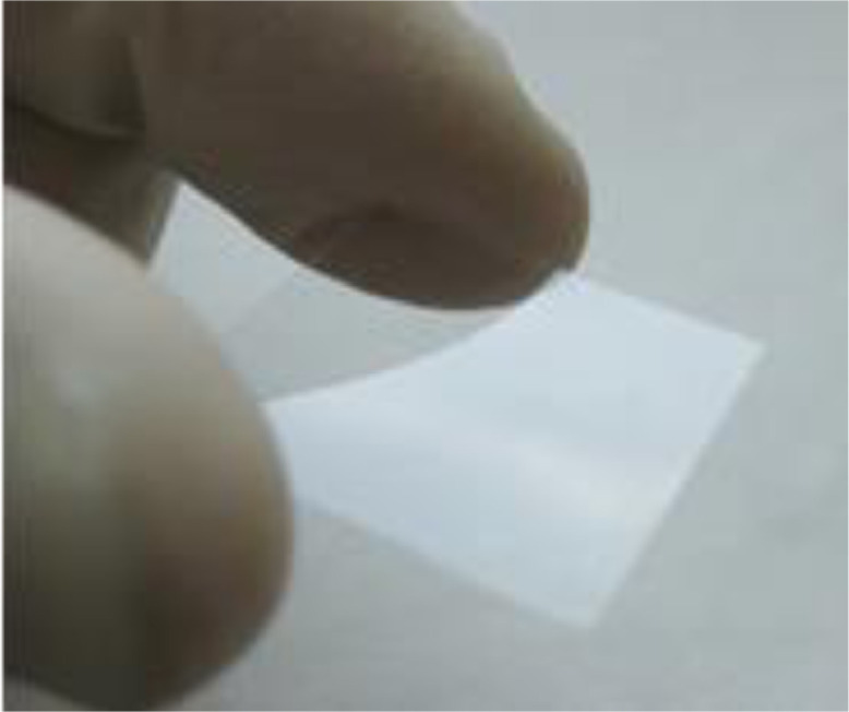
Photograph of a microscope cover glass plate used for fabrication of the single-used sensor chips.
Introducing free alkylamino groups to the glass surface
The cleaned microscope cover glass plates were put into a 3% solution of (3-Aminopropyl) triethoxysilane (APTES) in methanol and kept overnight at room temperature (RT). Then, they were washed three times with propanol-2 and dried. The aminated cover glasses were stored at RT before use [5,6].
Carboxylation of the glass plate surface
The aminated cover glass plates were washed three times with DMF, covered with a 15-mmol solution of succinic anhydride in DMF, and incubated for 2 h at RT. Finally, the modified microscope cover glass plates were thrice washed with DMF, dried, and stored at RT before use [7,8].
Immobilization of a molecular target onto carboxylated glass surface
The final modification of the glass plate surface was made as follows: 6% EDC solution in 0.1M MES buffer was applied to the carboxylated glass surface and kept for 15 min, then the surface was washed with PBS buffer. After that, a solution of OTA-BSA or hFABP (50 µg/mL in PBS) was applied, and the plate was incubated for 1 h at RT. Then the surface of the fabricated sensor chip was washed with PBS, exposed to 1 mL of 0.1M TRIS buffer for 30 min at RT, three times washed with water and dried.
Investigation of association and dissociation of antibodies
A solution of BSA (10 mg/mL in PBS) was pumped inside the SI instrument along the sensor chip with attached OTA-BSA or hFABP for 15 min. Then, the solution was changed for anti-OTA or anti-hFABP antibodies (25 µg/mL in PBS with BSA 10 mg/mL). This solution was pumped until the biolayer thickness reached a plateau. After that, a BSA solution (10 mg/mL in PBS) was pumped along the sensor chip for the next 1.5 h. The final step was necessary to initiate the dissociation of antigen-antibody [5,9,10]. During the procedure, the flow rate of solutions pumped along the glass sensor chip was constant – 6.7 μL/min.
Calculation of kinetic constants
The kinetic constants of interactions between the antigen on the sensor chip surface and free antibodies in a solution were calculated using a standard model for determination of kinetic constants [11], [12], [13], [14]. For the calculations, we used the sensograms (temporal dependences of SI signal) recorded in real-time by the SI instrument.
The fitting equation for the sensogram fragment corresponding to the antibody interaction with the antigen on the sensor chip surface:
where R(t) - is a temporal dependence of thickness of the molecular layer on the sensor chip surface, Rmax – maximal increase of thickness at the stage of antigen-antibody interaction, kon – kinetic constant of association, koff – kinetic constant of dissociation.
The fitting equation for the sensogram fragment during desorption of the antibodies from the antigen on the sensor chip surface:
Equilibrium constants were calculated as the ratio of the corresponding kinetic constants [15].
Method validation
As model biomolecules to be detected, the following substances were tested: ochratoxin A, which is one of the most dangerous mycotoxins naturally present in many vegetable raw materials [16], [17], [18]; and heart fatty acids binding protein, which is a cardiac marker used in differential diagnosis of acute myocardial infarction [19], [20], [21]. Fig. 3 exhibits the results of employment of the described above method for characterization of monoclonal antibodies to OTA and hFABP. As can be seen from the figures, the sensogram fragments related to both association and dissociation stages match well to the proposed theoretical model. The correlation between the experimental and theoretical data is not less than 95%.
Fig. 3.

Fitting the recorded sensogram fragments that correspond the processes of association and dissociation of antibodies to OTA and hFABP.
The calculated values of the kinetic constants of association (kon) and dissociation (koff) for monoclonal antibodies to OTA and hFABP, as well as the corresponding equilibrium constants (KA and KD) are shown in Table 1.
Table 1.
Calculated values of kinetic and equilibrium constants for monoclonal antibodies to OTA and hFABP.
| Complex | kon, M−1s−1 | koff, s−1 | KD, M | KA, M−1 |
|---|---|---|---|---|
| mAb — OTA-BSA | 4.54 * 103 | 3.32 * 10−4 | 7.31 * 10−8 | 1.37 * 107 |
| mAb – hFABP | 7.28 * 103 | 1.97 * 10−4 | 2.70 * 10−8 | 3.70 * 107 |
The presented here method was used for characterization and selection of immunoreagents for development of express high-sensitive detection of ochratoxin A in food by a lateral flow immunoassay based on magnetic biolabels [22]. Besides, the method can be used in developments of other rapid immunoassays, for example, point-of-care immunochromatographic tests based on magnetic particles [23], [24], [25] for differential diagnosis of acute myocardial infarction.
Acknowledgements
The authors thank Vladimir M. Voznyak and Leonid M. Vinokurov – for the help in obtaining anti-OTA antibodies and OTA-BSA conjugates, and Irina L. Nikitina – for assistance with the manuscript preparation and useful discussions. The research was funded by the Ministry of Science and Higher Education of the Russian Federation, contract No. 075-15-2022-315 for the organization and development of a World-class research center “Photonics”.
Declaration of Competing Interest
The authors declare that they have no known competing financial interests or personal relationships that could have appeared to influence the work reported in this paper.
Data availability
No data was used for the research described in the article.
References
- 1.Nikitin P.I., Gorshkov B.G., Valeiko M.V., Rogov S.I. Spectral-phase interference method for detecting biochemical reactions on a surface. Quantum Electron. 2000;30:1099–1104. doi: 10.1070/QE2000v030n12ABEH001876. [DOI] [Google Scholar]
- 2.Nikitin P.I., Valeiko M.V., Gorshkov B.G. New direct optical biosensors for multi-analyte detection. Sensors Actuators B Chem. 2003;90:46–51. doi: 10.1016/S0925-4005(03)00020-0. [DOI] [Google Scholar]
- 3.Ivanov A.E., Solodukhina N., Wahlgren M., Nilsson L., Vikhrov A.A., Nikitin M.P., Orlov A.V., Nikitin P.I., Kuzimenkova M.V., Zubov V.P. Reversible Conformational Transitions of a Polymer Brush Containing Boronic Acid and its Interaction with Mucin Glycoprotein. Macromol. Biosci. 2011;11:275–284. doi: 10.1002/mabi.201000295. [DOI] [PubMed] [Google Scholar]
- 4.Burenin A.G., Nikitin M.P., Orlov A.V., Ksenevich T.I., Nikitin P.I. Detection of pyrethroids by spectral correlation interferometry. Appl. Biochem. Microbiol. 2013;49:306–311. doi: 10.1134/S0003683813030058. [DOI] [PubMed] [Google Scholar]
- 5.Orlov A.V., Burenin A.G., Shipunova V.O., Lizunova A.A., Gorshkov B.G., Nikitin P.I. Development of immunoassays using interferometric real-time registration of their kinetics. Acta Naturae. 2014;6:85–95. doi: 10.32607/20758251-2014-6-1-85-95. [DOI] [PMC free article] [PubMed] [Google Scholar]
- 6.Jang L.-S., Liu H.-J. Fabrication of protein chips based on 3-aminopropyltriethoxysilane as a monolayer. Biomed. Microdevices. 2009;11:331–338. doi: 10.1007/s10544-008-9239-7. [DOI] [PubMed] [Google Scholar]
- 7.Burenin A.G., Urusov A.E., Betin A.V., Orlov A.V., Nikitin M.P., Ksenevich T.I., Gorshkov B.G., Zherdev A.V., Dzantiev B.B., Nikitin P.I. Direct immunosensing by spectral correlation interferometry: assay characteristics versus antibody immobilization chemistry. Anal. Bioanal. Chem. 2015;407:3955–3964. doi: 10.1007/s00216-015-8600-y. [DOI] [PubMed] [Google Scholar]
- 8.Lee M., Shin I. Fabrication of chemical microarrays by efficient immobilization of hydrazide-linked substances on epoxide-coated glass surfaces. Angew. Chemie Int. Ed. 2005;44:2881–2884. doi: 10.1002/anie.200462720. [DOI] [PubMed] [Google Scholar]
- 9.Orlov A.V., Nikitin M.P., Bragina V.A., Znoyko S.L., Zaikina M.N., Ksenevich T.I., Gorshkov B.G., Nikitin P.I. A new real-time method for investigation of affinity properties and binding kinetics of magnetic nanoparticles. J. Magnet. Magnet. Mater. 2015;380:231–235. doi: 10.1016/j.jmmm.2014.10.019. [DOI] [Google Scholar]
- 10.Linkuviene V., Talibov V.O., Danielson U.H., Matulis D. Introduction of intrinsic kinetics of protein–ligand interactions and their implications for drug design. J. Med. Chem. 2018;61:2292–2302. doi: 10.1021/acs.jmedchem.7b01408. [DOI] [PubMed] [Google Scholar]
- 11.Orlov A.V., Pushkarev A.V., Znoyko S.L., Novichikhin D.O., Bragina V.A., Gorshkov B.G., Nikitin P.I. Multiplex label-free biosensor for detection of autoantibodies in human serum: tool for new kinetics-based diagnostics of autoimmune diseases. Biosens. Bioelectron. 2020;159 doi: 10.1016/j.bios.2020.112187. [DOI] [PubMed] [Google Scholar]
- 12.Ivanov A.E., Pushkarev A.V., Orlov A.V., Nikitin M.P., Nikitin P.I. Interferometric detection of chloramphenicol via its immunochemical recognition at polymer-coated nano-corrugated surfaces. Sens. Actuat. B Chem. 2019;282:984–991. doi: 10.1016/j.snb.2018.11.043. [DOI] [Google Scholar]
- 13.Pushkarev A.V., Orlov A.V., Znoyko S.L., Bragina V.A., Nikitin P.I. Rapid and easy-to-use method for accurate characterization of target binding and kinetics of magnetic particle bioconjugates for biosensing. Sensors. 2021;21:2802. doi: 10.3390/s21082802. [DOI] [PMC free article] [PubMed] [Google Scholar]
- 14.Saftics A., Kurunczi S., Peter B., Szekacs I., Ramsden J.J., Horvath R. Data evaluation for surface-sensitive label-free methods to obtain real-time kinetic and structural information of thin films: A practical review with related software packages. Adv. Colloid Interface Sci. 2021;294 doi: 10.1016/j.cis.2021.102431. [DOI] [PubMed] [Google Scholar]
- 15.Pollard T.D. A guide to simple and informative binding assays. Mol. Biol. Cell. 2010;21:4061–4067. doi: 10.1091/mbc.e10-08-0683. [DOI] [PMC free article] [PubMed] [Google Scholar]
- 16.Nekrasov N., Yakunina N., Pushkarev A.V., Orlov A.V., Gadjanski I., Pesquera A., Centeno A., Zurutuza A., Nikitin P.I., Bobrinetskiy I. Spectral-phase interferometry detection of ochratoxin a via aptamer-functionalized graphene coated glass. Nanomaterials. 2021;11:226. doi: 10.3390/nano11010226. [DOI] [PMC free article] [PubMed] [Google Scholar]
- 17.Nekrasov N., Jaric S., Kireev D., Emelianov A.V., Orlov A.V., Gadjanski I., Nikitin P.I., Akinwande D., Bobrinetskiy I. Real-time detection of ochratoxin A in wine through insight of aptamer conformation in conjunction with graphene field-effect transistor. Biosensors and Bioelectronics. 2021;200 doi: 10.1016/j.bios.2021.113890. [DOI] [PubMed] [Google Scholar]
- 18.Iqbal S.Z., Rabbani T., Asi M.R., Jinap S. Assessment of aflatoxins, ochratoxin A and zearalenone in breakfast cereals. Food Chemistry. 2014;157:257–262. doi: 10.1016/j.foodchem.2014.01.129. [DOI] [PubMed] [Google Scholar]
- 19.Ye X., He Y., Wang S., Wong G.T., Irwin M.G., Xia Z. Heart-type fatty acid binding protein (H-FABP) as a biomarker for acute myocardial injury and long-term post-ischemic prognosis. Acta Pharmacol. Sin. 2018;39:1155–1163. doi: 10.1038/aps.2018.37. [DOI] [PMC free article] [PubMed] [Google Scholar]
- 20.Chaulin A.M., Duplyakov D.V. Biomarkers of acute myocardial infarction: diagnostic and prognostic value. Part 1. J. Clin. Pract. 2020;11:75–84. doi: 10.17816/clinpract34284. [DOI] [Google Scholar]
- 21.Rezar R., Jirak P., Gschwandtner M., Derler R., Felder T.K., Haslinger M., Kopp K., Seelmaier C., Granitz C., Hoppe U.C. Heart-type fatty acid-binding protein (H-FABP) and its role as a biomarker in heart failure: what do we know so far? J. Clin. Med. 2020;9:164. doi: 10.3390/jcm9010164. [DOI] [PMC free article] [PubMed] [Google Scholar]
- 22.Orlov A.V., Malkerov J.A., Novichikhin D.O., Znoyko S.L., Nikitin P.I. Express high-sensitive detection of ochratoxin A in food by a lateral flow immunoassay based on magnetic biolabels. Food Chem. 2022;383 doi: 10.1016/j.foodchem.2022.132427. [DOI] [PubMed] [Google Scholar]
- 23.Bragina V.A., Orlov A.V., Znoyko S.L., Pushkarev A.V., Novichikhin D.O., Guteneva N.V., Nikitin M.P., Gorshkov B.G., Nikitin P.I. Nanobiosensing based on optically selected antibodies and superparamagnetic labels for rapid and highly sensitive quantification of polyvalent hepatitis B surface antigen. Anal. Methods. 2021;13:2424–2433. doi: 10.1039/d1ay00354b. [DOI] [PubMed] [Google Scholar]
- 24.Guteneva N.V., Znoyko S.L., Orlov A.V., Nikitin M.P., Nikitin P.I. Rapid lateral flow assays based on the quantification of magnetic nanoparticle labels for multiplexed immunodetection of small molecules: application to the determination of drugs of abuse. Microchim. Acta. 2019;186:621. doi: 10.1007/s00604-019-3726-9. [DOI] [PubMed] [Google Scholar]
- 25.Znoyko S.L., Orlov A.V., Bragina V.A., Nikitin M.P., Nikitin P.I. Nanomagnetic lateral flow assay for high-precision quantification of diagnostically relevant concentrations of serum TSH. Talanta. 2020;216 doi: 10.1016/j.talanta.2020.120961. [DOI] [PubMed] [Google Scholar]
Associated Data
This section collects any data citations, data availability statements, or supplementary materials included in this article.
Data Availability Statement
No data was used for the research described in the article.