Abstract
Corneal tissue engineering has developed rapidly in recent years, with a large number of publications emerging worldwide. This study focused on exploring the global status and research trends in this field. Publications related to corneal tissue engineering from 1991 to 2021 were acquired from the Science Citation Index-Expanded (SCI-Expanded) of Web of Science (WoS). Firstly, the VOS viewer software was chosen for visualizing bibliometric networks, including bibliographic coupling analysis, co-citation analysis, co-authorship analysis, and co-occurrence analysis, and the CiteSpace software was used to detect burst keywords. Subsequently, the publication trends in corneal tissue engineering research were also predicted. In present study, 953 publications were selected and analyzed. The number of annual publications was increasing globally and was predicted to continue the current trend. While Japan ranked top 1 in terms of average citation, the USA contributed the most to the corneal tissue engineering research with highest number of citations and highest H-index. The journal of Biomaterials contributed the largest publication number. The top-ranked institutions were National University of Singapore and Singapore National Eye Center. Additionally, researches could be manually divided into four clusters: “biomaterial related research,” “cell related research,” “transplantation therapy,” and “mechanism research on biomaterials.” Specifically, the research topic “hydrogel” was predicted to be hotspots which may help researchers to explore new directions for future research.
Keywords: Bibliometrics, visualized study, global trend, cornea, tissue engineering
Introduction
The cornea is a clear and avascular tissue covering the anterior one sixth of the total surface of the eye. The main purposes of cornea are to protect the eye contents, provide 2/3 of the refractive power of the optical system and finally allow light to reach the retina.1 Various mechanical, thermal, or chemical trauma factors can damage the integrity and uniformity of the cornea. Corneal epithelium is the most anterior layer of cornea, which constantly renews itself due to the proliferation and migration of progenitor cells located at the boundary of cornea and sclera, and has good regeneration characteristics, which allows the self-healing of superficial corneal injury.2 However, deeper corneal damage can result in severe visual impairment, which is the fifth leading cause of blindness worldwide. More than 2.2 billion people worldwide are visually impaired or blind, according to the World Health Organization’s first global vision report released in November 2019. At least 1 billion of these patients are visually impaired, 4.2 million of which are owing to unresolved corneal opacity.3
At present, keratoplasty was the only approved therapeutic strategy for corneal injury.2,4 In 2012, it is estimated that 12.7 million people around the world are waiting for corneal transplants. Unfortunately, only 185,000 keratoplasty operations were performed worldwide in the same year, meeting the needs of only 1 in 70 patients.5 In addition, transplant rejection and postoperative complications significantly reduced the effectiveness of the operation.5,6 In order to provide alternatives to capture corneal shortages, lots of efforts have been made in the live corneal substitutes which are designed to mimic in vivo counterparts in terms of tissue structure and cellular phenotype.7 Tissue engineering technology includes three elements: scaffolds, seed cells, and cytokines.8 At present, tissue engineering technology has been developed to construct feasible corneal tissue equivalents from the engineering of epithelium, stroma, and endothelium, and to reconstruct natural innervation. These methods eventually lead to full-layer corneal tissue equivalent.9 Significant advances has provided effective cornea tissue substitutes and alternatives for in vitro research and clinical purposes.7 However, the global development trend of corneal tissue engineering has not been well reported. Therefore, it is necessary to summarize the current situation of corneal tissue engineering and predict the promising research hotspots and trends.
In scientific research, publishing is a vital index to measure the contribution of scientific research. Bibliometric analysis can offer information that based on bibliometric database and bibliometric characteristics, which can be chosen to analyze the trend of research activities qualitatively and quantitatively over a period of time.10 Bibliometric analysis is also performed to formulate policy and clinical practice guidelines.11 It has been successfully used to analyze the research trends of arthritis,12 diabetes,13 and thyroid cancer.14 However, to the best of our knowledge, the quantity and quality of corneal tissue engineering research have not been reported. Hence, the purpose of this study is to evaluate the current landscape and the future trend in the field of corneal tissue engineering by bibliometric analysis.
Materials and methods
Ethics statement
This study was based on published papers and did not involve human or animal experiments. So, this research did not require ethical approval.
Data source
Publication information is retrieved from the SCI-Expanded of Web of Science (WoS), which are considered as the optimal databases for bibliometrics.15
Search strategy
All the published papers were collected from WoS and the expiration date of the database was set to 31 December, 2021. In this study, the searching terms were shown below: theme = cornea OR corneal AND theme = tissue engineering AND publishing year = (1991.01.01–2021.12.31) AND Language = (English) AND Document types = (Article or Review). Additionally, the detailed information of certain countries or regions was refined by indexing country/region in the WoS.
Data collection
The inclusion criteria of publication were shown as follows: (1) the manuscript focused on the theme of corneal tissue engineering; (2) the document types are Article and Review; and (3) the papers must be written in English. The exclusion criteria were also shown as follows: (1) The themes were not related to corneal tissue engineering; (2) Articles were briefings, news, meeting abstracts, etc. All the records of publications, including year of publication, title, author names, affiliations, countries/regions, abstract, keywords, and name of publishing journals were downloaded and saved as .txt files from SCI-Expanded database and then imported into Excel 2019. Finally, GraphPad Prism 8.0 and Origin 2021 were chosen to analyze the data. Any problem emerged in present study has been solved by consulting experts.
Bibliometric analysis
The intrinsic function of WoS was to characterize the basic features of eligible publications. In addition, total publications of each year which was acquired from SCI-Expanded were firstly pictured by GraphPad Prism 8.0 and the relative research interest (RRI) was defined as the number of publications in one certain field by all field publications per year. The world map was performed by R software including python + numpy + scipy + matplotlib and the time curve of publications was depicted according to previous article.16 The data of publications from top 25 countries/regions were analyzed by GraphPad Prism 8.0. In addition, the total citation, average citation, and H-index level were also evaluated by GraphPad Prism 8.0. The H-index, indicating that a scholar has published H papers and they have been cited at least H times, was created to measure the impact of scientific research. Hence, it reflects both the number of publications and corresponding citations.17 Finally, high-contribution journals, institutions, funds, and authors of global publications about corneal tissue engineering were also analyzed by Origin 2021.
Visualized analysis
VOS viewer (Leiden University, The Netherlands) is a powerful software tool for mapping and visualizing bibliometric networks and thus was performed for the visualization in present study. These networks include journals, authors, countries, and individual publications, and they can be constructed based on bibliographic couplings, co-citations, co-authorship relationships, and co-occurrence of keywords. And CiteSpace was used to detect burst keywords for forecasting the possible hotspots and research frontiers in the future.
Results
Trend of global publications
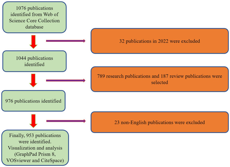
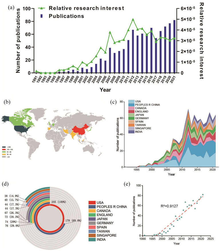
According to the inclusive criteria, 1076 articles were firstly included from 1991 to 2021. Finally, 953 articles were selected for analysis by excluding conditions (Figure 1). When evaluating the number of publications per year, most papers were published in 2021 (82, 8.6%). From 1991 to 2021, the trend of global publications generally increased steadily year by year. In addition, RRI in the field has declined over the past few years (Figure 2(a)). Totally, 52 countries or regions made contributions in publications in this field (Figure 2(b)). The United States published the most papers (255, 26.76%), followed by China (178), Canada (76), England (74), and Japan (72). We could clearly see that the publishing number of USA and China increased dramatically since the year of 2012 (Figure 2(c)). The USA contributed mostly in this research topic as China, Canada, England only counts for 69.8%, 29.8%, and 29%, respectively (Figure 2(d)). In order to predict the future global publications trend, a linear fitting model was used to create a time curve of the number of publications. We can see from Figure 2(e) that the fitting curve was Y = 3.2623 × X − 6514.9131 (R2 = 0.9127), indicating that the number of global publications in the coming years might increase at a stationary rate.
Figure 1.
Flow diagram of the study identification and inclusion process.
Figure 2.
Global trends and countries/regions contributing to the research field of corneal tissue engineering. (a) The annual number of publications related to corneal tissue engineering. (b) A world map depicting distribution of corneal tissue engineering. (c) The annual number of publications in the top 10 most productive countries from 1991 to 2021. (d) The sum of corneal tissue engineering related publications in the top 10 countries/regions. (e) Model fitting curves of global trends in publications of corneal tissue engineering (Y = 3.2623 × X − 6514.9131, R2 = 0.9127, where X indicates the publishing year).
Quality of publications of different countries and regions
In terms of WoS database analysis, we counted the total citations, H-index and average citations of each country/region. Publications from the USA had the highest total citation frequencies (14,071). Japan ranked second in total citation frequency (4753), followed by China (4165), Canada (3759), and England (3207; Figure 3(a)). In regards to the H-index analysis, the relative publications from the USA had the highest H-index (60), followed by Canada (35), Japan (34), China (34), and England (28; Figure 3(b)). As to the average citation frequency detection, publications from Japan showed the highest average citation frequencies (66.01). Sweden ranked second in average citation frequency (58.63), followed by the United States (55.84), Scotland (50.36), and Canada (49.46) (Figure 3(c)).
Figure 3.
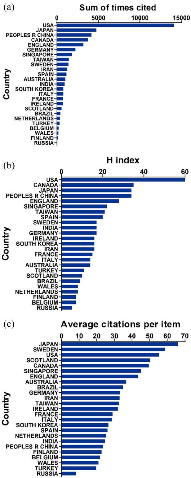
Total citation, H index, and citation frequency levels of different countries/regions. (a) The top 25 countries/regions of total citations of corneal tissue engineering. (b) The top 25 countries/regions of the H-index of organoids in corneal tissue engineering. (c) The top 25 countries/regions of the average citations per paper of corneal tissue engineering.
Global publications analysis of leading journals, institutions, funding, and authors
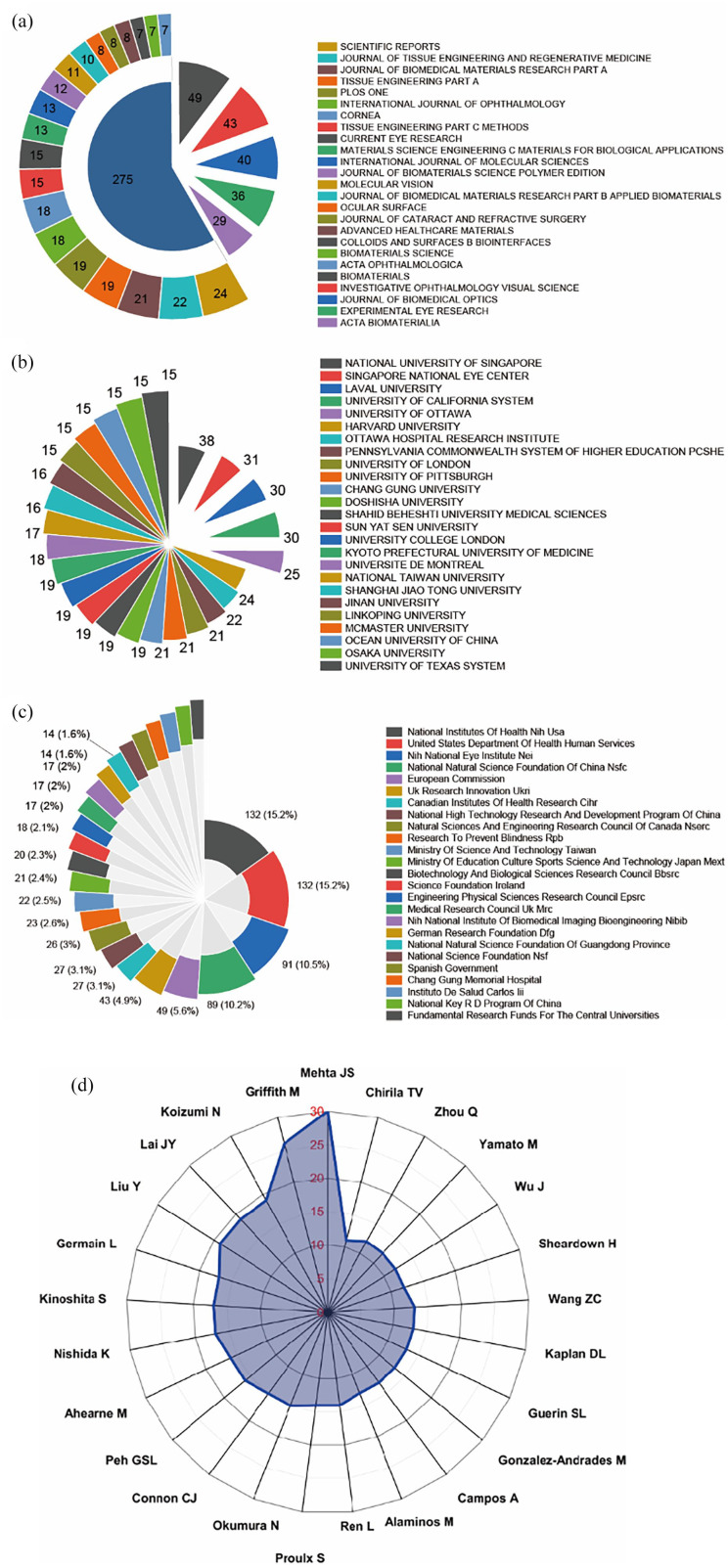
Biomaterials (impact factor [IF] = 15.304, 2021) published the most with 49 publications. There were 43 publications in Investigative Ophthalmology Visual Science (IF = 4.925, 2021), 40 publications in Journal of Biomedical Optics (IF = 3.758, 2021), 36 articles in Experimental Eye Research (IF = 3.770, 2021), and 29 publications in Acta Biomaterialia (IF = 10.633, 2021). The top 25 journals with the most publications were listed in Figure 4(a).
Figure 4.
High-contribution journals, institutions, funds, and authors of global publications about corneal tissue engineering. (a) The top 25 journals with most publications on corneal tissue engineering. (b) The top 25 institutions with most publications on corneal tissue engineering. (c) The top 25 funding sources with most publications on corneal tissue engineering. (d) The top 25 authors with most publications on corneal tissue engineering.
The top 25 contributive institutions were listed in Figure 4(b). National Unniversity of Singapore published the most (38 publications), Singapore National Eye Center ranked second (31 publications), while Laval University ranked third (30 publications).
The top 25 funding sources were shown in Figure 4(c). In total, 132 publications were funded by National Institutes of Health (NIH) or United States Department of Health Human Services (Tied for first), 91 publications were funded by NIH National Eye Institute (ranked second), and 89 publications were funded by National Natural Science Foundation of China (NSFC) (ranked third).
The top 25 authors published 391 publications, which accounted for 41.03% of all publications in this field. Mehta JS contributed the most research with 30 publications, followed by Griffith M with 26 publications, and Koizumi N with 18 publications (Figure 4(d)).
Bibliographic coupling analysis of leading author, institution, country, and journal
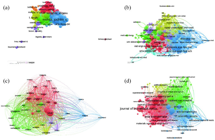
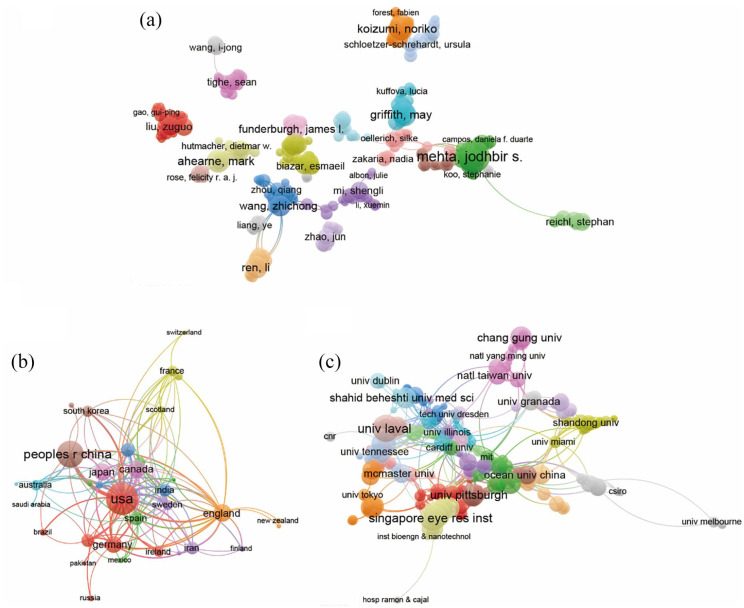
Publications (the minimum number of documents per author was defined as exceeds 2) were produced from 789 authors and further analyzed by VOS viewer. As shown in Figure 5(a), the top 5 productive authors are shown below: Mehta, Jodhbir S (total link strength equals to 69,724 times), Koizumi, Noriko (total link strength equals to 50,238 times), Ahearne, Mark (total link strength equals to 50,033 times), Germain, Lucie (total link strength equals to 44,706 times), and Okumara, Naoki (total link strength equals to 43,583 times).
Figure 5.
Mapping of bibliographic coupling analysis related corneal tissue engineering. (a) Mapping of the 1089 authors on corneal tissue engineering. (b) Mapping of the 831 institutions on corneal tissue engineering. (c) Mapping of the 62 countries on corneal tissue engineering. (d) Mapping of the 350 identified journals on corneal tissue engineering. The line between different points represents that the authors/institutions/countries/journals had establish a similarity relationship. The thicker the line, the closer the link between the authors/institutions/countries/journals.
Papers (the minimum number of documents used by an organization was defined as more than two and the maximum number of organizations used by each document was defined not more than 25) were identified in the 340 institutions and analyzed using VOS viewer (Figure 5(b)). The top 5 institutions with largest total link strength were shown as follows: Singapore Eye Res Inst (total link strength equals to 46,545 times), Singapore Natl Eye Ctr (total link strength equals to 41,507 times), Laval Univ (total link strength equals to 35,328 times), Doshisha Univ (total link strength equals to 27,399 times), and Shahid Beheshti Univ Med Sci (total link strength equals to 27,038 times).
Publications (the minimum number of documents of each country was defined as more than 2) originating from 40 countries were analyzed via VOS viewer (Figure 5(c)). The top 5 countries with large total link strength were as follows: USA (total link strength equals to 112,911 times), China (total link strength equals to 80,256 times), England (total link strength equals to 50,733 times), Japan (total link strength equals to 47,315 times), and Canada (total link strength equals to 40,790 times).
The bibliographic coupling was used to analyze the similarity relationship between documents. Firstly, VOS viewer was performed to analyzed the name of journals in total publications. There are 123 identified journals appeared in total link strength which were shown in Figure 5(d). The top 5 journals with larger total link strength were shown as follows: Investigative ophthalmology & Visual Science (Impact Factor, IF = 4.925, 2021, total link strength equals to17,306 times), Experimental Eye Research (Impact Factor, IF = 3.770, 2021, total link strength equals to 17,297 times), Biomaterials (Impact Factor, IF = 15.304, 2021, total link strength equals to 16,843 times), Acta Biomaterialia (Impact Factor, IF = 10.633, 2021, total link strength equals to 11,182 times), and Journal of Tissue Engineering and Regenerative Medicine (Impact Factor, IF = 4.323, 2021, total link strength equals to 9483 times).
Co-citation analysis of leading authors, journals, and references
The co-citation analysis was to consider the relatedness of the items based on the numbers they were co-cited. Total of 841 authors with the minimum of 10 documents were analyzed using VOS viewer (Figure 6(a)). The top 5 publications with largest total link strength were shown below: Mimura, T (total link strength equals to 13,887 times), Lai, JY (total link strength equals to 11,832 times), Dua, HS (total link strength equals to 10,747 times), Joyce, NC (total link strength equals to 10,586 times), and Okumura, N (total link strength equals to 10,356 times).
Figure 6.
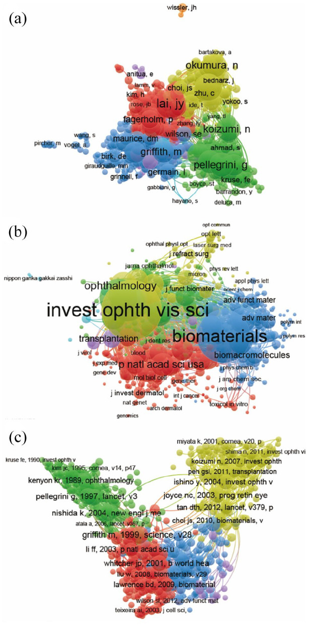
Mapping of co-citation related to corneal tissue engineering. (a) Mapping of the co-cited authors related to this field. (The 576 points with different colors represent the 576 identified authors.) (b) Mapping of the co-cited journals related to this field. (The 2109 points with different colors represent the 2109 identified journals.) (c) Mapping of the co-cited references related to this field. (The 3064 points with different colors represent the 3064 cited references.). The point sizes represent the citation frequency. The line between different points indicates that they were cited in one paper. The shorter the line, the closer the link between two papers. The same color of the points represents the same research area they belong to.
The journal names of co-citation analysis were analyzed using VOS viewer and the journal with minimum number of citations over 10 were finally defined. As illustrated in Figure 6(b), 617 journals were shown in the total link strength analysis. The top 5 journals with best total link strength were shown as follows: Investigative ophthalmology & Visual Science (total link strength equals to 330,804 times), Biomaterials (total link strength equals to 282,271 times), Cornea (total link strength equals to 136,806 times), Experimental Eye Research (total link strength equals to 126,508 times), and Acta Biomaterialia (total link strength equals to 85,450 times).
The purpose of reference co-citation analysis is to reveal the relatedness of items based on the total cited number and there are 658 references were analyzed via VOS viewer (Figure 6(c)). The top 5 articles with greatest total link strength were as follows: Pellegrini G, 1997, LANCET, v349 (total link strength equals to 2487 times); Griffith M, 1999, SCIENCE, v286 (total link strength equals to 2463 times); Nishida K, 2004, New England J Med, v351 (total link strength equals to 2409 times); Ishino Y, 2004, Invest Ophth & Vis Sci, v45 (total link strength equals to 2264 times); and Tsai RJ, 2000, New England J Med, v343 (total link strength equals to 2190 times).
Co-authorship analysis of leading authors, countries, and institutions
Co-authorship analysis was performed to evaluate the items relatedness based on the total number of co-authored papers. A total of 789 authors with more than two documents were analyzed using the VOS viewer, and the results are shown in Figure 7(a). The top 5 authors with larger total link strength were as follows: Mehta JS (total link strength equals to 118 times), Wang ZC (total link strength equals to 87 times), Griffith M (total link strength equals to 81 times), Peh GSL (total link strength equals to 74 times), and Germain L (total link strength equals to 70 times).
Figure 7.
Visualized images of co-authorship analysis of global research about corneal tissue engineering. (a) Mapping of the 328-author co-authorship analysis on corneal tissue engineering research. (b) Mapping of the 46-country co-authorship analysis on corneal tissue engineering research. (c) Mapping of the 458-institution co-authorship analysis on corneal tissue engineering research.
There are 40 countries with over two papers were chosen and analyzed using VOS viewer and the results were depicted in Figure 7(c). The top 5 ranked countries with biggest total link strength were shown below: USA (total link strength equals to 148 times), England (total link strength equals to 56 times), Canada (total link strength equals to 55 times), China (total link strength equals to 46 times), and Sweden (total link strength equals to 46 times).
In addition, there are 340 institutions with more than two documents were analyzed using VOS viewer (Figure 7(c)). The top 5 institutions with greatest total link strength were shown below: Singapore Eye Res Inst (total link strength equals to 92 times), Singapore Natl Eye Ctr (total link strength equals to 92 times), Duke Nus Grad Med Sch (total link strength equals to 58 times), Univ Ottawa (total link strength equals to 56 times), and Natl Univ Singapore (total link strength equals to 49 times).
Co-occurrence analysis of keywords
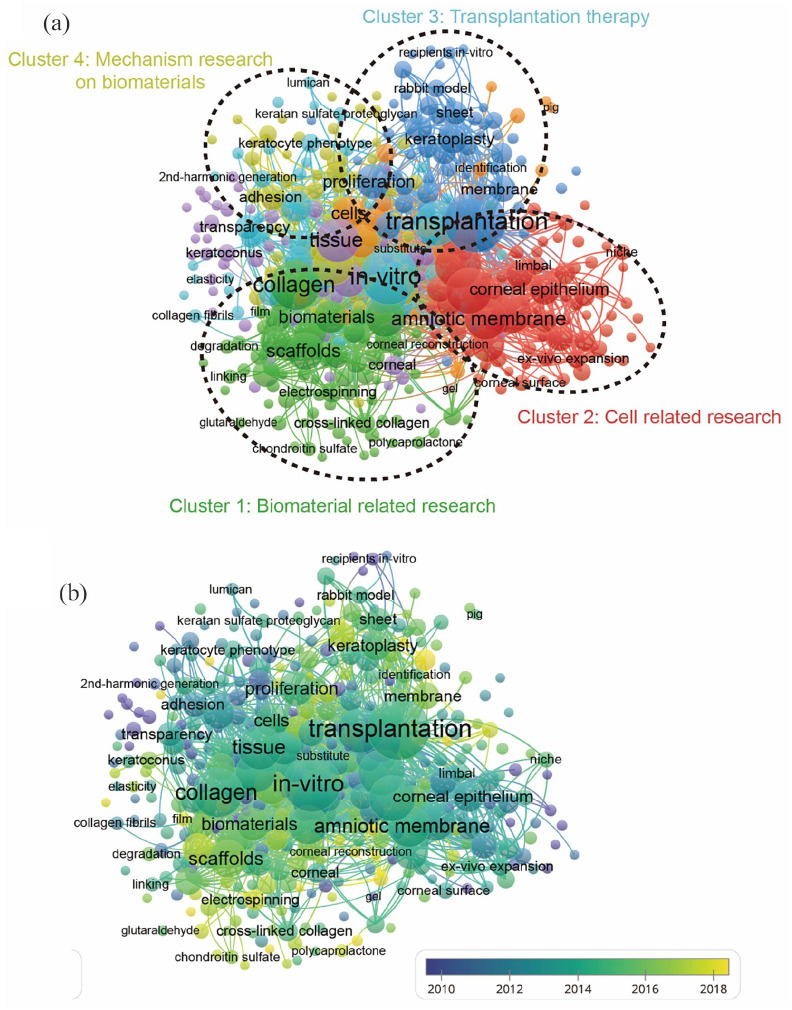
The objective of co-occurrence analysis is to investigate popular directions and area of researches, and it also plays a vital role in monitoring the developments in scientific research. Keywords, which was defined as the words used more five times in titles/abstracts in all papers, were chosen and analyzed via VOS viewer. The 390 identified keywords were mainly classified into four clusters as follows: cluster 1: biomaterial related research (green), cluster 2: cell related research (red), cluster 3: transplantation therapy (blue), cluster 4: mechanism research on biomaterials (yellow) (Figure 8(a)). These results exhibited the most prominent research topics in corneal tissue engineering research so far. In the “biomaterial related research” cluster, the primary keywords were: collagen, biomaterials, and scaffolds. For the “cell related research” cluster, the frequently used keywords were: amniotic membrane, corneal epithelium, and ex-vivo expansion. As for the “transplantation therapy” cluster, the main used keywords were: transplantation, keratoplasty, and sheet. Furthermore, in the “mechanism research on biomaterials” cluster, the dominantly used keywords were: proliferation, adhesion, and cells. These results exhibited that the most prominent fields of corneal tissue engineering included the above mentioned four directions.
Figure 8.
Visualization of co-occurrence analysis based on corneal tissue engineering. (a) Mapping of keywords in the research on corneal tissue engineering; the frequency is represented by point size; The keywords of research fields were divided into four clusters by different colors: biomaterial related research (green), cell related research (red), transplantation therapy (blue), and mechanism research on biomaterials (yellow). (b) Visualization of keywords distribution; The yellow color represents an earlier appearance and blue point appeared later.
Additionally, the keywords were coded with different colors by VOS viewer based on average times that they appeared in all the published papers (Figure 8(b)). The color in blue meant that the keywords appeared earlier, whereas the color in yellow indicating later appearance. As shown in Figure 8(b), the trends of most studies in the four clusters did not change dramatically, meaning that every research fields would be evenly concerned in the future.
Focus shift and research frontiers
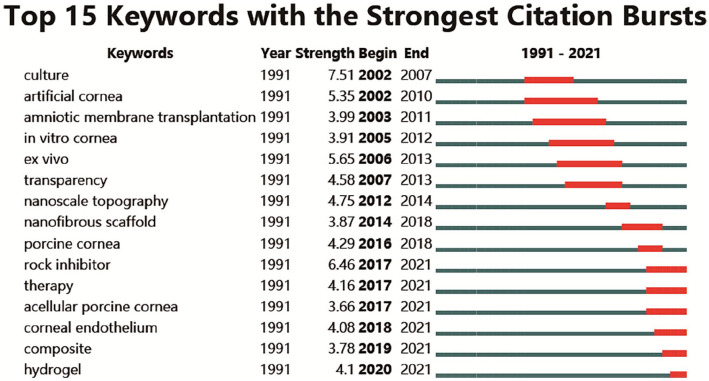
Keywords with intense bursts in a short period can act as a sensitive indicator to reflect the research focus. Recent burst keywords provide researchers the possible research frontiers in a short future. A keyword burst map was generated by CiteSpace, where the strength and the beginning or ending year of burst was shown (Figure 9). The strength reveals the burst intensity, and the burst year indicates the transformation of the research focus and its duration. Early studies focused on the cell (“culture” begin in 2002) and “artificial corneal” begin in 2002. Afterwards, “in vitro cornea” begin in 2005, “nanoscale topography” begin in 2012, “nanofibrous scaffold” begin in 2014 generalized the research focus transformation over the past 20 years. In recent years, “corneal endothelium” (begin in 2018), “composite” (begin in 2019), and “hydrogel” (begin in 2020) appeared and kept bursting till 2021.
Figure 9.
Top 15 keywords with the strongest citation bursts. Keywords with the strongest citation bursts in the scientific literature were analyzed and visualized in the keyword’s bursts map. Lines in red stood for the burst detection years. Keywords with red lines extending to the latest year can indicate the research frontiers in a short period of time in the future.
Discussion
Trends in corneal tissue engineering
Bibliometrics and visual analysis can reveal the current situation in the search field and make predictions.16 Therefore, the purpose of this research is to evaluate corneal tissue engineering in the field of open access from the aspects of authors, donor countries, journal, institutions, and research focus. Recently, the progress of corneal tissue engineering has become an exciting and steady developing research field.18 As shown in this study, there has been a steady increase in the number of publications published each year. However, in the past few years, related research interest has declined. Based on present data, we have also predicted the number of future publications. More in-depth corneal tissue engineering studies will be published in the coming years. The current optimistic results will also allow researchers to conduct further high-quality studies.
The quality and standing of global publications
The total citations and H-index represent the academic influence and publishing quality of one county.19 Based on the total number of published papers, the total number of citations and the H-index, the USA makes the largest contribution to global research and it is arguably the leadership in this area. China ranks second in the world in the total number of publications. However, it ranked third and fourth in terms of total citation frequency and H-index, respectively. Discrepancy in the quantity and quality of publications in China may be an important reason. China’s academic evaluation system has always focused on the quantity rather than the quality of published papers.
More research on corneal tissue engineering has been published in Biomaterials, Investigative Ophthalmology & Visual Science, Journal of Biomedical Optics and Experimental Eye Research. The journals in the list (Figure 4(a)) are likely to be the main publishing channels for future discoveries in this area. Further researches in this area may appear at the top of the list.
The top 3 research institutes with the largest number of articles are the leading institutions of corneal tissue engineering research. This is not consistent with the leading position of the top three countries in global publications, which may be closely related to the large number of corneal transplants in Singapore. Authors who have published more research results in this field are also listed. This suggests that further studies of these authors deserve more attention in order to obtain the latest advances in corneal tissue engineering.
In this study, the similar relationship among journals, institutions, and countries is established by bibliographic coupling analysis. Bibliographic coupling is defined as two works citing a common third work in their bibliography. These data show that Investigative Ophthalmology & Visual Science is the most relevant journal, while USA is the leading country in this field. The purpose of co-citation analysis is to investigate the influence of research by calculating the number of times cited together. The current results show that the milestone research in corneal tissue engineering has a large total citation frequency. In addition, Investigative Ophthalmology & Visual Science is the most frequently cited journals in this field. Co-authorship analysis is used to assess cooperation among authors, institutions, and countries. Results with higher total link strength indicate that the countries/institutions/authors are more willing to cooperate with others.
The research focus of corneal tissue engineering
On the basis of co-occurrence analysis, we explored the development direction and hot topics in this field. All keywords included in the titles and abstracts of the study were analyzed to create a map of the co-occurrence network. Four research directions can be observed from the co-occurrence map (Figure 8(a)), including “biomaterial related research,” “cell related research,” “transplantation therapy,” and “mechanism research on biomaterials.” Although this result is in line with the common sense in this field, this study can make the future research direction clearer. At the center of the co-occurrence map, as is shown obviously, keywords including “transplantation,” “in-vitro,” “collagen,” and “amniotic membrane,” etc. have a largest weight. Therefore, further high-quality researches of corneal tissue engineering studies in these four directions are still needed. The overlay visualization map was assigned colors by VOS viewer according to the average times the keywords appeared in the papers.20 This method is of great significance for the monitoring of research directions. The color bar indicates how fractions are mapped to colors. In the overlay visualization shown in Figure 8(b), the color represents the year of publication. According to the results, the topics of early published articles mainly focused on the “transparency” of grafts (purple for early stage), “transplantation” (green), and gradually transferred to the field of biomaterials: “collagen,” “biomaterials,” “cross-linked,” “electrospinning” (yellow for recent). Burst keywords revealed the research hotspots and their transformation from cell research to scaffold and biomaterial study. Particularly, burst keywords that continue to the present indicate the potential trends and possible frontiers in the field of corneal tissue engineering (Figure 9). The latest burst keywords include “acellular porcine cornea,” “corneal endothelium,” “composite,” and “hydrogel.” So, studies on these aspects might indicate the frontier of the corneal tissue engineering field.
Studies on corneal regeneration, especially the development of tissue engineering techniques to restore or regenerate cornea, have been widely emerged.7,9,18,21 A 3D bioprinting is an emerging method for the fabrication of biological grade cornea. A bioprinter can combine different biomaterials such as keratocytes, collagens, stem cells, plasma, and other molecules in the matrix with high spatial resolution. A major advantage of 3D printed scaffolds is the delivery of precision medicine based on fulfilling patient-specific needs.21–24 Further researches are deeply required to develop truly biomimetic corneal regenerative therapies and then translate to clinical practice.
Strengths and limitations
The publications derived from SCI-expanded of WoS were explored in present study to acquire reliable and objective results. Bibliometric and visualized analysis were achieved by variety of software (including Prism 8, Origin 2021, VOS viewer, andCiteSpace), and we evaluated the status and trends of studies about corneal tissue engineering reliably and objectively. However, several limitations still exist in our study. It is well known that publications from different databases such as WoS, Pubmed, Embase, and the Cochrane Library are varied. Therefore, we may miss some publications due to database bias. Besides, due to limitation of searching strategy in English from SCI-expanded database, the Non-English language literatures could have been omitted, leading to language bias. Our search strategy only included entire tissues, so we cannot get further analysis of single layer of corneal tissue. Owing to the constant updates of the target database, slight differences may exist in the real world and the present results. Finally, there is no uniform standard for parameter settings in VOS viewer, thus, the outputs of cluster analysis may differ slightly under different settings.
Conclusion
This study showed the global status and trends in corneal tissue engineering research. The USA was the largest contributor to studies and had the leading position in global research in this field. The journal Biomaterials attracted the most publications related to this area. We predicted that more researches about corneal tissue engineering will be published in the coming years. An overall analysis of this field from the perspective of biomaterial, cell, transplantation therapy, mechanism related researches might be the latest research directions. Particularly, biomaterials especially hydrogel might get more attention and be the next popular hotspot in the future.
Footnotes
The author(s) declared no potential conflicts of interest with respect to the research, authorship, and/or publication of this article.
Funding: The author(s) disclosed receipt of the following financial support for the research, authorship, and/or publication of this article: This work was supported by the National Natural Science Foundation of China (82002364 MXZ), Natural Science Foundation of Hunan Province (2021JJ40509 to MXZ), Project for Clinical Research of Hunan Provincial Health Commission (20201956 to MXZ), and China Scholarship Council (202106370071 to BWZ).
ORCID iD: Bo-Wen Zheng  https://orcid.org/0000-0002-9308-3794
References
- 1. Sridhar M-S. Anatomy of cornea and ocular surface. Indian J Ophthalmol 2018; 66(2): 190–194. [DOI] [PMC free article] [PubMed] [Google Scholar]
- 2. Gonzalez G, Sasamoto Y, Ksander B-R, et al. Limbal stem cells: identity, developmental origin, and therapeutic potential. Wiley Interdiscip Rev Dev Biol 2018;7(2). doi:10.1002/wdev.303. [DOI] [PMC free article] [PubMed] [Google Scholar]
- 3. Fernandes T-M, Alves M-C, Diniz C-O, et al. Artificial cornea transplantation and visual rehabilitation: an integrative review. Arq Bras Oftalmol. Epub ahead of print 23 September 2022. DOI: 10.5935/0004-2749.2021-0350. [DOI] [PMC free article] [PubMed] [Google Scholar]
- 4. Mobaraki M, Abbasi R, Vandchali S-O, et al. Corneal repair and regeneration: current concepts and future directions. Front Bioeng Biotechnol 2019; 7: 135. [DOI] [PMC free article] [PubMed] [Google Scholar]
- 5. Gain P, Jullienne R, He Z, et al. Global survey of corneal transplantation and eye banking. JAMA Ophthalmol 2016; 134(2): 167–73. [DOI] [PubMed] [Google Scholar]
- 6. Qazi Y, Hamrah P. Corneal allograft rejection: immunopathogenesis to therapeutics. J Clin Cell Immunol 2013; 2013(Suppl. 9): 006. [DOI] [PMC free article] [PubMed] [Google Scholar]
- 7. Guérin L-P, Le-bel G, Desjardins P, et al. The human tissue-engineered cornea (hTEC): recent progress. Int J Mol Sci 2021; 22(3): 1291. [DOI] [PMC free article] [PubMed] [Google Scholar]
- 8. Berthiaume F, Maguire T-J, Yarmush M-L. Tissue engineering and regenerative medicine: history, progress, and challenges. Annu Rev Chem Biomol Eng 2011; 2: 403–430. [DOI] [PubMed] [Google Scholar]
- 9. Ghezzi C-E, Rnjak-Kovacina J, Kaplan D-L. Corneal tissue engineering: recent advances and future perspectives. Tissue Eng Part B Rev 2015; 21(3): 278–287. [DOI] [PMC free article] [PubMed] [Google Scholar]
- 10. Pu Q-H, Lyu Q-J, Su H-Y. Bibliometric analysis of scientific publications in transplantation journals from Mainland China, Japan, South Korea and Taiwan between 2006 and 2015. BMJ Open 2016; 6(8): e011623. [DOI] [PMC free article] [PubMed] [Google Scholar]
- 11. Avcu G, Bal Z-S, Duyu M, et al. Thanks to trauma: a delayed diagnosis of pott disease. Pediatr Emerg Care 2015; 31: e17–e18. [DOI] [PubMed] [Google Scholar]
- 12. Kai W, Dan X, Dong S, et al. The global state of research in nonsurgical treatment of knee osteoarthritis: a bibliometric and visualized study. BMC Musculoskelet Disord 2019; 20: 407. [DOI] [PMC free article] [PubMed] [Google Scholar]
- 13. Gao Y, Wang Y, Zhai X, et al. Publication trends of research on diabetes mellitus and T cells (1997–2016): A 20-year bibliometric study. PLOS ONE 2017; 12(9): e0184869. [DOI] [PMC free article] [PubMed] [Google Scholar]
- 14. Wang H, Yu Y, Yu Y, et al. Bibliometric insights in advances of anaplastic thyroid cancer: research landscapes, turning points, and global trends. Front Oncol 2021; 11: 769807. [DOI] [PMC free article] [PubMed] [Google Scholar]
- 15. Aggarwal A, Lewison G, Idir S, et al. The state of lung cancer research: a global analysis. J Thorac Oncol 2016; 11(7): 1040–1050. [DOI] [PubMed] [Google Scholar]
- 16. Xing D, Zhao Y, Dong S, et al. Global research trends in stem cells for osteoarthritis: a bibliometric and visualized study. Int J Rheu Dis 2018; 21(7): 1372–1384. [DOI] [PubMed] [Google Scholar]
- 17. Bertoli-Barsotti L, Lando T. A theoretical model of the relationship between the h-index and other simple citation indicators. Scientometrics 2017, 111(3): 1415–1448. [DOI] [PMC free article] [PubMed] [Google Scholar]
- 18. Oh J-Y, Kim E, Yun Y-I, et al. Mesenchymal stromal cells for corneal transplantation: literature review and suggestions for successful clinical trials. Ocul Surf 2021; 20: 185–194. [DOI] [PMC free article] [PubMed] [Google Scholar]
- 19. Bastian S, Ippolito J-A, Lopez S-A, et al. The use of the h-index in academic orthopaedic surgery. J Bone Joint Surg Am 2017; 99(4): e14. [DOI] [PubMed] [Google Scholar]
- 20. Hesketh K-R, Law C, Bedford H, et al. Co-occurrence of health conditions during childhood: longitudinal findings from the UK millennium cohort study (MCS). PLOS ONE 2016; 11(6): e0156868. [DOI] [PMC free article] [PubMed] [Google Scholar]
- 21. Mohan R-R, Kempuraj D, D’souza S, et al. Corneal stromal repair and regeneration. Prog Retin Eye Res. Epub ahead of print 29 May 2022. DOI: 10.1016/j.preteyeres.2022.101090. [DOI] [PubMed] [Google Scholar]
- 22. Bektas C-K, Hasirci V. Cell loaded 3D bioprinted GelMA hydrogels for corneal stroma engineering. Biomater Sci 2019; 8 (1): 438–449. [DOI] [PubMed] [Google Scholar]
- 23. Mahdavi S-S, Abdekhodaie M-J, Kumar H, et al. Stereolithography 3D bioprinting method for fabrication of human corneal stroma equivalent. Ann Biomed Eng 2020; 48(7): 1955–1970. [DOI] [PubMed] [Google Scholar]
- 24. Isaacson A, Swioklo S, Connon C-J. 3D bioprinting of a corneal stroma equivalent. Exp Eye Re 2018; 173: 188–193. [DOI] [PMC free article] [PubMed] [Google Scholar]


