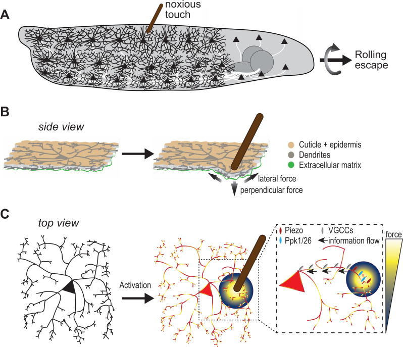
Figure 1. Sensing noxious touch in Drosophila larvae.
(A) Schematic of a Drosophila larva with nociceptive cells called c4da neurons covering its entire body. The cd4a neurons can sense noxious touch (here provided by a small diameter probe) resulting in the larva undergoing a rolling escape response. (B) Schematic side view of c4da neurons in the larval body sandwiched between the epidermal cell layer (beige) and the extracellular matrix (green). The noxious touch of the probe deforms the body wall including c4da dendrites and generates perpendicular and lateral forces (indicated by arrows) (C) The force applied by the probe (yellow-blue color code) activates c4da neurons by triggering two mechanosensory channels – Ppk1/Ppk26 (cyan) and Piezo (red) – located on its dendrites. Voltage-gated calcium channels (grey) then allow information to reach the soma so they can elicit a response in the c4da neuron soma.

