. 2022 Oct 6;11:e76574. doi: 10.7554/eLife.76574

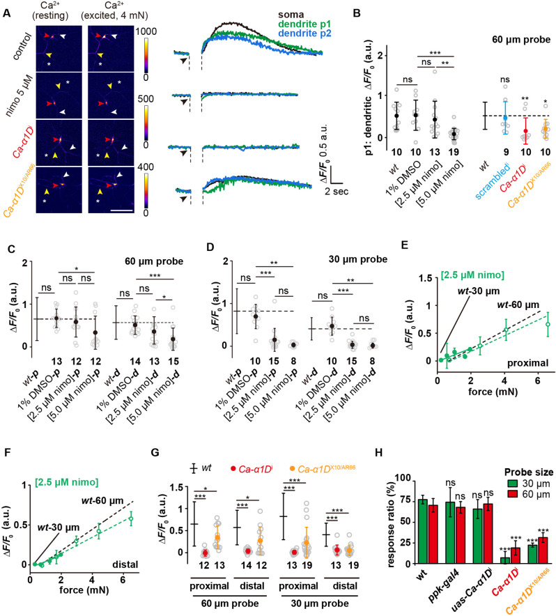
Figure 6. Active signal propagation in the dendrites of c4da.

(A) Representative images and curves showing the responses (ΔF/F0) of the soma (red arrowhead), the homolateral dendrite (position p1: yellow arrowhead) and contralateral dendrite (position p2: white arrowhead) of c4da-wt, nimodipine-treated c4da-wt, c4da-Ca-α1Di and Ca-α1DX10/AR66 to the stimuli (4 mN, 60 μm probe). The asterisk indicated the force application point. Scale bar: 50 μm. C4da-wt: ppk-gal4/+; ppk-cd4-tdtom/20×uas-ivs-gcamp6s. Ca-α1Di: ppk-gal4/20×uas-ivs-gcamp6s; ppk-cd4-tdtom/ uas-Ca-α1Di. Ca-α1DX10/AR66: Ca-α1DX10/ Ca-α1DAR66; ppk-gal4/20×uas-ivs-gcamp6s. (B) Statistical quantification of calcium increases in the homolateral dendrites in response to the stimuli (4 mN, 60 μm probe). The genotypes were the same as those shown in panel (A). Mann Whitney U test was used for the comparison between c4da-wt and the mutants. Kruskal Wallis test was used for the comparison among the groups with drug treatment. ***: p<0.001. **: p<0.01. ns: no significance. (C) and (D) The responses (ΔF/F0) of nimodipine treated c4da-wt to the proximal and distal stimuli from the probes of different sizes. (C): 60 μm probe stimuli. (D) : 30 μm probe stimuli. Kruskal Wallis test was used. ***: p<0.001. **: p<0.01. *: p<0.05. ns: no significance. (E) and (F) The responses (ΔF/F0) of c4da-wt (n=10 cells) treated with 2.5 μM nimodipine to the proximal (E) and distal (F) stimuli delivered using the probes of two different sizes. Solid lines: 30 μm probe. Dashed lines: 60 μm probe. (G) The responses (ΔF/F0) of c4da-Ca-α1Di and c4da-Ca-α1DX10/AR66 to the stimuli applied at different dendritic regions (proximal and distal) and delivered using the probes of two different sizes (60 μm: 4 mN, 30 μm: 1.5 mN). (H) The behavioral responses of the c4da-Ca-α1Di larvae to mechanical poking using the probes of two different sizes. wt: w1118. ppk-gal4: ppk-gal4; +/+. uas-Ca-α1Di: +/+; uas-Ca-α1Di. Ca-α1Di: ppk-gal4/+; uas-Ca-α1Di /+. Ca-α1DX10/AR66: Ca-α1DX10/ Ca-α1DAR66; +/+. uas-Ca-α1Di larvae: 30 μm probe (n=61 larvae from three experiments), 60 μm probe (n=61 larvae from three experiments). Ca-α1Di larvae: 30 μm probe (n=56 larvae from three experiments), 60 μm probe (n=60 larvae from three experiments). Ca-α1DX10/AR66 larvae: 30 μm probe (n=43 larvae from three experiments), 60 μm probe (n=47 larvae from three experiments). One-way ANOVA test was used for the comparison among different genotypes. ***: p<0.001. ns: no significance. In panels (B), (C), (D), (G) and (H), data were presented as mean ± std. In panels (B), (C), (D) and (G), the numbers of cells were indicated below the scattered data points. In panels (E) and (F), data were presented as mean ± sem. In panels (C), (D), (E), (F), (G) and (H), the corresponding data from c4da-wt were provided for comparison.

Figure 6—source data 1. Numerical data for Figure 6.

Figure 6.

Figure 6—figure supplement 1. Statistics quantification of the calcium increase in the contralateral dendrites in response to the force stimuli.
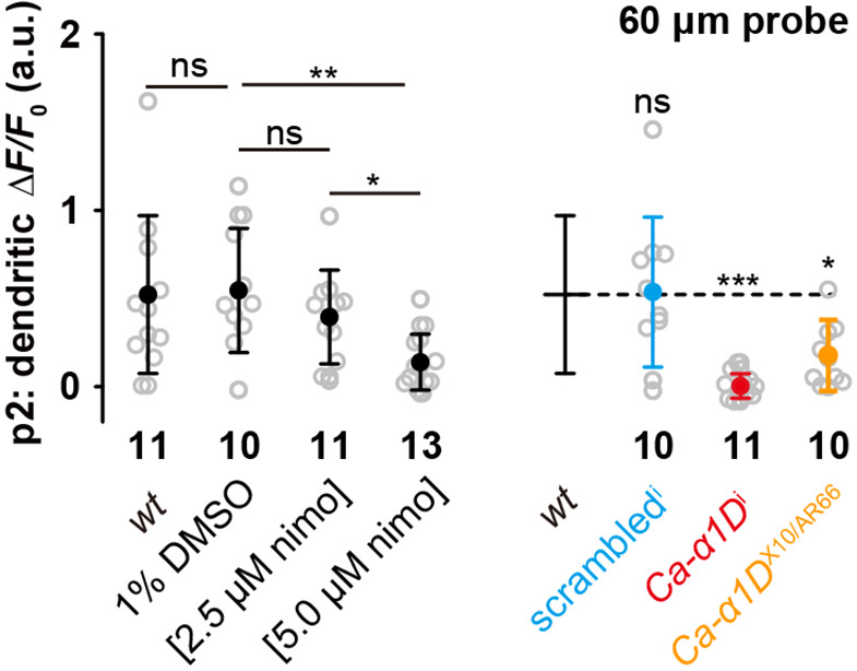
Figure 6—figure supplement 1.

C4da-wt: ppk-gal4/+; ppk-cd4-tdtom/20×uas-ivs-gcamp6s. Scrambledi: ppk-gal4/20×uas-ivs-gcamp6s, ppk-cd4-tdtom/uas-Scrambledi. Ca-α1Di: ppk-gal4/20×uas-ivs-gcamp6s; ppk-cd4-tdtom/uas-Ca-α1Di. Ca-α1DX10/AR66: Ca-α1DX10/Ca-α1DAR66; ppk-gal4/20×uas-ivs-gcamp6s. The force stimuli (4 mN) were applied using a 60 μm probe. The numbers of cells were indicated below the scattered data points. Mann Whitney U test was used for the comparison between c4da-wt and the mutants. Kruskal Wallis test was used for the comparison among the groups with drug treatment. *: p<0.05. **: p<0.01. **: p<0.001. ns: no significance.
Figure 6—figure supplement 1—source data 1. Numerical data for Figure 6—figure supplement 1.
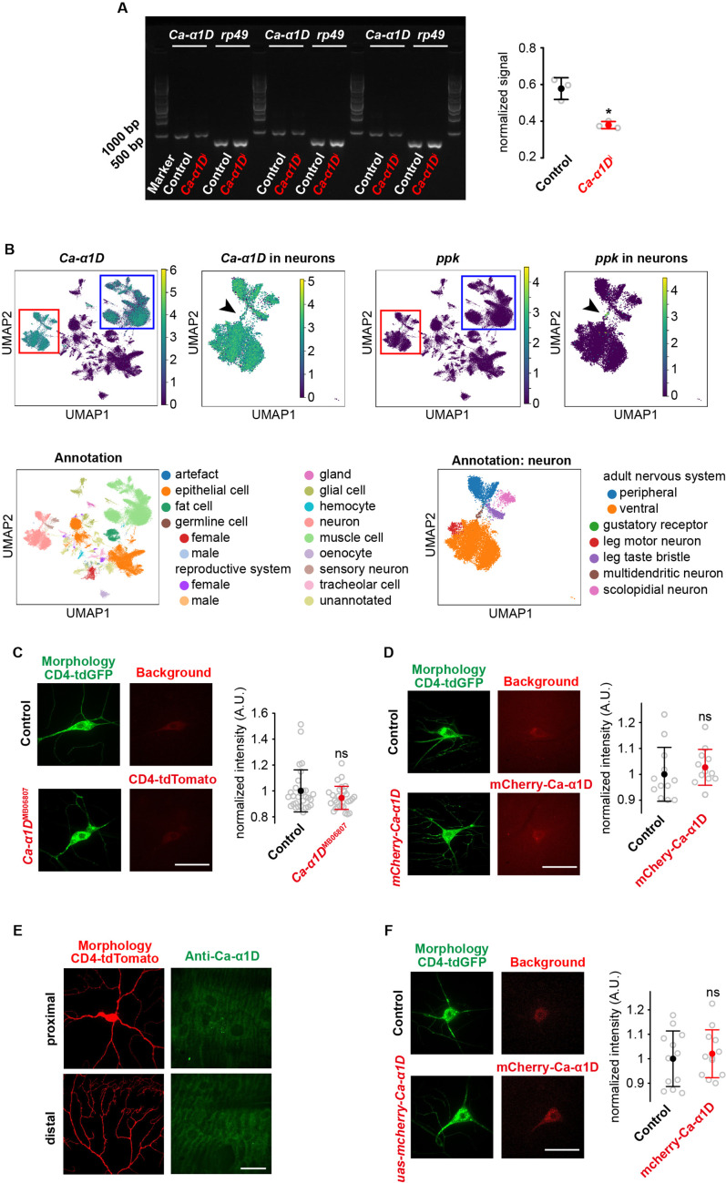
Figure 6—figure supplement 2. The expression and localization of Ca-α1D in vivo.

Figure 6—figure supplement 2.

(A) The expression level of Ca-α1D in wild-type and Ca-α1Di was examined using RT-PCR. Because Ca-α1D expressed in various types of neurons and the number of c4da in the whole animal was small, we knocked down the expression of Ca-α1D in the whole animal using tub-gal4. Using RT-PCR, we found that the expression level of Ca-α1D was significantly reduced in the Ca-α1Di strain. The expression of rp49 was used as the reference for normalization. Note that the same strain was also used as a knockdown mutant in other functional studies (Terada et al., 2016; Basak et al., 2021). (B) The expression pattern of Ca-α1D analyzed based on the FlyCellAtlas (Li et al., 2022) single-cell RNA-sequencing datasets We found that Ca-α1D mainly expressed in excitable cells, such as neurons (red square) and muscle cells (blue square). In particular, it expressed in the multidendritic neurons that specifically expressed ppk1 (black arrowhead). (C) To confirm the expression of Ca-α1D in c4da, we examined the Mi{ET1}Ca-α1DMB06807 line. However, no significant signal was observed. Control: ppk-cd4-tdgfp/+; uas-cd4-tdtom/+. Ca-α1DMB06807: Mi{ET1}Ca-α1DMB06807/ppk-cd4-tdgfp; uas-cd4-tdtom/+. n=30 cells for each genotype. (D) We generated a transgenic strain that expressed mCherry-Ca-α1D under the endogenous promoter of Ca-α1D. However, no signal was observed (n=12 cells). Control: ppk-cd4-tdgfp/+; +/+. mcherry-Ca-α1D: ppk-cd4-tdgfp/+;mcherry-Ca-α1D. (E) We used a previously reported antibody (see Materials and Methods) to examine the localization of Ca-α1D. However, no signal was observed. (F) We generated a transgenic strain to express mCherry-Ca-α1D under the control of the ppk-gal4 strain. However, no signal was observed (n=12 cells). Control: ppk-cd4-tdgfp/+; ppk-gal4/+. uas-mcherry-Ca-α1D: ppk-cd4-tdgfp/+; 10×uas-mcherry-Ca-α1D/ppk-gal4. In panel C, D and F, scale bar: 30 μm. In panel (E), scale bar: 100 μm. In panel (A, C, D and F), data were presented as mean ± std. Student’s t test was used for statistical analysis. *: p<0.05; ns: no significance.
Figure 6—figure supplement 2—source data 1. Gel image and numerical data for Figure 6—figure supplement 2.