Abstract
Introduction
The incidence of oral cavity squamous cell carcinoma (OSCC) continues to rise. OSCC is associated with a low average survival rate, and most patients have a poor disease prognosis because of delayed diagnosis. We used machine learning techniques to predict high-risk cases of OSCC by using salivary autoantibody levels and demographic and behavioral data.
Methods
We collected the salivary samples of patients recruited from a teaching hospital between September 2008 and December 2012. Ten salivary autoantibodies, sex, age, smoking, alcohol consumption, and betel nut chewing were used to build prediction models for identifying patients with a high risk of OSCC. The machine learning algorithms applied in the study were logistic regression, random forest, support vector machine with the radial basis function kernel, eXtreme Gradient Boosting (XGBoost), and a stacking model. We evaluated the performance of the models by using the area under the receiver operating characteristic curve (AUC), with simulations conducted 100 times.
Results
A total of 337 participants were enrolled in this study. The best predictive model was constructed using a stacking algorithm with original forms of age and logarithmic levels of autoantibodies (AUC = 0.795 ± 0.055). Adding autoantibody levels as a data source significantly improved the prediction capability (from 0.698 ± 0.06 to 0.795 ± 0.055, p < 0.001).
Conclusions
We successfully established a prediction model for high-risk cases of OSCC. This model can be applied clinically through an online calculator to provide additional personalized information for OSCC diagnosis, thereby reducing the disease morbidity and mortality rates.
Supplementary Information
The online version contains supplementary material available at 10.1186/s12903-022-02607-2.
Keywords: Oral cavity squamous cell carcinoma, Autoantibodies, Biomarker, Machine learning
Introduction
Oral cancer incidence is increasing globally, and this form of cancer is associated with a low average survival rate [1–3]. Developed countries have higher rates of oral cancer incidence, whereas less-developed countries have higher rates of disease mortality [4]. Oral cavity squamous cell carcinoma (OSCC) accounts for over 90% of oral cancer cases [5]. In more than 50% of patients, the OSCC diagnosis is delayed, and over half of patients are in an advanced stage (overall pathological stage III–IV) of the disease by the time of diagnosis [6]. In the past few decades, the effectiveness of OSCC detection has not improved considerably [7]. In Taiwan, over 40% of patients received a diagnosis of OSCC at a late stage [8], leading to poor disease prognosis and treatment failure [9, 10]. Therefore, an accurate diagnosis of OSCC, especially at its early stage, is crucial to improve the survival rate [11].
Oral potentially malignant disorders (OPMDs) consist of premalignant lesions that often progress to OSCC [12, 13]. Currently, conventional oral examination (COE) is the classical method for premalignant epithelium and oral cancer detection; however, differentiating OPMD and lesions with no risk of cancer by using COE remains challenging [14]. People lack knowledge about OPMD symptoms, and even clinicians may miss the signs of OPMD [2]. Therefore, developing an OPMD diagnostic tool to complement the COE conducted by clinicians can help patients to receive appropriate treatments in time and can help prevent malignant transformation.
Autoantibodies are antibodies produced against substances formed by a person’s own body and are expressed at low concentrations in healthy cells and at abnormally high concentrations in tumor cells [15]. Autoantibodies are potential biomarkers of breast [16], lung [17], colon [18], head and neck [19], esophageal [20, 21], and prostate [22] cancers. Several autoantibodies have been reported as OSCC biomarker candidates [23, 24]. Each of these reported autoantibodies exhibits a limited sensitivity or specificity for detecting OSCC, and whether a combined panel of these autoantibodies would be more effective than a single autoantibody in diagnosing the disease remains uncertain. Therefore, further verifying the efficacy of using these biomarkers together with demographic and behavioral data to identify patients with a high risk of OSCC is necessary.
Machine learning is a branch of artificial intelligence (AI) that enables computers to learn from previous data and make predictions [25]. Machine learning techniques are beneficial and are commonly applied to several cancers for predicting diagnosis, recurrence, metastasis, and prognosis [26–30]. We used machine learning models to predict high-risk cases of OSCC by using salivary autoantibody levels and demographic and behavioral data and evaluated the efficacy of salivary biomarkers in detecting OSCC.
Methods
Study participants and design
Participants were recruited from a teaching hospital, namely Chi-Mei Medical Center (Liouying, Taiwan), from September 2008 to December 2012 [23]. Inclusion criteria were Taiwanese adults > 30 years of age with current or previous habitual behaviors, such as smoking or betel nut chewing, following the oral cancer screen project launched by the Health Promotion Administration, Taiwan. Participants received a visual oral cavity examination by a trained dentist or physician and were divided into high-risk (patients with OSCC and high-risk OPMD, as confirmed by biopsy) and low-risk (patients with low-risk OPMD and healthy) groups on the basis of the criteria described in a previous study [23]. Salivary samples were collected at the time of recruitment, and saliva processing and autoantibody detection were performed in 2018. The experimental procedure of autoantibody detection was described in a previous study [23]. Before collection of saliva specimens, volunteers avoided smoking, eating, and drinking for at least 2 h. To remove cell debris, collected saliva samples were centrifuged at 3000×g for 15 min at 4 °C. The supernatants were immediately treated with a mixture of protease inhibitors (2 μL/mL; Cat. No. P8340, Sigma-Aldrich, Burlington, MA, USA), aliquoted into a volume of 100 μL, and then stored at a − 80 °C freezer. To avoid protein aggregation and degradation, saliva samples with more than one freeze–thaw cycle were not used. Using the strict protocol of collection and storage, the property and quality of saliva samples were preserved [24]. We evaluated the salivary autoantibody levels and selected 9 oral cancer-related proteins, ANXA2, CA2, HSPA5, ISG15, KNG1, MMP1, MMP3, PRDX2, SPARC, identified in a previous study [23] and p53 as the biomarker candidates of OSCC. Demographic and behavioral data, including sex, age, smoking, alcohol consumption, and betel nut chewing, were obtained. All participants signed the informed consent form before undergoing screening and treatment. The study was approved by the Institutional Review Board of Chi-Mei Medical Center (No. 10012-L02). Model reporting followed the TRIPOD (transparent reporting of a multivariable prediction model for individual prediction or diagnosis) guidelines [31].
Data preprocessing for model development
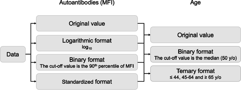
For continuous variables, namely age and the mean fluorescence intensity (MFI) values of autoantibodies, we used the original values to develop prediction models. Moreover, we converted the MFI value into a binary, logarithmic, or standardized format and age into a binary or ternary format to evaluate the effect of using different forms of data (Fig. 1). In the training set, the 90th percentile of the MFI value was set as the cutoff value for transforming the MFI value into the binary format. The median age (50 years) was set as the cutoff value for transforming age into the binary format; for transforming age into the ternary format, the cutoff values were set as ≤ 44, 45–64, and ≥ 65 years, based on the risk of oral cancer [32]. There were no missing values in the dataset.
Fig. 1.
Flow chart of median fluorescent intensity (MFI) and age variable processing
Development of prediction models
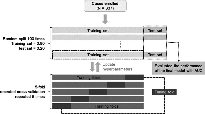
We employed logistic regression (LR), random forest (RF), support vector machine (SVM) with the radial basis function (RBF) kernel, eXtreme Gradient Boosting (XGBoost), and a stacking model containing all the aforementioned models (i.e., LR, RF, SVM, and XGBoost) to develop the risk prediction models for OSCC. We performed a five-fold cross-validation that was repeated five times to optimize the model parameters during each round of model development (Fig. 2). All tuning parameters and the range of tuning for model development are listed in Additional file 1: Table S1.
Fig. 2.
Flow chart of prediction model development and evaluation
LR is a statistical model that uses a logistic function to model a binary dependent variable [33]. An SVM with an RBF kernel constructs a classification model for a two-group issue; the data from two groups are separated using a hyperplane and transformed into a higher dimension [34]. RF is a bagging ensemble algorithm that uses random feature selection and consists of multiple classification trees [35]. XGBoost is an ensemble algorithm that optimizes distributed libraries under the gradient boosting framework and can predict a model from weak prediction models, usually decision trees. Thus, XGBoost uses a regularized model to control overfitting, and this gives it better performance [36]. Model stacking is an ensemble method that combines the prediction results of other models (LR, RF, SVM, and XGBoost) to generate a new ensemble model. Stacking combination was performed using lasso regression, a linear regression that uses the L1 penalty for both fitting and penalization of coefficients. Lasso regression performs both feature selection and regularization to improve prediction accuracy and model interpretability [37]. The caret [38], tidymodels [39], and stacks [40] packages in R software (R Core Team, Vienna, Austria) were used to develop and evaluate the predictive models.
Evaluation of prediction models
The machine learning models for distinguishing high-risk and low-risk groups were trained with 80% of the data and tested using the remaining 20% of the data. To better estimate the performance of prediction models, we randomly split the training–test dataset 100 times to construct and evaluate 100 models for each algorithm (Fig. 2). All prediction models were then evaluated using the test sets. The area under the receiver operating characteristic curve (AUC) was used to evaluate model performance. The optimal model was used to build an online calculator. We applied the International Journal of Medical Informatics (IJMEDI) checklist for medical AI assessment (Additional file 1: Table S2) [41].
Effectiveness of autoantibodies as biomarkers for OSCC risk prediction
Variable selection was performed using the permutation-based variable importance measure, which is based on the hypothesis that if a variable is important, the model’s performance will worsen after permuting the values of the variable. The larger the change in performance is, the more important is the variable. We used 1 − AUC as the loss metric; the higher the value of this metric is, the higher is its importance as a predictive variable [42]. The DALEX [42] and DALEXtra [43] packages were used to explain the model and evaluate the variable importance.
To evaluate the effectiveness of autoantibodies and demographic and behavioral data in identifying patients with high OSCC risk, we compared the performance of prediction models that used both autoantibodies and demographic and behavioral data with those that used patient characteristics alone.
Statistical analysis
Continuous variables are expressed as medians (interquartile range) for skewed distributions and were analyzed using the Mann–Whitney U test to determine the differences between the two groups. Categorical variables are presented as percentages and were calculated using the chi-square or Fisher’s exact test. The scale function, converting each original value into a z-score, in R was used for standardization. The AUC values of prediction models, from the 100 times of performance evaluations, were compared using repeated-measures ANOVA (rANOVA), and the Holm–Bonferroni post hoc test was used to compare processing strategies and algorithms. All statistical tests were two-sided, and statistical significance was defined as p < 0.05. Statistical analyses were performed using R 4.1.2.
Results
Patient characteristics and salivary IgA autoantibody levels
A total of 337 participants were enrolled in this study. The baseline characteristics and the salivary levels of 10 autoantibodies are listed in Table 1. Among participants, 331 (98.2%) were male, 306 (90.8%) smoked, 118 (35.0%) consumed alcohol, and 271 (85.4%) chewed betel nut; the median age was 50.4 years (IQR = 12). Participants were stratified into high-risk (n = 190; 107 OSCC cases and 83 high-risk OPMD cases) and low-risk (n = 147; 55 low-risk OPMD cases and 92 healthy individuals) groups. Participants in the high-risk group were older (51.8 vs. 49.2 years, p = 0.003), consumed more alcohol (42.6% vs. 25.2%, p = 0.001), chewed more betel nut (92.1% vs. 65.3%, p < 0.001), and exhibited higher levels of all salivary autoantibodies (p < 0.005) except for anti-ANXA2 (p = 0.253).
Table 1.
Patient characteristics and salivary autoantibody levels
| Overall | High-risk | Low-risk | p-value | |
|---|---|---|---|---|
| No. of cases (%) | 337 | 190 (56.4) | 147 (43.6) | – |
| Sex, male, n (%) | 331 (98.2) | 188 (98.9) | 143 (97.3) | 0.41a |
| Age, range | 31–82 | 32–82 | 31–78 | 0.003b |
| Years, median (IQR) | 50.4 (17) | 51.8 (15) | 49.2 (19) | |
| Smoking, n (%) | 306 (90.8) | 176 (92.6) | 130 (88.4) | 0.258c |
| Alcohol consumption, n (%) | 118 (35.0) | 81 (42.6) | 37 (25.2) | 0.001c |
| Betel nut chewing, n (%) | 271 (80.4) | 175 (92.1) | 96 (65.3) | < 0.001c |
| Autoantibodies, MFI, median (IQR) | ||||
| Anti-ANXA2 | 2727.2 (2611.4) | 2791.8 (3079.5) | 2600.1 (2244.6) | 0.253b |
| Anti-CA2 | 1191.2 (1232.8) | 1446.0 (1509.2) | 1020.0 (947.3) | < 0.001b |
| Anti-HSPA5 | 607.8 (722.8) | 757.5 (892.1) | 457.1 (484.0) | < 0.001b |
| Anti-ISG15 | 1273.7 (1299.0) | 1506.0 (1635.8) | 1021.4 (1004.4) | < 0.001b |
| Anti-KNG1 | 4176.5 (4720.2) | 4814.0 (6782.6) | 3379.6 (2981.0) | < 0.001b |
| Anti-MMP1 | 1426.3 (1214.5) | 1647.8 (1523.4) | 1239.3 (953.8) | < 0.001b |
| Anti-MMP3 | 3907.2 (548.4) | 4018.6 (777.1) | 3828.9 (405.2) | < 0.001b |
| Anti-p53 | 1616.7 (1782.1) | 1814.1 (2432.3) | 1485.1 (1437.4) | 0.003b |
| Anti-PRDX2 | 1038.0 (1286.3) | 1336.4 (1525.1) | 813.0 (865.5) | < 0.001b |
| Anti-SPARC | 707.0 (724.8) | 833.9 (956.5) | 555.2 (507.2) | < 0.001b |
IQR interquartile range, MFI median fluorescence intensity
p-values of high-risk and low-risk groups were compared
aFisher’s exact test
bMann–Whitney U test
cChi-square test
Comparison of prediction model performances
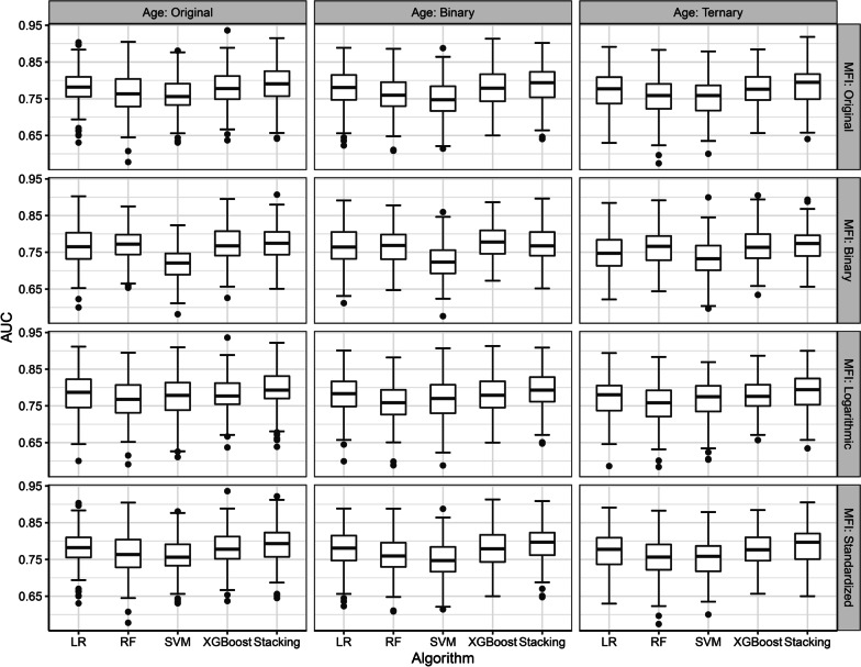
First, we evaluated different data processing strategies and five machine learning models for distinguishing high-risk from low-risk patients; the AUCs, from the test sets, are listed in Fig. 3 and Additional file 1: Table S3. The AUCs from the training sets are listed in Additional file 1: Table S4. The optimal machine learning algorithm for predicting high-risk OSCC cases, based on the AUC from the test sets, was the stacking method; rANOVA and post hoc analysis revealed significant differences among other machine learning algorithms (p < 0.001, Table 2). When building a model with the stacking method, the best data processing strategy was to use age in the original format and logarithmic autoantibody levels (AUC = 0.795 ± 0.055). Compared with transforming autoantibody levels to the binary format, transforming autoantibody levels to the logarithmic format resulted in significant improvement in prediction performance (p < 0.05, Additional file 1: Table S5). However, no significant difference in prediction performance was observed between transforming autoantibody levels to the logarithmic format and transforming autoantibody levels to the standardized format.
Fig. 3.
Performance of risk prediction models by using 12 data processing strategies. LR: logistic regression; RF: random forest; SVM: support vector machine with a radial basis function kernel; XGBoost: eXtreme Gradient Boosting
Table 2.
Holm–Bonferroni post hoc test across machine learning models performed using age in the original format and logarithmic autoantibody levels
| Algorithm (AUC) | LR 0.784 ± 0.057 | RF 0.764 ± 0.057 | SVM 0.772 ± 0.059 | XGBoost 0.778 ± 0.051 | Stacking 0.795 ± 0.055 |
|---|---|---|---|---|---|
| RF | < 0.001 | – | – | – | – |
| SVM | 0.006 | 0.188 | – | – | – |
| XGBoost | 0.270 | 0.002 | 0.270 | – | – |
| Stacking | 0.001 | < 0.001 | < 0.001 | < 0.001 | – |
LR logistic regression, RF random forest, SVM support vector machine with a radial basis function kernel, XGBoost eXtreme Gradient Boosting, Stacking a stacking model that contained all models, SD standard deviation
The calibration curves are provided in Additional file 1: Fig. S1, and the Brier scores were 0.125, 0.123, 0.151, 0.140, and 0.127 for the stacking, XGBoost, RF, SVM, and LR models, respectively. The lift chart is provided in Additional file 1: Fig. S2; the lift value in the highest risk group (top 5%) was 1.79 for all models, indicating that the positive predictive value in the highest risk group, as identified by the stacking model, was approximately two times higher than the average positive predictive value. The IJMEDI checklist is provided in Additional file 1: Table S2. An online calculator developed based on the optimal model is depicted in Additional file 1: Fig. S3.
Autoantibodies as biomarkers for OSCC risk prediction
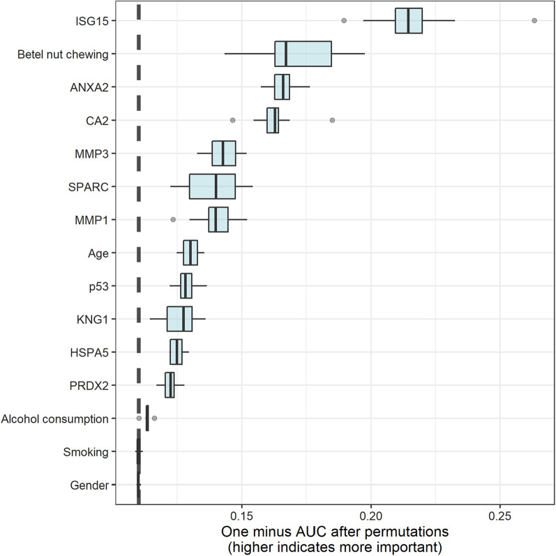
Variable importance, as identified by the stacking model, is provided in Fig. 4. Importance was calculated explicitly for each variable in the dataset, allowing variables to be ranked and compared with each other. Higher variable importance indicates that the variable contributes more to the AUC. The top five highest-ranking important variables in the stacking model were anti-ISG15, betel nut chewing, anti-ANXA2, anti-CA2, and anti-MMP3.
Fig. 4.
Variable importance in the stacking model
The AUC of the models developed through the stacking approach, using demographic and behavioral data together with age in the original format and logarithmic autoantibody levels, was 0.698 ± 0.060. Therefore, adding autoantibodies as biomarkers for identifying patients with a high risk of OCSS improved prediction performance by 13.9% (0.698 vs. 0.795).
Discussion
We used machine learning models to successfully predict high-risk cases of OSCC and evaluated the efficiency of salivary autoantibody levels to detect patients with a high risk of OSCC accurately and reliably.
Oral visual inspection is the main method for evaluating the risk of OSCC; however, differentiating OPMD and lesions with no risk of cancer progression through oral inspection remains challenging for clinicians [14]. Compared with the levels of other biomarkers, autoantibody levels are usually steady; therefore, they are easily detected using reagents available in the market [44]. In addition, autoantibodies exhibit an enduring response to tumor-associated antigens [45]. Therefore, tumor-associated autoantibodies are clinically useful and can serve as biomarkers for OSCC screening. We demonstrated that evaluating salivary autoantibody levels can help physicians identify the risk of OSCC early. Our results revealed that the salivary levels of CA2 [46], HSPA5 [47], ISG15 [48], KNG1 [49], MMP1 [50], MMP3 [51, 52], p53 [53], PRDX2 [54], and SPARC [55] were significantly elevated in the high-risk group compared with the low-risk group, a result similar to those of previous studies (Table 1). The data revealed that the elevated salivary autoantibody levels were related to OSCC progression. Moreover, four of the five most important variables in the stacking model were autoantibodies (Fig. 4). Thus, using salivary autoantibody levels to assist OSCC screening is a promising strategy.
Although salivary autoantibody levels exhibited strong performance in detecting high-risk OSCC cases, traditional risk factors, such as betel nut chewing, are still crucial for estimating the risk of OSCC (Fig. 4). In previous studies, the majority of patients with OSCC had habitual behaviors, such as betel nut chewing and drinking alcohol [56–58]. Adding salivary autoantibody levels to these traditional risk factors increased the model capacities.
The salivary samples were collected from 2008 to 2012, and the autoantibody detection was performed in 2018. Previous studies revealed a statistical decrease in concentration of salivary immunoglobulin A and hormones as storage time increased [59, 60]. However, before long-term storage, the salivary samples used in this study were centrifuged, treated with a protease inhibitor mixture, and stored at a − 80 °C freezer to avoid protein degradation. This strict protocol can ensure the quality of saliva samples [61]. More importantly, detection of salivary anti-p53 levels has been carried out in our previous study in 2014 [24]. Part of saliva samples used in 2014 is the same to that in the present study. For each identical patient or healthy control, salivary level of anti-p53 in the present study is similar to that acquired in 2014, indicating that the salivary properties might be appropriately preserved in terms of salivary immunoglobulin.
There are several limitations to this study. First, we recruited participants from a single institution in Taiwan; therefore, external validation is not available and our results may not be generalizable to other regions. Although we performed an internal evaluation with 100 randomly split training–test datasets to minimize model evaluation bias, multiple-center studies may be required to increase model generalizability. Second, we only included a small number of cases; therefore, future studies should include a large sample size collected from multiple centers. The application of autoantibodies as biomarkers should be validated by performing a cohort study to evaluate the efficiency of autoantibodies in diagnosing patients with early-stage OSCC. Finally, we evaluated the levels of only 10 salivary autoantibodies [23]; however, other potential salivary protein biomarkers should be applied to detect high-risk OSCC cases [49]. Moreover, other factors, such as human papillomavirus infection, ultraviolet light, poor nutrition, and genetic syndromes [62], may increase the risk of OSCC and should be included in future studies.
Conclusion
We successfully established a prediction model for high-risk cases of OSCC. Combining the online calculator, which was developed on the basis of the proposed model, with a common clinical visual exam would help in the early diagnosis of OSCC, thereby reducing the disease morbidity and mortality rates.
Supplementary Information
Additional file 1. The supporting information including Supplemental Tables S1–S5 and Supplemental Figures S1–S3.
Acknowledgements
This manuscript was edited by Wallace Academic Editing.
Abbreviations
- OSCC
Oral cavity squamous cell carcinoma
- OPMDs
Oral potentially malignant disorder
- COE
Conventional oral examination
- MFI
Mean fluorescence intensity
- LR
Logistic regression
- RF
Random forest
- SVM
Support vector machine
- XGBoost
EXtreme Gradient Boosting
- AUC
Area under the receiver operating characteristic curve
- rANOVA
Repeated-measures ANOVA
Author contributions
YJT and YC had full access to all the data in the study and takes responsibility for the integrity of the data and the accuracy of the data analysis. YJT and YCW analyzed/interpreted the data and performed experiments. YJT and YCW designed the study and wrote the paper. PCH and CCW reviewed/edited the manuscript for important intellectual content and provided administrative, technical, or material support. YJT and CCW obtained finding and supervised the study. The sponsors had no role in the design and conduct of the study; collection, management, analysis, and interpretation of the data; preparation, review, or approval of the manuscript; and decision to submit the manuscript for publication. All authors read and approved the final manuscript.
Funding
Dr. Tseng was funded by grants from Ministry of Science and Technology, Taiwan (MOST 111-2636-E-A49-014 and 111-2628-E-A49-026-MY3). Dr. Wu was funded by grants from Ministry of Science and Technology, Taiwan (MOST 108-2320-B-182-030-MY3) and from Chang Gung Memorial Hospital (BMRPC77).
Availability of data and materials
Authors' data use agreement for the dataset does not permit public posting of this patient information. The R codes for generating analysis results were available at: https://doi.org/10.57770/QTYQZS.
Declarations
Ethics approval and consent to participate
All experiments were performed in accordance with the guidelines and regulations and all participants signed the informed consent form before screening and permitting the use of saliva samples collected before treatment. Guidelines, regulations and informed consent form are approved by the Institutional Review Board of Chi-Mei Medical Center (No. 10012-L02).
Consent for publication
Not applicable.
Competing interests
The authors declare that they have no competing of interest.
Footnotes
Publisher's Note
Springer Nature remains neutral with regard to jurisdictional claims in published maps and institutional affiliations.
References
- 1.Siegel RL, Miller KD, Jemal A. Cancer statistics, 2020. CA Cancer J Clin. 2020;70:7–30. doi: 10.3322/caac.21590. [DOI] [PubMed] [Google Scholar]
- 2.Warnakulasuriya S, Kujan O, Aguirre-Urizar JM, Bagan JV, González-Moles MÁ, Kerr AR, et al. Oral potentially malignant disorders: a consensus report from an international seminar on nomenclature and classification, convened by the WHO Collaborating Centre for Oral Cancer. Oral Dis. 2021;27:1862–1880. doi: 10.1111/odi.13704. [DOI] [PubMed] [Google Scholar]
- 3.Miranda-Filho A, Bray F. Global patterns and trends in cancers of the lip, tongue and mouth. Oral Oncol. 2020;102:104551. doi: 10.1016/j.oraloncology.2019.104551. [DOI] [PubMed] [Google Scholar]
- 4.Kuruvilla J, Nayar K. Distribution pattern and its correlation for oral cancer rate and human development rank for countries: an ecological approach. Contemp Clin Dent. 2021;12:9–13. doi: 10.4103/ccd.ccd_1_20. [DOI] [PMC free article] [PubMed] [Google Scholar]
- 5.Johnson NW, Jayasekara P, Amarasinghe AA, Hemantha K. Squamous cell carcinoma and precursor lesions of the oral cavity: Epidemiology and aetiology. Periodontol. 2000 doi: 10.1111/j.1600-0757.2011.00401.x. [DOI] [PubMed] [Google Scholar]
- 6.Warnakulasuriya S, Diz Dios P, Lanfranchi H, Jacobson J, Hua H, Rapidis A. Understanding gaps in the oral cancer continuum and developing strategies to improve outcomes. In: WHO, Global Oral Cancer Forum—Working Group. 2016.
- 7.Warnakulasuriya S. Global epidemiology of oral and oropharyngeal cancer. Oral Oncol. 2009;45:309–316. doi: 10.1016/j.oraloncology.2008.06.002. [DOI] [PubMed] [Google Scholar]
- 8.Taiwan Cancer Registry Annual Report (2018). Available online: https://www.hpa.gov.tw/Pages/ashx/File.ashx?FilePath=~/File/Attach/13498/File_15611.pdf. 2020.
- 9.Chen YJ, Chang JTC, Liao CT, Wang HM, Yen TC, Chiu CC, et al. Head and neck cancer in the betel quid chewing area: Recent advances in molecular carcinogenesis. Cancer Sci. 2008;99:1507–1514. doi: 10.1111/j.1349-7006.2008.00863.x. [DOI] [PMC free article] [PubMed] [Google Scholar]
- 10.Liu SY, Lu CL, Chiou CT, Yen CY, Liaw GA, Chen YC, et al. Surgical outcomes and prognostic factors of oral cancer associated with betel quid chewing and tobacco smoking in Taiwan. Oral Oncol. 2010;46:276–282. doi: 10.1016/j.oraloncology.2010.01.008. [DOI] [PubMed] [Google Scholar]
- 11.Warnakulasuriya S, Dios PD, Lanfranchi H, Jacobson JJ, Honghua, Rapidis A. Global Oral Cancer Forum (Group 2) Understanding gaps in the oral cancer continuum and developing strategies to improve outcomes. 2016.
- 12.Mortazavi H, Baharvand M, Mehdipour M. Oral potentially malignant disorders: an overview of more than 20 entities. J Dent Res Dent Clin Dent Prospect. 2014;8:6–14. doi: 10.5681/joddd.2014.002. [DOI] [PMC free article] [PubMed] [Google Scholar]
- 13.George A, Sreenivasan BS, Sunil S, Varghese SS, Thomas J, Gopakumar D, et al. Potentially malignant disorders of oral cavity. Oral Maxillofac Pathol J. 2011;2:95–100. [Google Scholar]
- 14.van der Waal I. Potentially malignant disorders of the oral and oropharyngeal mucosa; terminology, classification and present concepts of management. Oral Oncol. 2009;45:317–323. doi: 10.1016/j.oraloncology.2008.05.016. [DOI] [PubMed] [Google Scholar]
- 15.Zaenker P, Ziman MR. Serologic autoantibodies as diagnostic cancer biomarkers: a review. Cancer Epidemiol Biomark Prev. 2013;22:2161–2181. doi: 10.1158/1055-9965.EPI-13-0621. [DOI] [PubMed] [Google Scholar]
- 16.Qiu J, Keyser B, Lin ZT, Wu T. Autoantibodies as potential biomarkers in breast cancer. Biosensors. 2018 doi: 10.3390/bios8030067. [DOI] [PMC free article] [PubMed] [Google Scholar]
- 17.Chapman CJ, Thorpe AJ, Murray A, Parsy-Kowalska CB, Allen J, Stafford KM, et al. Immunobiomarkers in small cell lung cancer: potential early cancer signals. Clin Cancer Res. 2011;17:1474–1480. doi: 10.1158/1078-0432.CCR-10-1363. [DOI] [PubMed] [Google Scholar]
- 18.Scanlan MJ, Chen Y-T, Williamson B, Gure AO, Stockert E, Gordan JD, et al. Characterization of human colon cancer antigens recognized by autologous antibodies. Int J Cancer. 1998;76:652–658. doi: 10.1002/(SICI)1097-0215(19980529)76:5<652::AID-IJC7>3.0.CO;2-P. [DOI] [PubMed] [Google Scholar]
- 19.Smith EM, Rubenstein LM, Ritchie JM, Lee JH, Haugen TH, Hamsikova E, et al. Does pretreatment seropositivity to human papillomavirus have prognostic significance for head and neck cancers? Cancer Epidemiol Biomark Prev. 2008;17:2087–2096. doi: 10.1158/1055-9965.EPI-08-0054. [DOI] [PMC free article] [PubMed] [Google Scholar]
- 20.Tan C, Qian X, Guan Z, Yang B, Ge Y, Wang F, et al. Potential biomarkers for esophageal cancer. Springerplus. 2016;5:467. doi: 10.1186/s40064-016-2119-3. [DOI] [PMC free article] [PubMed] [Google Scholar]
- 21.Xu Y-W, Peng Y-H, Chen B, Wu Z-Y, Wu J-Y, Shen J-H, et al. Autoantibodies as potential biomarkers for the early detection of esophageal squamous cell carcinoma. Am J Gastroenterol. 2014;109:36–45. doi: 10.1038/ajg.2013.384. [DOI] [PMC free article] [PubMed] [Google Scholar]
- 22.Wright JL, Lange PH. Newer potential biomarkers in prostate cancer. Rev Urol. 2007;9:207–213. [PMC free article] [PubMed] [Google Scholar]
- 23.Yu J-S, Chen Y-T, Chiang W-F, Hsiao Y-C, Chu LJ, See L-C, et al. Saliva protein biomarkers to detect oral squamous cell carcinoma in a high-risk population in Taiwan. Proc Natl Acad Sci. 2016;113:11549–11554. doi: 10.1073/pnas.1612368113. [DOI] [PMC free article] [PubMed] [Google Scholar]
- 24.Wu CC, Chang YT, Chang KP, Liu YL, Liu HP, Lee IL, et al. Salivary auto-antibodies as noninvasive diagnostic markers of oral cavity squamous cell carcinoma. Cancer Epidemiol Biomark Prev. 2014;23:1569–1578. doi: 10.1158/1055-9965.EPI-13-1269. [DOI] [PubMed] [Google Scholar]
- 25.Bur AM, Shew M, New J. Artificial intelligence for the otolaryngologist: a state of the art review. Otolaryngol Head Neck Surg. 2019 doi: 10.1177/0194599819827507. [DOI] [PubMed] [Google Scholar]
- 26.Cruz JA, Wishart DS. Applications of machine learning in cancer prediction and prognosis. Cancer Inform. 2006;2:59–77. doi: 10.1177/117693510600200030. [DOI] [PMC free article] [PubMed] [Google Scholar]
- 27.Tseng Y-J, Wang H-Y, Lin T-W, Lu J-J, Hsieh C-H, Liao C-T. Development of a machine learning model for survival risk stratification of patients with advanced oral cancer. JAMA Netw Open. 2020;3:e2011768–e2011768. doi: 10.1001/jamanetworkopen.2020.11768. [DOI] [PMC free article] [PubMed] [Google Scholar]
- 28.Tseng YJ, Huang CE, Wen CN, Lai PY, Wu MH, Sun YC, et al. Predicting breast cancer metastasis by using serum biomarkers and clinicopathological data with machine learning technologies. Int J Med Inform. 2019;128:79–86. doi: 10.1016/j.ijmedinf.2019.05.003. [DOI] [PubMed] [Google Scholar]
- 29.Huang S, Yang J, Fong S, Zhao Q. Artificial intelligence in cancer diagnosis and prognosis: opportunities and challenges. Cancer Lett. 2020;471:61–71. doi: 10.1016/j.canlet.2019.12.007. [DOI] [PubMed] [Google Scholar]
- 30.Alabi RO, Youssef O, Pirinen M, Elmusrati M, Mäkitie AA, Leivo I, et al. Machine learning in oral squamous cell carcinoma: Current status, clinical concerns and prospects for future: a systematic review. Artif Intell Med. 2021;115:102060. doi: 10.1016/j.artmed.2021.102060. [DOI] [PubMed] [Google Scholar]
- 31.Collins GS, Reitsma JB, Altman DG, Moons KGM. Transparent reporting of a multivariable prediction model for individual prognosis or diagnosis (TRIPOD): The TRIPOD Statement. BMC Med. 2015;13:1. [DOI] [PMC free article] [PubMed]
- 32.Hung LC, Kung PT, Lung CH, Tsai MH, Liu SA, Chiu LT, et al. Assessment of the risk of oral cancer incidence in a high-risk population and establishment of a predictive model for oral cancer incidence using a population-based cohort in Taiwan. Int J Environ Res Public Health. 2020;17:665. doi: 10.3390/ijerph17020665. [DOI] [PMC free article] [PubMed] [Google Scholar]
- 33.Cramer JS. The origins of logistic regression. SSRN Electron J. 2005 doi: 10.2139/ssrn.360300. [DOI] [Google Scholar]
- 34.Cortes C, Vapnik V. Support-vector networks. Mach Learn. 1995;20:273–297. doi: 10.1007/BF00994018. [DOI] [Google Scholar]
- 35.Breiman L. Random forests. Mach Learn. 2001;45:5–32. doi: 10.1023/A:1010933404324. [DOI] [Google Scholar]
- 36.Chen T, Guestrin C. XGBoost. In: Proceedings of the 22nd ACM SIGKDD international conference on knowledge discovery and data mining. New York: ACM; 2016. p. 785–94.
- 37.Tibshirani R. Regression shrinkage and selection via the lasso. J Roy Stat Soc Ser B. 1996;58:267–288. [Google Scholar]
- 38.Kuhn M. Building predictive models in R Using the caret package. J Stat Softw. 2008;28:1–26. doi: 10.18637/jss.v028.i05. [DOI] [Google Scholar]
- 39.Kuhn M, Wickham H. Tidymodels: a collection of packages for modeling and machine learning using tidyverse principles. 2020.
- 40.Couch S, Kuhn M. stacks: Tidy model stacking. 2022.
- 41.Cabitza F, Campagner A. The need to separate the wheat from the chaff in medical informatics: Introducing a comprehensive checklist for the (self)-assessment of medical AI studies. Int J Med Inform. 2021;153:104510. doi: 10.1016/j.ijmedinf.2021.104510. [DOI] [PubMed] [Google Scholar]
- 42.Biecek P. DALEX: explainers for complex predictive models in R. J Mach Learn Res. 2018;19:1–5. [Google Scholar]
- 43.Maksymiuk S, Gosiewska A, Biecek P. Landscape of R packages for eXplainable Artificial Intelligence. 2020.
- 44.Anderson KS, Sibani S, Wallstrom G, Mendoza EA, Raphael J, Hainsworth E, et al. Protein microarray signature of autoantibody biomarkers for the early detection of breast cancer. J Proteome Res. 2011;10:85–96. doi: 10.1021/pr100686b. [DOI] [PMC free article] [PubMed] [Google Scholar]
- 45.Murphy MA, O’Leary JJ, Cahill DJ. Assessment of the humoral immune response to cancer. J Proteomics. 2012;75:4573–4579. doi: 10.1016/j.jprot.2012.01.021. [DOI] [PubMed] [Google Scholar]
- 46.Liu C-M, Lin Y-M, Yeh K-T, Chen M-K, Chang J-H, Chen C-J, et al. Expression of carbonic anhydrases I/II and the correlation to clinical aspects of oral squamous cell carcinoma analyzed using tissue microarray. J Oral Pathol Med. 2012;41:533–539. doi: 10.1111/j.1600-0714.2012.01135.x. [DOI] [PubMed] [Google Scholar]
- 47.Kang BH, Shu CW, Chao JK, Lee CH, Fu TY, Liou HH, et al. HSPD1 repressed E-cadherin expression to promote cell invasion and migration for poor prognosis in oral squamous cell carcinoma. Sci Rep. 2019 doi: 10.1038/s41598-019-45489-1. [DOI] [PMC free article] [PubMed] [Google Scholar]
- 48.Zhang Q, He Y, Nie M, Cai W. Roles of miR-138 and ISG15 in oral squamous cell carcinoma. Exp Ther Med. 2017;14:2329–2334. doi: 10.3892/etm.2017.4720. [DOI] [PMC free article] [PubMed] [Google Scholar]
- 49.Chen Y, Chiang W, Hsiao Y, Julie L, See L, Wu C, et al. Erratum: Saliva protein biomarkers to detect oral squamous cell carcinoma in a high-risk population in Taiwan (Proc Natl Acad Sci USA (2016) 113:41 (11549–11554) DOI: 10.1073/pnas.1612368113). Proc Natl Acad Sci U S A. 2016;113:E7139. [DOI] [PMC free article] [PubMed]
- 50.Chang YT, Chu LJ, Liu YC, Chen CJ, Wu SF, Chen CH, et al. Verification of saliva matrix metalloproteinase-1 as a strong diagnostic marker of oral cavity cancer. Cancers. 2020 doi: 10.3390/cancers12082273. [DOI] [PMC free article] [PubMed] [Google Scholar]
- 51.Stott-Miller M, Houck JR, Lohavanichbutr P, Méndez E, Upton MP, Futran ND, et al. Tumor and salivary matrix metalloproteinase levels are strong diagnostic markers of oral squamous cell carcinoma. Cancer Epidemiol Biomark Prev. 2011;20:2628–2636. doi: 10.1158/1055-9965.EPI-11-0503. [DOI] [PMC free article] [PubMed] [Google Scholar]
- 52.Tadbir AA, Purshahidi S, Ebrahimi H, Khademi B, Malekzadeh M, Mardani M, et al. Serum level of MMP-3 in patients with oral squamous cell carcinoma: lack of association with clinico-pathological features. Asian Pac J Cancer Prev. 2012;13:4545–4548. doi: 10.7314/APJCP.2012.13.9.4545. [DOI] [PubMed] [Google Scholar]
- 53.Shinohara S, Kikuchi M, Tona R, Kanazawa Y, Kishimoto I, Harada H, et al. Prognostic impact of p16 and p53 expression in oropharyngeal squamous cell carcinomas. Jpn J Clin Oncol. 2014 doi: 10.1093/jjco/hyt223. [DOI] [PubMed] [Google Scholar]
- 54.Chuerduangphui J, Ekalaksananan T, Heawchaiyaphum C, Vatanasapt P, Pientong C. Peroxiredoxin 2 is highly expressed in human oral squamous cell carcinoma cells and is upregulated by human papillomavirus oncoproteins and arecoline, promoting proliferation. PLoS ONE. 2020 doi: 10.1371/journal.pone.0242465. [DOI] [PMC free article] [PubMed] [Google Scholar]
- 55.Jing Y, Jin Y, Wang Y, Chen S, Zhang X, Song Y, et al. SPARC promotes the proliferation and metastasis of oral squamous cell carcinoma by PI3K/AKT/PDGFB/PDGFRβ axis. J Cell Physiol. 2019 doi: 10.1002/jcp.28205. [DOI] [PubMed] [Google Scholar]
- 56.Lo W-L, Kao S-Y, Chi L-Y, Wong Y-K, Chang RC-S. Outcomes of oral squamous cell carcinoma in Taiwan after surgical therapy: factors affecting survival. J Oral Maxillofac Surg. 2003;61:751–758. doi: 10.1016/S0278-2391(03)00149-6. [DOI] [PubMed] [Google Scholar]
- 57.Lee J-J, Jeng J-H, Wang H-M, Chang H-H, Chiang C-P, Kuo Y-S, et al. Univariate and multivariate analysis of prognostic significance of betel quid chewing in squamous cell carcinoma of buccal mucosa in Taiwan. J Surg Oncol. 2005;91:41–47. doi: 10.1002/jso.20276. [DOI] [PubMed] [Google Scholar]
- 58.Zuckerman A. IARC monographs on the evaluation of carcinogenic risks to humans. IARC Monogr Eval Carcinog Risks Hum. 2010;93:9–38. [Google Scholar]
- 59.Toone RJ, Peacock OJ, Smith AA, Thompson D, Drawer S, Cook C, et al. Measurement of steroid hormones in saliva: effects of sample storage condition. Scand J Clin Lab Invest. 2013;73:615–621. doi: 10.3109/00365513.2013.835862. [DOI] [PubMed] [Google Scholar]
- 60.Presser E, Simuyandi M, Brown J. The effects of storage time and temperature on recovery of salivary secretory immunoglobulin A. Am J Hum Biol. 2014;26:417–420. doi: 10.1002/ajhb.22525. [DOI] [PubMed] [Google Scholar]
- 61.Wu CC, Chu HW, Hsu CW, Chang KP, Liu HP. Saliva proteome profiling reveals potential salivary biomarkers for detection of oral cavity squamous cell carcinoma. Proteomics. 2015;15:3394–3404. doi: 10.1002/pmic.201500157. [DOI] [PubMed] [Google Scholar]
- 62.Conway DI, Purkayastha M, Chestnutt IG. The changing epidemiology of oral cancer: definitions, trends, and risk factors. Br Dent J. 2018;225:867–873. doi: 10.1038/sj.bdj.2018.922. [DOI] [PubMed] [Google Scholar]
Associated Data
This section collects any data citations, data availability statements, or supplementary materials included in this article.
Supplementary Materials
Additional file 1. The supporting information including Supplemental Tables S1–S5 and Supplemental Figures S1–S3.
Data Availability Statement
Authors' data use agreement for the dataset does not permit public posting of this patient information. The R codes for generating analysis results were available at: https://doi.org/10.57770/QTYQZS.


