Abstract
Pleurotus species isolated in vitro were studied to determine the effect of different media on their production of secondary metabolites, antimicrobial, and antioxidant activity. The different metabolites among Pleurotus samples covered a total of 58 pathways. Comparisons were made between the metabolic profiles of Pleurotus spp. mycelia grown in two substrates: Potato-dextrose-agar-PDA, used as control (S1), and PDA enriched with 0.5 % of wheat straw (S2). The main finding was that the metabolic pathways are strongly influenced by the chemical composition of the growth substrate. The antibacterial effects were particularly evident against Escherichia coli, whereas Arthroderma curreyi (CCF 5207) and Trichophyton rubrum (CCF 4933) were the dermatophytes more sensitive to the mushroom extracts. The present study supports more in-depth investigations, aimed at evaluating the influence of growth substrate on Pleurotus spp. antimicrobial and antioxidant properties.
Keywords: Pleurotus species, metabolomics, antimicrobial effect, phenolic compounds
1. Introduction
The genus Pleurotus (Fr.) P. Kumm. (Pleurotaceae, Basidiomycota) includes some of the main cultivated edible mushrooms in the world due to their gastronomic, nutritional, and medicinal properties, such as anti-inflammatory [1], antioxidant [2,3], antidiabetic [4], antitumor [5], and immunomodulating [6]. Due to their ability to improve protein content and quality, as well as the valuable health benefits of myco-chemicals or bioactive components present in these mushrooms [7,8,9], Pleurotus spp. can also be used to supplement different processed products such as bread and dairy foods. Pleurotus genus includes white rot fungi distributed all over the globe [10] due to their capacity to colonize and use accessible lignocellulosic materials and wastes, that have been considered suitable for bioconversion of agro-wastes into food and feed in developing countries [11,12,13,14,15]. They play an important role in managing organic waste whose disposal is problematic, e.g., those deriving from olive-oil production (i.e., olive pruning residues, olive mill wastes, and olive leaves) and wineries (e.g., grape marc) [16,17]
Compared to the most commonly cultivated mushrooms, such as Agaricus bisporus, Pleurotus species have the advantage of very simple cultivation; in fact, it could be sufficient to use a non-composted straw, chopped and soaked in water [18]. Based on the CABI Index Fungorum (http://www.indexfungorum.org/names/Names.asp; accessed 6 February 2022), the genus Pleurotus currently consists of about 217 accepted species all of which are edible and appreciated for their taste, aroma, and texture, as well as the health-enhancing bioactive potentials [19]. P. ostreatus is the most popular among Pleurotus mushrooms. It is a cosmopolitan species growing on dead wood of many broad-leaved and coniferous trees. Its cultivation is widespread throughout the world on a remarkable spectrum of lignocellulosic substrates such as maize straw, maize cob, palm kernel cake, sawdust, spent grain, rice bran, cereal grasses, sugarcane bagasse, coffee residues, coir waste, and cardboard industrial waste, etc. [15].
Much of the research on Pleurotus species has to date focused on the potential of various lignocellulosic by-products (e.g., cereal grasses, sugarcane bagasse, coffee residues, coir waste, and cardboard industrial waste) to support satisfactory mushroom yields [20,21,22], extraction of secondary metabolites (alkaloids, flavonoids, betalain among others) and pharmaceutical, biotechnological and food applications [23,24].
However, there is some research concerning the composition of fruiting bodies cultivated on various agro-wastes, and no data exist on the effect of cultural media on the mycelium content in bioactive compounds and their functional properties. The growth of mycelium is strongly influenced by many factors such as culture media, temperature, carbon and nitrogen sources, grain sources, and sources of lignocellulosic substrate [25,26]. Manipulating mycelium growth conditions is a common strategy used by pharmaceutical companies to improve the diversity of secondary metabolites of therapeutic interest [27].
The main objectives of the present work were (a) to investigate the suitability of two different cultural media for mycelium growth, and (b) to investigate how the culture medium affects the antimicrobial and antioxidant activity in the mycelial stage. Mass spectrometry (MS)-based metabolomic profiles coupled with multivariate statistical analysis will help determine the effects of different culture media as a function of metabolomic and transcriptomic disparity in Pleurotus spp. mycelia [28]. Additionally, the quantitative analysis of phenolic compounds has been carried out, as well.
2. Materials and Methods
2.1. Mushroom Material
The fruiting bodies of P. columbinus, P. ostreatus, P. nebrodensis, and P. eryngii species (P. eryngii var. thapsiae, P. eryngii var. ferulae, P. eryngii var. elaeoselini) were collected on different dates and in different locations (Table 1) and immediately transferred to the laboratory to obtain pure mycelial cultures.
Table 1.
Sample ID and data relating to the location and date of collection of Pleurotus species studied.
| Sample ID | Species | Locality | Date |
|---|---|---|---|
| PC1 | Pleurotus columbinus Quél. | Cascata delle Marmore (TR) | Apr 2019 |
| PE1 | Pleurotus eryngii var. thapsiae Venturella, Zervakis & Saitta | Madonie (Piano Zucchi) Palermo | Oct 2019 |
| PO1 | Pleurotus ostreatus (Jacq.) P. Kumm. | Cima di Tuoro (PG) | Sep 2017 |
| PN | Pleurotus nebrodensis (Inzenga) Quél. | Monte Malletto (Etna, CT) | May 2021 |
| PE2 | Pleurotus eryngii (DC.) Quél. | Etna Sciare S. Venera Maletto | Nov 2021 |
| PE3 | Pleurotus eryngii var. ferulae (Lanzi) Sacc. | Isola Polvese (PG) | Jan 2017 |
| PO2 | Pleurotus ostreatus (Jacq.) P. Kumm. | Castel Porziano (RM) | Nov 2018 |
| PO3 | Pleurotus ostreatus (Jacq.) P. Kumm. | Castel Porziano (RM) | Nov 2018 |
| PO4 | Pleurotus ostreatus (Jacq.) P. Kumm. | Rivotorto (PG) | Oct 2020 |
| PE4 | Pleurotus eryngii var. elaeoselini Venturella, Zervakis & La Rocca | Vallone dei Sieli, Motta sant’Anastasia (Catania) | Nov 2017 |
| PO5 | Pleurotus ostreatus (Jacq.) P. Kumm. | Monte Peglia (S. Venanzo, TR) | Nov 2017 |
| PO6 | Pleurotus ostreatus (Jacq.) P. Kumm. | Monte Subasio (PG) | May 2017 |
Briefly, for the isolation of mycelia, context pieces (5 × 5 × 5 mm) were excised aseptically from the context of fresh Basidiomycota and transferred to Petri dishes containing Rose Bengal Chloramphenicol agar (Sigma-Aldrich, Milan, Italy) under sterile conditions. Petri dishes inoculated with 3–4 explants were then incubated at 25 °C in the dark for 7 days.
Basidiomata identification was carried out by morphological and molecular analysis [29,30] in consideration of the available data on the occurrence of Pleurotus species in Italy [31].
2.2. Molecular Identification
To test the morphological identification (Table 1), the ITS region of the nrDNA was used as a fingerprint marker, as suggested for the wide majority of fungi [32] and successfully applied in previous works by the authors themselves [33,34,35]
The total genomic DNA was extracted using the ZR Fungal/Bacterial DNA Kit (Euroclone S.p.A., Milan, Italy). The genomic DNA quality and quantity were evaluated with BIORAD (Milan, Italy) model 200/2.0 Power Supply gel electrophoresis [0.8% agarose gel in 1× TBE buffer (89 mM Tris, 89 mM boric acid, 2 mM EDTA, pH 7.6)] in the presence of SafeView Nucleic Acid Stain (NBS Biologicals, Huntingdon, UK) and a MassRuler DNA Ladder Mix (Thermo Scientific, Vilnius, Lithuania), and visualized with Safe ImagerTM 2.0 Blue Light Trans illuminator Invitrogen (Parma, Italy). DNA samples were subsequently diluted with up to 10 μg/μL nuclease-free water before PCR amplification. The internal transcribed spacer (ITS) region of the nrDNA was amplified by ITS1F and ITS4 primers. SimpliAmp Thermal Cycler Applied Biosystems (Monza, Italy) was programmed as follows: one cycle of denaturation at 95 °C for 2.5 min; 35 cycles of denaturation at 95 °C for 20 s, annealing at 55 °C for 20 s and extension at 72 °C for 45 s; one final extension cycle at 72 °C for 7 min. Electrophoresis of PCR amplicons was carried out on 1.2% agarose gel as described above. The PCR amplified ITS fragment was purified using the ExoSapIT PCR Cleanup reagent (Thermo Fisher, Monza, Italy) and then sequenced by Macrogen Europe (Netherlands) (Table 2).
Table 2.
GenBank sequences and identity percentages with different samples of Pleurotus species studied.
| Species | Sample ID | Base Pair | Correspondence with Genbank Seq. | % Identity | Accession no. |
|---|---|---|---|---|---|
| Pleurotus columbinus | PC1 | 621 | Pleurotus columbinus | 100 | MG282482.1 |
| Pleurotus eryngii var. thapsiae | PE1 | 345 | Pleurotus eryngii | 99.42 | MH517527.1 |
| Pleurotus ostreatus | PO1 | 568 | Pleurotus ostreatus | 100 | MT644908.1 |
| Pleurotus nebrodensis | PN | 616 | Pleurotus nebrodensis | 99.51 | KF743821.1 |
| Pleurotus eryngii var. ferulae | PE3 | 511 | Pleurotus eryngii var. ferulae | 99.42 | AB286153.1 |
| Pleurotus ostreatus | PO2 | 641 | Pleurotus pulmonarius | 100 | MN239983.1 |
| Pleurotus ostreatus | PO3 | 670 | Pleurotus ostreatus | 100 | GU186818.1 |
| Pleurotus eryngii var. elaeoselini | PE4 | 636 | Pleurotus eryngii | 100 | OPE241308.1 |
| Pleurotus ostreatus | PO5 | 613 | Pleurotus ostreatus | 99.19 | MT644908.1 |
| Pleurotus ostreatus | PO6 | 612 | Pleurotus pulmonarius | 99.19 | MH810334.1 |
2.3. Preparation of Samples
The in vitro culture of Pleurotus spp. was performed in the following solid media: (1) Potato-dextrose-agar-PDA, used as control (S1), and (2) PDA enriched with 0.5% of wheat straw (S2). Each medium in flasks was autoclaved at 121 °C for 20 min and subsequently dispensed into 15 100 mm Petri dishes. The mycelium discs (1 cm diameter) of each Pleurotus mushroom were placed in Petri dishes containing each culture medium (20 mL) under aseptic condition and incubated at 25 °C in the darkness. After 15 days of growth (when mycelium reached maximum radial growth in the PDA medium) the mycelium was recovered from the medium. All samples, realized in duplicate, were lyophilized (FreeZone 4.5 model 7750031, Labconco, Kansas, MO, USA), quantified, and reduced to a fine-dried powder (Supplementary Material: Tables S1 and S2). Preparation of mycelia extract: the lyophilized mycelia were extracted for 30 min with distilled and deionized water under ultrasonic agitation.
2.4. Untargeted LC-MS/MS-Based Metabolomics and Statistical Analysis
Untargeted LC/MS QTOF analysis was performed using a 1260 Infinity II LC System coupled with an Agilent 6530 Q-TOF spectrometer (Agilent Technologies, Santa Clara, CA USA). The LC consists of a quaternary pump, a thermostated column compartment, and an autosampler. Separation was carried out on an Agilent InfinityLab Poroshell 120 HILIC-Z, 2.1 × 150 mm, 2.7 µm at 25 °C, and 0.25 mL/min flow. The mobile phase consisted of a mixture of water (A) and water/ACN 15:85 (B) both containing a concentration of 10 mM ammonium acetate. Gradient was: time 0–3 min isocratic at A 2%, B 98%; time from 3 to 11 min: linear-gradient to A 30%, B 70; time 11–12 min linear gradient to A 60%, B 40%; time from 12 to 16 min: linear-gradient to A 95%, B 5%; time 16–18 min isocratic at A 95%, B 5%; time 18 min: stop run.
Spectrometric data were acquired in the 40–1700 m/z range both in negative and positive polarity. The Agilent JetStream source operated as follows: Gas Temp (N2) 200 °C, Drying Gas 10 L/min, Nebulizer 50 psi, Sheath Gas temp: 300 °C at 12 L/min.
Raw data were processed using MS-DIAL software (4.48) [36] to perform peak-picking, alignment, and peak integration. The MS signal threshold was set at 1000 counts. In the end, a data matrix was obtained reporting the accurate mass and area of each peak revealed in each sample analyzed.
The putative annotation of metabolites and the prediction of metabolic pathways was performed using the mummichog algorithm [37], implemented in the ‘MS Peaks to Pathways’ module of Metaboanalyst 5.0 [38]. It considers any possible adducts and different ionic polarities and classifies the peaks annotated on the basis of the t-test. In this case, the list of putative compounds was mapped onto the KEGG library of Saccaromices cerevisiae. ANOVA and Functional Meta-Analysis were also performed with MetaboAnalyst. For statistical analysis, samples were normalized by the median, followed by pareto scaling.
2.5. HPLC-DAD-MS Determination of Phenolic Compounds
The HPLC apparatus consisted of two PU-2080 PLUS chromatographic pumps, a DG-2080-54 line degasser, a mix-2080-32 mixer, UV, diode array (DAD) and detectors, a mass spectrometer (MS) detector (expression compact mass spectrometer), Advion, Ithaca, NY 14850, USA), an AS-2057 PLUS autosampler, and a CO-2060 PLUS column thermostat (all from Jasco, Tokyo, Japan). Integration was performed by ChromNAV2 Chromatography software. Before the injection in the HPLC apparatus, the extracts were centrifuged at 3500× g for 15 min, and the supernatant was diluted to 20 mg/mL. The extracts were analyzed for phenol quantitative determination using a reversed-phase HPLC-DAD-MS in gradient elution mode (Table 3). The separation was conducted within 60 mins of the chromatographic run, starting from the following separation conditions: 95% water with 0.1% formic acid, and 5% methanol with 0.1% formic acid (Table 4). The separation was performed on an Infinity lab Poroshell 120-SB reverse phase column (C18, 150 × 4.6 mm i.d., 2.7 μm) (Agilent, Santa Clara, CA, USA). The column temperature was set at 30 °C. Quantitative determination of phenolic compounds was performed via a DAD detector, at 254 nm. Quantification was done through 7-point calibration curves, with linearity coefficients (R2) > 0.999, in the concentration range of 2–140 µg/mL. The limits of detection were lower than 1 µg/mL for all assayed analytes. The area under the curve from HPLC chromatograms was used to quantify the analyte concentrations in the extracts. The extracts were also qualitatively analyzed with an MS detector in negative ion mode. MS signal identification was realized through comparison with a standard solution and MS spectra present in the MassBank Europe database.
Table 3.
Gradient elution conditions of the HPLC-DAD analyses for the identification and quantification of polyphenolic compounds.
| Time (Min.) | Composition A% (Water+Formic Acid 0.1%) | Flow (mL/min) |
|---|---|---|
| 1 | 97 | 0.6 |
| 5 | 77 | 0.6 |
| 12 | 73 | 0.6 |
| 18 | 57 | 0.6 |
| 25 | 52 | 0.6 |
| 32 | 50 | 0.6 |
| 34 | 50 | 0.6 |
| 37 | 35 | 0.6 |
| 40 | 5 | 0.6 |
| 47 | 5 | 0.6 |
| 48 | 97 | 0.6 |
| 60 | 97 | 0.6 |
Table 4.
Phenolic compounds analyzed.
| Peak Name | Retention Time | |
|---|---|---|
| 1 | Gallic acid | 8.80 |
| 2 | 3-Hydroxytyrosol | 11.71 |
| 3 | Caftaric acid | 12.93 |
| 4 | Catechin | 14.80 |
| 5 | 4-Hydroxybenzoic acid | 16.20 |
| 6 | Loganic acid | 16.60 |
| 7 | Chlorogenic acid | 16.81 |
| 8 | Vanillic acid | 18.60 |
| 9 | Caffeic acid | 19.00 |
| 10 | Epicatechin | 19.41 |
| 11 | Syringic acid | 20.05 |
| 12 | p-Coumaric acid | 23.06 |
| 13 | t-Ferulic acid | 24.00 |
| 14 | Benzoic acid | 26.38 |
| 15 | Hyperoside | 26.92 |
| 16 | Rutin | 27.16 |
| 17 | Isoquercetin | 27.29 |
| 18 | Resveratrol | 27.70 |
| 19 | Rosmarinic acid | 28.53 |
| 20 | t-Cinnamic acid | 34.39 |
| 21 | Quercetin | 35.89 |
| 22 | Hesperetin | 39.38 |
| 23 | Kaempferol | 41.74 |
| 24 | Carvacrol | 44.69 |
| 25 | Thymol | 44.92 |
| 26 | Flavone | 45.60 |
| 27 | 3-Hydroxyflavone | 46.05 |
| 28 | Emodin | 47.70 |
2.6. Scavenging Effects
The scavenging effect of mushroom extracts on DPPH and ABTS radicals was evaluated as previously reported [33].
2.7. Antimicrobial Effects
The in vitro antimicrobial activity of extracts was assessed against the following Gram-negative and Gram-positive bacterial strains: Escherichia coli (ATCC 10536), E. coli (PeruMycA 2), E. coli (PeruMycA 3), Bacillus cereus (PeruMycA 4), B. subtilis (PeruMyc 6), Salmonella typhy (PeruMyc 7), Pseudomonas aeruginosa (ATCC 15442), and Staphylococcus aureus (ATCC 6538). Furthermore, the same extracts were assayed for the antifungal assays against different yeasts, dermatophyte, and fungal pool species: Candida albicans (YEPGA 6183), C. tropicalis (YEPGA 6184), C. albicans (YEPGA 6379), C. parapsilopsis (YEPGA 6551), Arthroderma crocatum (CCF 5300), A. curreyi (CCF 5207), A. gypseum (CCF 6261), A. quadrifidum (CCF 5792), A. insingulare (CCF 5417), A. quadrifidum (CCF 5792), Trichophyton mentagrophytes (CCF 4823), T. mentagrophytes (CCF 5930), T. rubrum (CCF 4933), and T. tonsurans (CCF 4834). Details are reported in our previous paper [2].
3. Results and Discussion
3.1. Mushroom Identification
The exact characterization and identification of medicinal mushrooms were fundamental for exploiting their full potential in the food and pharmaceutical industries [18].
The morphological characteristics of Pleurotus spp. (Table S1) fruiting bodies corresponded to those reported in the literature [29].
The taxonomic affiliation of the mushroom strains was performed by targeting the ITS region of the ribosomal DNA. Additionally, a BLAST search confirmed that our samples belong to P. columbinus, P. eryngii var. thapsiae, P. ostreatus, P. nebrodensis, P. eryngii var. ferulae, and P. eryngii var. elaeoselini, as it showed a close match with deposited sequences of these species.
3.2. Untargeted LC-MS/MS-Based Metabolomics
In this study, the metabolomic profile of Pleurotus spp. was evaluated through mass spectrometry ultra-performance liquid chromatography-mass spectrometry (UHPLC)-QTOF method. The different metabolites in all Pleurotus samples covered a total of 58 pathways, including biotin metabolism, pantothenate and CoA biosynthesis, tryptophan metabolism, arginine biosynthesis, valine, leucine and isoleucine degradation, glutathione metabolism, one carbon pool by folate, vitamin B6 metabolism, sulfur metabolism, and riboflavin metabolism (Table 5).
Table 5.
Regulated (+) or unregulated (-) metabolic pathway between Pleurotus samples as an effect of the different growth substrates.
| ID Sample | PC1 | PE1 | PO1 | PN | PE2 | PE3 | PO2 | PO3 | PO4 | PE4 | PO5 | PO6 | KEGG Pathway Map |
|---|---|---|---|---|---|---|---|---|---|---|---|---|---|
| Metabolic Pathway * | |||||||||||||
| Aminoacid metabolism | |||||||||||||
| Valine, leucine, and isoleucine biosynthesis | + | - | - | - | + | + | + | - | - | + | + | - | map00290 |
| Glycine, serine, and threonine metabolism | + | - | - | - | + | - | - | - | + | + | - | - | map00260 |
| Tyrosine metabolism | - | - | + | + | - | - | - | - | - | - | - | - | map00350 |
| Tryptophan metabolism | - | - | - | + | + | + | - | + | - | + | - | + | map00380 |
| Arginine biosynthesis | + | + | + | + | + | + | - | + | - | + | - | + | map00220 |
| Valine, leucine and isoleucine degradation | - | - | - | - | + | + | + | + | - | + | + | - | map00280 |
| Lysine biosynthesis | - | - | - | - | + | - | + | + | - | + | + | - | map00300 |
| Phenylalanine metabolism | - | - | + | + | + | + | - | - | - | + | - | + | map00360 |
| Thiamine metabolism | - | + | - | + | + | + | - | + | - | + | - | - | map00730 |
| Histidine metabolism | - | - | - | + | + | + | - | - | + | + | - | + | map00340 |
| Cysteine and methionine metabolism | + | + | - | - | + | + | - | + | - | + | - | + | map00270 |
| Phenylalanine, tyrosine, and tryptophan biosynthesis | - | + | - | - | + | + | - | - | - | + | - | + | map00400 |
| Arginine and proline metabolism | + | + | + | - | + | + | + | + | + | + | + | + | map00330 |
| Glutathione metabolism | + | - | + | - | + | + | - | - | + | + | - | - | map00480 |
| beta-Alanine metabolism | - | - | + | - | + | + | - | - | + | - | - | - | map00410 |
| Lysine degradation | - | - | - | - | + | - | + | + | - | + | - | - | map00310 |
| Alanine, aspartate and glutamate metabolism | + | - | - | - | - | + | - | + | + | + | - | + | map00250 |
| Amino sugar and nucleotide sugar metabolism | |||||||||||||
| Amino sugar and nucleotide sugar metabolism | - | + | - | - | + | - | - | - | - | + | - | - | map00520 |
| Glycolysis or Gluconeogenesis | - | + | - | - | + | + | - | - | - | - | - | - | map00010 |
| Pentose phosphate pathway | - | - | + | - | + | - | - | - | - | + | + | - | map00030 |
| Fructose and mannose metabolism | - | - | - | - | + | + | - | - | - | - | - | - | map00051 |
| Galactose metabolism | - | - | - | - | + | - | - | - | - | + | - | - | map00052 |
| Pentose and glucuronate interconversions | - | - | - | - | + | - | - | - | - | - | - | - | map00040 |
| N-Glycan biosynthesis | - | - | - | - | + | - | - | - | - | - | - | - | map00510 |
| Starch and sucrose metabolism | - | - | - | - | + | - | - | - | - | + | - | - | map00500 |
| Nucleotide metabolism | |||||||||||||
| Purine metabolism | + | + | - | - | + | + | - | - | + | + | - | + | map00230 |
| Pyrimidine metabolism | + | - | - | + | + | + | - | + | + | + | - | + | map00240 |
| Aminoacyl-tRNA biosynthesis | + | - | - | + | + | + | - | - | + | + | - | + | map00970 |
| Lipid metabolism | |||||||||||||
| Inositol phosphate metabolism | - | - | - | - | + | - | - | - | - | - | - | map00562 | |
| Phosphatidylinositol signaling system | - | - | - | - | + | - | - | - | - | - | - | - | map04070 |
| Glycerophospholipid metabolism | - | - | - | - | + | + | - | - | - | + | - | - | map00564 |
| Sphingolipid metabolism | - | - | - | - | + | + | - | - | - | + | - | - | map00600 |
| Vitamin metabolism | |||||||||||||
| Nicotinate and nicotinamide metabolism | - | - | + | - | + | - | - | - | + | + | - | - | map00760 |
| Cyanoamino acid metabolism | - | - | - | - | - | - | - | - | - | + | - | - | map00460 |
| Riboflavin metabolism | - | - | - | - | - | - | - | - | - | + | - | - | map00740 |
| Vitamin B6 metabolism | - | - | - | - | + | - | - | - | - | + | - | + | map00750 |
| Pantothenate and CoA biosynthesis | - | - | + | - | + | + | + | - | - | + | + | + | map00770 |
| Folate biosynthesis | + | + | - | - | + | + | - | - | + | + | - | - | map00790 |
| One carbon pool by folate | + | - | + | - | + | + | - | - | + | + | - | - | map00670 |
| Fatty acid metabolism | |||||||||||||
| Biosynthesis of unsaturated fatty acids | - | - | - | + | + | - | - | - | - | + | - | - | map01040 |
| Arachidonic acid metabolism | - | - | + | - | + | + | - | - | - | + | - | - | map00590 |
| Steroid biosynthesis | - | - | - | - | + | + | - | - | + | + | - | + | map00905 |
| Terpenoid metabolism | |||||||||||||
| Terpenoid backbone biosynthesis | - | + | - | - | + | + | - | - | + | + | - | + | map00909 |
| Sesquiterpenoid and triterpenoid biosynthesis | - | - | - | - | - | + | - | - | + | + | - | + | map00909 |
| Other metabolic pathways | |||||||||||||
| Porphyrin and chlorophyll metabolism | - | - | - | - | + | - | - | - | - | + | - | - | map00860 |
| Pyruvate metabolism | - | + | + | - | + | + | - | - | - | + | - | - | map00620 |
| Citrate cycle (TCA cycle) | - | + | - | - | - | + | - | - | - | map00020 | |||
| Glyoxylate and dicarboxylate metabolism | - | - | - | - | - | + | - | - | + | - | - | - | map00630 |
| Butanoate metabolism | - | + | - | - | + | - | + | + | + | + | - | - | map00650 |
| Phosphonate and phosphinate metabolism | + | - | - | - | + | - | - | - | - | - | - | - | map00440 |
| Monobactam biosynthesis | - | - | - | - | + | - | - | - | - | - | - | - | map00261 |
| Methane metabolism | + | - | - | - | + | - | - | - | + | + | - | - | map00680 |
| Nitrogen metabolism | - | - | - | - | - | + | - | - | + | - | - | - | map00910 |
* metabolic pathway absent (-), metabolic pathway present (+).
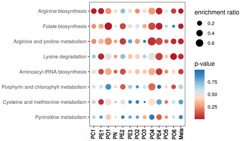
A comparative investigation for exploring the effect of different substrates on the metabolic profile was made between Pleurotus spp. mycelia were grown in substrate S2 with respect to substrate S1 taken as reference (Figure 1 and Figure 2). The most evident thing was that the metabolic pathways were strongly influenced by the substrate. Some differences can be tentatively explained. For example, folate biosynthesis was greater for sample PE4 grown on substrate S2. Indeed, this substrate contained, among others, wheat straw which had a good content of vitamin B12 and folic acid [39]. Similar evidence was noted for the metabolic pathway of arginine and proline metabolism which was increased in sample PE2 grown on substrate S2. The same substrate was able to activate the arginine and proline pathways, compared to substrate S1.
Figure 1.
Comparative investigation for exploring the effect of different substrates on the metabolic profile was made between Pleurotus spp. mycelia grown in substrates S2. Substrate S1 was taken as a reference and data were calculated as mean differences compared to S1 (calibrator of the relative quantification). In the figure, red indicates a higher probability of metabolic pathway activation, whereas blue suggests a minor one.
Figure 2.
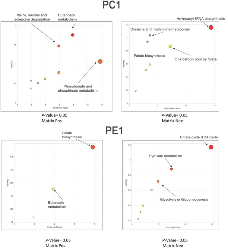
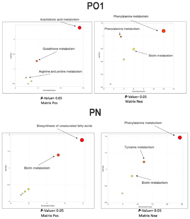
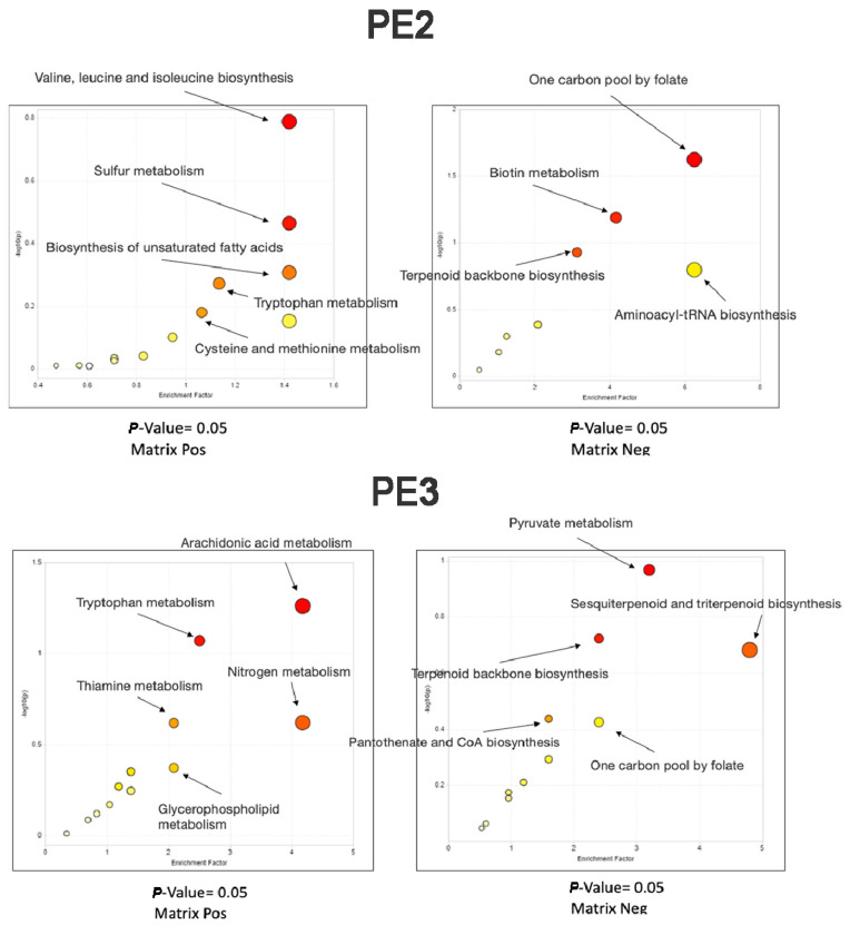
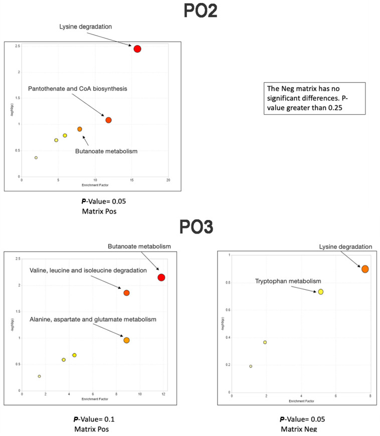
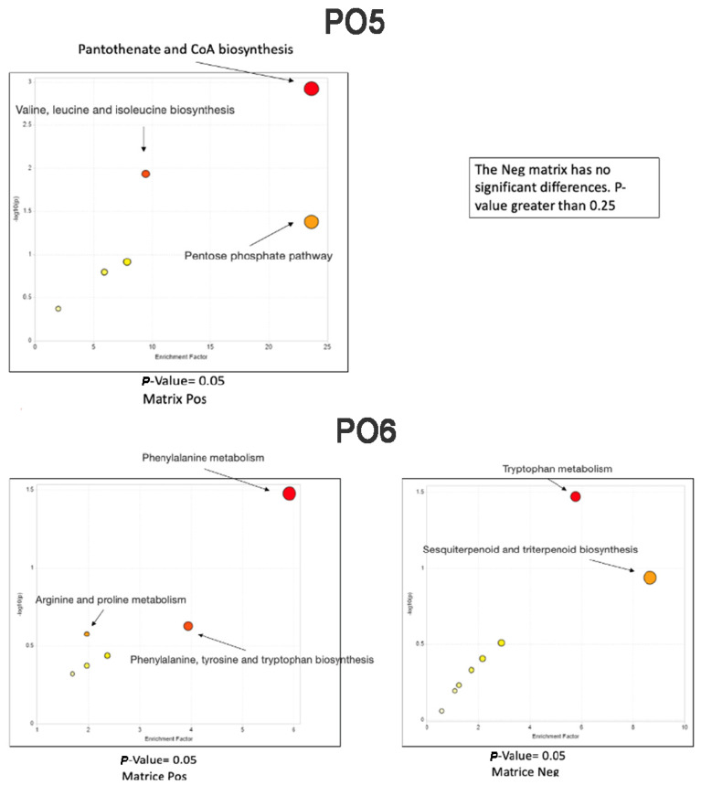
Mummichog Pathway Activity Profile: Comparison of metabolic networks present in samples in both polarities.
In these figures, the pathways revealed by the functional analysis are represented by colored circles, whose abscissas correspond to the enrichment factor and ordinates to the -log of the p-Value. Also, the size of the circle represents the enrichment factor, the color from yellow to red is proportional to the -log of the p-value. The most regulated pathways are in the upper right-hand corner. One carbon pool by folate pathway was particularly expressed in samples PE2 and PO4, rather in the other Pleurotus samples. By contrast, the folate biosynthesis pathway was expressed at higher levels in PE1 and PE4 samples, whereas the pantothenate and CoA biosynthesis pathway were particularly high in the PO5 sample.
3.3. Phenolic Composition of The Extracts
The extracts were also investigated through HPLC-DAD-MS to determine the composition of phenolic compounds.
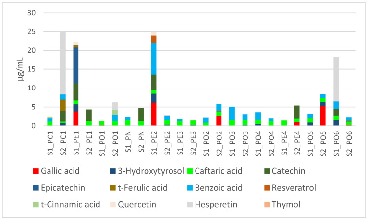
Among all tested extracts, 28 compounds have been identified (Supplementary Material: Table S3 and chromatograms). Only caftaric acid was quantified in all extracts (Figure 3).
Figure 3.
Cumulative distribution of the most abundant phytochemicals in the tested extracts.
Among these phytochemicals, the combination of caftaric acid and benzoic acid was recorded in 9 extracts, while PE1-S2, PN-S2, and PE4-S2 are characterized by the presence of only caftaric acid and catechin. Anyway, there was no effect of the substrate on the qualitative or quantitative composition.
Regarding P. ostreatus, caftaric acid and benzoic acid were the prominent compounds; by contrast in P. eryngii, with the only exception of the sample PE3, catechin is the main phytochemical. In P. nebrodensis (PN), the presence of caftaric acid was not influenced by the substrate composition, while S2 increased the catechin level. In P. columbinus (PC1) cultivated in substrate S2, there was a significant increase in total phenols, as also witnessed by the elevated level of the flavonoid hesperitin. We cannot exclude a possible influence of substrate S2 on the flavonoid biosynthesis pathway (KEGG map 00941), in P. columbinus.
As a final remark, the presence of phenolic compounds in these samples was also an index of potential scavenging/reducing and enzyme inhibition properties [40]. Additionally, phenolic compounds have also been demonstrated to exert antimicrobial effects [41,42]. In this context, the phenolic composition of the extracts was consistent with subsequent investigations on antioxidant, enzyme inhibition, and antimicrobial effects.
3.4. Antimicrobial Activity
The antimicrobial activity of the extracts is shown in Table 6, Table 7, Table 8, Table 9, Table 10 and Table 11, also in comparison with reference antimicrobial drugs, namely ciprofloxacin, fluconazole, and griseofulvin. All extracts from Pleurotus mycelia displayed antimicrobial activity in the concentration range of 1.56 to 200 μg mL−1. Regarding the yeasts, C. parapsilosis (YEPGA 6551) was the most sensitive strain to the PN-PO6 extracts (S2), with MIC ranges of 7.78–>200 μg mL−1, while C. albicans (YEPGA 6183) showed the least sensitivity to the mushroom extracts. The results of the growth inhibition of yeast strains highlighted, albeit partially, the major activity of the extract derived from the S2 growth substrate. With reference to bacteria, the strongest inhibition was observed for the Pleurotus extracts PC1 and PO5 (S1) [MIC <2.47–>200 μg mL−1 against E. coli (ATCC 10536) and B. cereus PeryMycA 2]. Collectively, Gram-bacterial strains (E. coli PeruMyc 2 and 3, S. typhi 7, and P. aeruginosa ATCC 15442) were less sensitive to mushroom extracts than that of Gram+ ones, as already observed for F. torulosa [43]. All results from the tested extracts showed active inhibition of dermatophytes growth. Regarding A. curreyi (CCF 5207), A. insingulare, and T. rubrum (CCF 4933), they were the most sensitive fungal species to all mushroom extracts, with MIC range between 31.49 and 158.74 μg mL−1. Values of MIC < 100 μg mL−1 were considered as an index of high antimicrobial activity [44].
Table 6.
Minimal inhibitory concentrations (MICs) of Pleurotus mycelia (S1) extracts against bacteria isolates.
| MIC (µg mL−1) * | ||||||||
|---|---|---|---|---|---|---|---|---|
|
Escherichia
coli |
Escherichia
coli |
Escherichia
coli |
Bacillus
cereus |
Pseudomonas
aeruginosa |
Bacillus
subtilis |
Salmonella
typhi |
Staphylococcus
aureus |
|
| Bacteria | (ATCC 10536) | (PeruMycA 2) | (PeruMycA 3) | (PeruMycA 4) | (ATCC 15442) | (PeruMycA 6) | (PeruMycA 7) | (ATCC 6538) |
| PC1 | 125.99 (100–200) | 2.47 (1.56–3.12) | >200 | 7.87 (6.26–12.5) | >200 | >200 | >200 | >200 |
| PE1 | 9.92 (6.25–12.5) | 3.93 (3.12–6.25) | >200 | 4.92 (3.125–6.25) | 3.93 (3.12–6.25) | 4.96 (3.125–6.25) | >200 | 19.84 (12.5–25) |
| PO1 | 79.37 (50–100) | 79.37 (50–100) | 158.74 | >200 | >200 | >200 | >200 | >200 |
| PN | 2.47 (1.56–3.12) | 3.93 (3.12–6.25) | >200 | 7.87 (6.25–12.5) | 7.87 (6.25–12.5) | 3.93 (3.12–6.25) | >200 | 7.87 (6.25–12.5) |
| PE2 | 3.93 (3.12–6.25) | 3.93 (3.12–6.25) | >200 | 3.93 (3.12–6.25) | 7.87 (6.25–12.5) | 3.93 (3.12–6.25) | >200 | 16.74 (12.5–25) |
| PE3 | 9.92 (6.25–12.5) | 7.87 (6.25–12.5) | >200 | 2.47 (1.56–3.12) | 3.93 (3.12–6.25) | 7.87 (6.25–12.5) | >200 | 9.92 (6.25–12.5) |
| PO2 | 39.68 (25–50) | 79.37 (50–100) | >200 | 158.74 (100–200) | 158.74 (100–200) | >200 | >200 | >200 |
| PO3 | 158.74 (100–200) | 3.93 (3.12–6.25) | >200 | 79.37 (50–100) | >200 | >200 | >200 | >200 |
| PO4 | 39.68 (25–50) | 7.87 (6.25–12.5) | >200 | 15.74 (12.5–25) | >200 | >200 | >200 | >200 |
| PE4 | 2.47 (1.56–3.12) | 3.93 (3.12–6.25) | >200 | 3.93 (3.12–6.25) | 7.87 (6.25–12.5) | <6.25 | >200 | 9.92 (6.25–12.5) |
| PO5 | 3.93 (3.12–6.25) | >200 | >200 | 125.99 (100–200) | >200 | >200 | >200 | >200 |
| PO6 | 1.96 (1.56–3.12) | 31.49 (25–50) | >200 | 125.99 (100–200) | >200 | >200 | >200 | >200 |
| Ciprofloxacin (µg mL−1 ) | 31.49 (25–50) | 9.92 (6.25–12.5) | 79.37 (50–100) | 125.99 (100–200) | 125.99 (100–200) | 125.99 (100–200) | 79.37 (50–100) | 200- > 200 |
* Mic values are reported as geometric means of three independent replicates (n = 3). MIC range concentrations are reported within brackets.
Table 7.
Minimal inhibitory concentrations (MICs) of Pleurotus mycelia (S2) extracts against bacteria isolates.
| MIC (µg mL−1) * | ||||||||
|---|---|---|---|---|---|---|---|---|
|
Escherichia
coli |
Escherichia
coli |
Escherichia
coli |
Bacillus
cereus |
Pseudomonas
aeruginosa |
Bacillus
subtilis |
Salmonella
typhy |
Staphylococcus
aureus |
|
| Bacteria | (ATCC 10536) | (PeruMycA 2) | (PeruMycA 3) | (PeruMycA 4) | (ATCC 15442) | (PeruMycA 6) | (PeruMycA 7) | (ATCC 6538) |
| PC1 | 79.37 (50–100) | 125.99 (100–200) | 125.99 (100–200) | 125.99 (100–200) | 125.99 (100–200) | 125.99 (100–200) | 79.37 (50–100) | 125.99 (100–200) |
| PE1 | 3.93 (3.125–6.25) | 15.75 (12.5–25) | >200 | 3.93 (3.125–6.25) | 62.99 (50–100) | 31.49 (25–50 | >200 | 158.74 (100–200) |
| PO1 | 158.74 (100–200) | 79.37 (100–200) | 158.74 (100–200) | 158.74 (100–200) | 125.99 (100–200) | 158.74 (100–200) | 39.68 (25–50) | 158.74 (100–200) |
| PN | 4.96 (3.125–6.25) | 9.92 (6.25–12.5) | >200 | 3.93 (3.125–6.25) | 79.37 (50–100) | 15.75 (12.5–25) | >200 | 125.99 (100–200) |
| PE2 | 4.96 (3.125–6.25) | 7.87 (6.25–12.5) | >200 | 9.92 (6.25–12.5) | 39.68 (25–50) | 9.92 (6.25–12.5) | >200 | 62.99 (50–100) |
| PE3 | 7.87 (6.25–12.5) | 9.92 (6.25–12.5) | >200 | 39.68 (25–50) | 62.99 (50–100) | 15.75 (12.5–25) | >200 | 158.74 (100–200) |
| PO2 | 125.99 (100–200) | 158.74 (100–200) | 79.37 (50–100) | 62.99 (50–100) | 125.99 (100–200) | 158.74 (100–200) | 125.99 (100–200) | 62.99 (50–100) |
| PO3 | 125.99 (100–200) | 125.99 (100–200) | 79.37 (50–100) | 125.99 (100–200) | 158.74 (100–200) | 158.74 (100–200) | 125.99 (100–200) | 79.37 (50–100) |
| PO4 | 158.74 (100–200) | 158.74 (100–200) | 125.99 (100–200) | 125.99 (100–200) | 79.37 (50–100) | >200 | 79.37 (50–100) | 62.99 (50–100) |
| PE4 | 3.93 (3.125–6.25) | 3.93 (3.125–6.25) | >200 | 15.75 (12.5–25) | 62.99 (50–100) | 19.84 (12.5–25) | >200 | 62.99 (50–100) |
| PO5 | 158.74 (100–200) | >200 | >200 | 158.74 (100–200) | >200 | >200 | >200 | >200 |
| PO6 | 79.37 (50–100) | 62.99 (50–100) | >200 | 158.74 (100–200) | >200 | >200 | >200 | >200 |
| Ciprofloxacin (µg mL−1) | 31.49 (25–50) | 9.92 (6.25–12.5) | 79.37 (50–100) | 125.99 (100–200) | 125.99 (100–200) | 125.99 (100–200) | 79.37 (50–100) | 200- > 200 |
* Mic values are reported as geometric means of three independent replicates (n = 3). MIC range concentrations are reported within brackets.
Table 8.
Minimal inhibitory concentrations of Pleurotus mycelia (S1) extracts against yeast isolates.
| MIC (µg mL−1) * | ||||
|---|---|---|---|---|
| Candida tropicalis | Candida albicans | Candida parapsilosis | Candida albicans | |
| Yeast Strain | (YEPGA 6184) | (YEPGA 6379) | (YEPGA 6551) | (YEPGA 6183) |
| PC1 | 158.74 (100–200) | 158.74 (100–200) | 158.74 (100–200) | >200 |
| PE1 | >200 | 158.74 (100–200) | 15.74 (12.5–25) | >200 |
| PO1 | >200 | 158.74 (100–200) | 158.74 (100–200) | 158.74 (100–200) |
| PN | >200 | >200 | 7.87 (6.25–12.5) | >200 |
| PE2 | >200 | >200 | 9.92 (6.25–12.5) | >200 |
| PE3 | >200 | >200 | 7.87 (6.25–12.5) | >200 |
| PO2 | 158.74 (100–200) | >200 | 125.99 (100–200) | >200 |
| PO3 | 158.74 (100–200) | >200 | >200 | 158.74 (100–200) |
| PO4 | 158.74 (100–200) | >200 | >200 | >200 |
| PE4 | >200 | >200 | 7.87 (6.25–12.5) | >200 |
| PO5 | 158.74 (100–200) | >200 | 158.74 (100–200) | >200 |
| PO6 | >200 | 158.74 (100–200) | 158.74 (100–200) | 158.74 (100–200) |
| Fluconazole (µg mL−1) | 2 | 1 | 4 | 2 |
* Mic values are reported as geometric means of three independent replicates (n = 3). MIC range concentrations are reported within brackets.
Table 9.
Minimal inhibitory concentrations of Pleurotus mycelia (S2) extracts against yeast isolates.
| MIC (µg mL−1) * | ||||
|---|---|---|---|---|
| Candida tropicalis | Candida albicans | Candida parapsilosis | Candida albicans | |
| Yeast Strain | (YEPGA 6184) | (YEPGA 6379) | (YEPGA 6551) | (YEPGA 6183) |
| PC1 | 79.37 | >200 | 39.68 (25–50) | >200 |
| PE1 | 158.74 (100–200) | 158.74 (100–200) | 15.74 | >200 |
| PO1 | >200 | >200 | 62.99 (50–100) | >200 |
| PN | >200 | >200 | 7.87 (6.25–12.5) | >200 |
| PE2 | >200 | >200 | 7.87 (6.25–12.5) | >200 |
| PE3 | >200 | >200 | 7.87 (6.25–12.5) | >200 |
| PO2 | 158.74 (100–200) | 158.74 (100–200) | 158.74 (100–200) | >200 |
| PO3 | 158.74 (100–200) | >200 | >200 | 62.99 (50–100) |
| PO4 | 158.74 (100–200) | >200 | 79.37 (50–100) | >200 |
| PE4 | >200 | >200 | 9.92 (6.25–12.5) | >200 |
| PO5 | 158.74 (100–200) | 158.74 (100–200) | 125.99 (100–200) | >200 |
| PO6 | 158.74 (100–200) | 158.74 (100–200) | >200 | 158.74 (100–200) |
| Fluconazole (µg mL−1) | 2 | 1 | 4 | 2 |
* Mic values are reported as geometric means of three independent replicates (n = 3). MIC range concentrations are reported within brackets.
Table 10.
Minimal inhibitory concentrations (MICs) of Pleurotus mycelia (S1) extracts against dermatophyte isolates.
| MIC (µg mL−1) * | ||||||||
|---|---|---|---|---|---|---|---|---|
|
Trichophyton
mentagrophytes |
Trichophyton
tonsurans |
Trichophyton
rubrum |
Arthroderma
quadrifidum |
Trichophyton
mentagrophytes |
Arthroderma
gypseum |
Arthroderma
curreyi |
Arthroderma
insingulare |
|
| Dermatophyte | (CCF 4823) | (CCF 4834) | (CCF 4933) | (CCF 5792) | (CCF 5930) | (CCF 6261) | (CCF 5207) | (CCF 5417) |
| PC1 | >200 | >200 | 158.74 (100–200) | 125.99 (100–200) | 125.99 (100–200) | 125.99 (100–200) | 62.99 (50–100) | 79.37 (50–100) |
| PE1 | >200 | >200 | 158.74 (100–200) | 79.37 (50–100) | 158.74 (100–200) | >200 | 79.37 (50–100) | 125.99 (100–200) |
| PO1 | >200 | 158.74 (100–200) | 125.99 (100–200) | 125.99 (100–200) | 125.99 (100–200) | >200 | 62.99 (50–100) | 79.37 (50–100) |
| PN | 125.99 (100–200) | 158.74 (100–200) | 125.99 (100–200) | 79.37 (50–100) | 158.74 (100–200) | >200 | >200 | >200 |
| PE2 | 125.99 (100–200) | >200 | 158.74 (100–200) | >200 | >200 | 125.99 (100–200) | 79.37 (50–100) | 79.37 (50–100) |
| PE3 | 79.37 (50–100) | 158.74 (100–200) | 125.99 (100–200) | 125.99 (100–200) | 158.74 (100–200) | 158.74 (100–200) | 79.37 (50–100) | 79.37 (50–100) |
| PO2 | 79.37 (50–100) | >200 | 125.99 (100–200) | 79.37 (50–100) | 158.74 (100–200) | >200 | 31.49 (25–50) | 62.99 (50–100) |
| PO3 | 158.74 (100–200) | 158.74 (100–200) | >200 | 125.99 (100–200) | >200 | >200 | 79.37 (50–100) | 125.99 (100–200) |
| PO4 | >200 | >200 | 158.74 (100–200) | 125.99 (100–200) | 158.74 (100–200) | >200 | 79.37 (50–100) | 125.99 (100–200) |
| PE4 | 158.74 (100–200) | 125.99 (100–200) | 79.37 (50–100) | >200 | 125.99 (100–200) | 158.74 (100–200) | 125.99 (100–200) | 79.37 (50–100) |
| PO5 | >200 | >200 | >200 | >200 | >200 | >200 | 62.99 (50–100) | 79.37 (50–100) |
| PO6 | 79.37 (50–100) | >200 | 79.37 (50–100) | 125.99 (100–200) | 79.37 (50–100) | 158.74 (100–200) | 31.49 (25–50) | 39.68 (25–50) |
| Griseofulvin µg mL−1 | 2.52 (2–4) | 0.198 (0.125–0.25) | 1.26 (1–2) | >8 | 3.174 (2–4) | 1.587 (1–2) | >8 | >8 |
* Mic values are reported as geometric means of three independent replicates (n = 3). MIC range concentrations are reported within brackets.
Table 11.
Minimal inhibitory concentrations (MICs) of Pleurotus mycelia (S2) extracts against dermatophyte isolates.
| MIC (µg mL−1) * | ||||||||
|---|---|---|---|---|---|---|---|---|
|
Trichophyton
mentagrophytes |
Trichophyton
tonsurans |
Trichophyton
rubrum |
Arthroderma
quadrifidum |
Trichophyton
mentagrophytes |
Arthroderma
gypseum |
Arthroderma
curreyi |
Arthroderma
insingulare |
|
| Dermatophyte | (CCF 4823) | (CCF 4834) | (CCF 4933) | (CCF 5792) | (CCF 5930) | (CCF 6261) | (CCF 5207) | (CCF 5417) |
| PC1 | 125.99 (100–200) | 158.74 (100–200) | 158.74 (100–200) | 158.74 (100–200) | >200 | >200 | 31.49 (25–50) | >200 |
| PE1 | 62.99 (50–10) | 158.74 (100–200) | 39.68 (25–50) | 39.68 (25–50) | >200 | 62.99 (50–100) | 39.68 (25–50) | 62.99 (50–100) |
| PO1 | >200 | 125.99 (100–200) | 158.74 (100–200) | >200 | 125.99 (100–200) | 158.74 (100–200) | 39.68 (25–50) | 125.99 (100–200) |
| PN | 125.99 (100–200) | 79.37 (50–100) | 31.49 (25–50) | >200 | 158.74 (100–200) | 158.74 (100–200) | 62.99 (50–100) | >200 |
| PE2 | 158.74 (100–200) | 125.99 (100–200) | 79.37 (50–100) | 125.99 (100–200) | >200 | 125.99 (100–200) | 62.99 (50–100) | 79.37 (50–100) |
| PE3 | >200 | 158.74 (100–200) | 125.99 (100–200) | 158.74 (100–200) | >200 | 158.74 (100–200) | 125.99 (100–200) | 125.99 (100–200) |
| PO2 | 158.74 (100–200) | >200 | 125.99 (100–200) | 125.99 (100–200) | 158.74 (100–200) | >200 | 158.74 (100–200) | 79.37 (50–100) |
| PO3 | >200 | >200 | 79.37 (50–100) | 125.99 (100–200) | >200 | >200 | 79.37 (50–100) | 79.37 (50–100) |
| PO4 | 158.74 (100–200) | 158.74 (100–200) | 125.99 (100–200) | 125.99 (100–200) | 158.74 (100–200) | >200 | 79.37 (50–100) | 62.99 (50–100) |
| PE4 | >200 | >200 | 79.37 (50–100) | >200 | 125.99 (100–200) | 79.37 (50–100) | 62.99 (50–100) | 125.99 (100–200) |
| PO5 | >200 | 158.74 (100–200) | 158.74 (100–200) | 158.74 (100–200) | >200 | >200 | 62.99 (50–100) | 158.74 (100–200) |
| PO6 | >200 | >200 | 125.99 (100–200) | 125.99 (100–200) | >200 | >200 | 125.99 (100–200) | 79.37 (50–100) |
| Griseofulvin (µg mL−1) | 2.52 (2–4) | 0.198 (0.125–0.25) | 1.26 (1–2) | >8 | 3.174 (2–4) | 1.587 (1–2) | >8 | >8 |
* Mic values are reported as geometric means of three independent replicates (n = 3). MIC range concentrations are reported within brackets.
The antimicrobial activity of Pleurotus samples can be hypothesized, albeit partially, as due to the presence of the so-called CP compounds (derivatives of phosphonate and phosphinate with substitution of alkyl group for hydrogen of phosphorus-hydrogen bonds), monobactams, that are beta-lactam antibiotics (containing a monocyclic beta-lactam nucleus) and sesquiterpenoid and triterpenoid, for which the following metabolic pathways have been highlighted: phosphonate and phosphinate metabolism (PC1 and PE2 samples), monobactam biosynthesis (PE2) and sesquiterpenoid and triterpenoid biosynthesis (PE3, PO4, PE4, and PO6), respectively. The presence of these metabolic pathways has never been indicated in the mycelia of Pleurotus spp. Many C-P compounds are known bioactive substances used in medicine (antibiotics) and agriculture (herbicide) such as fosfomycin, FR-33289, rhizocticin, and bialaphos, while monobactams are beta-lactam antibiotics containing a monocyclic beta-lactam nucleus. Sesquiterpenoid and triterpenoid (present in PE3, PO4, PE4, and PO6 samples) are a group of terpenoids consisting of three isoprene units known for their antimicrobial capacity [45].
3.5. Antiradical Activity
Regarding the antiradical activity, experimental data were normalized and expressed as EC50 values (μg mL−1) for each mushroom extract and Trolox, which was used as a reference antioxidant compound. The antiradical properties were investigated through both DPPH and ABTS, which are common assays used for measuring the intrinsic antioxidant properties of extracts. The results of these tests are shown in Table 12. Values for DPPH radical scavenging activity varied between 886 and 4871 (μg mL−1), with the higher potency demonstrated by sample PE3 cultivated in substrate S1. Values for ABTS radical scavenging activity varied between 87 and 377 (μg mL−1), and the best activity was shown by the samples PE2 in both substrates, with mean values in the range 87–94 μg mL−1 referred to the EC50; thus, suggesting that the substrates can affect the sample properties in different, that cannot be generalized. For instance, in the DDPH test, samples PC1, PO2, PO3, PO4, and PO5 were not influenced by the change of growth substrate. Whilst in the ABTS test, the substrate always influenced the intrinsic activity, with both stimulating or inhibiting antiradical effects. We cannot exclude that the discrepancies observed between DPPH and ABTS tests can be regarded as the differences between ABTS and DPPH radicals. Indeed, ABTS has been described to be more accurate compared with DPPH, when applied to samples rich in hydrophilic, lipophilic, and highly pigmented antioxidant compounds [46].
Table 12.
Antiradical properties of the tested Pleurotus extracts.
| DPPH Test | ABTS Test | |||
|---|---|---|---|---|
| Sample | EC50 | EC50 | ||
| µg mL−1 | Trolox eq. | µg mL−1 | Trolox eq. | |
| PC1-S1 | 3248 ± 388 | 650 ± 78 | 152 ± 18 | 38 ± 5 |
| PE1-S1 | 4871 ± 538 | 974 ± 108 | 103 ± 12 | 26 ± 3 |
| PO1-S1 | 4871 ± 525 | 974 ± 105 | 133 ± 16 | 33 ± 4 |
| PN-S1 | 1392 ± 168 | 278 ± 34 | 97 ± 12 | 24 ± 3 |
| PE2-S1 | 2436 ± 294 | 487 ± 59 | 87 ± 10 | 22 ± 3 |
| PE3-S1 | 886 ± 103 | 177 ± 21 | 135 ± 16 | 34 ± 4 |
| PO2-S1 | 3248 ± 353 | 650 ± 71 | 189 ± 22 | 47 ± 6 |
| PO3-S1 | 3248 ± 368 | 650 ± 74 | 374 ± 41 | 87 ± 10 |
| PO4-S1 | 3248 ± 360 | 650 ± 72 | 170 ± 20 | 43 ± 5 |
| PE4-S1 | 1949 ± 235 | 390 ± 47 | 271 ± 32 | 68 ± 8 |
| PO5-S1 | 2436 ± 292 | 487 ± 58 | 101 ± 12 | 25 ± 3 |
| PO6-S1 | 1949 ± 277 | 390 ± 46 | 131 ± 16 | 33 ± 4 |
| PC1-S2 | 3248 ± 375 | 650 ± 75 | 234 ± 28 | 59 ± 7 |
| PE1-S2 | 1949 ± 218 | 390 ± 44 | 189 ± 22 | 47 ± 6 |
| PO1-S2 | 2436 ± 279 | 487 ± 56 | 100 ± 12 | 25 ± 3 |
| PN-S2 | 1392 ± 152 | 278 ± 30 | 167 ± 20 | 42 ± 5 |
| PE2-S2 | 1392 ± 154 | 278 ± 31 | 94 ± 11 | 24 ± 3 |
| PE3-S2 | 1624 ± 178 | 354 ± 36 | 91 ± 11 | 23 ± 3 |
| PO2-S2 | 3248 ± 390 | 650 ± 78 | 147 ± 17 | 37 ± 4 |
| PO3-S2 | 3248 ± 397 | 650 ± 79 | 255 ± 30 | 64 ± 8 |
| PO4-S2 | 3248 ± 391 | 650 ± 78 | 377 ± 45 | 94 ± 11 |
| PE4-S2 | 2436 ± 289 | 487 ± 58 | 263 ± 31 | 66 ± 8 |
| PO5-S2 | 2436 ± 287 | 487 ± 57 | 119 ± 14 | 30 ± 4 |
| PO6-S2 | 3248 ± 386 | 650 ± 77 | 206 ± 22 | 52 ± 6 |
4. Conclusions
With the development of technology, liquid chromatography coupled to a mass-spectrometry approach can be widely applied in metabolomic studies currently, having a wide detected range and high specificity and sensitivity [47]. In the present study, this method was used to analyze the metabolic profiling of Pleurotus species mycelia, which showed satisfactory data quality. The literature about the characterization of Pleurotus metabolic pathways is unexpectedly poor, even for P. ostreatus and a few other species. It is almost missing as concerns P. columbinus, since it has been only recently accepted as an independent species. The present findings support further investigations aimed at evaluating the influence of growth substrate on Pleurotus spp. antimicrobial and antioxidant properties. The extracts from Pleurotus revealed valuable sources of primary and secondary metabolites, thus suggesting potential applications in the formulation of food supplements, above all in terms of antioxidant and antimicrobial properties.
Regarding the antimicrobial effects, the results from the present study did not point out the optimal substrate for the cultivation of fungi. However, the effect of the substrate was present and should be deeply considered in view of the production of antioxidant extracts from Pleurotus species.
As a concluding remark, in view of a modern concept of sustainability, waste products of agronomical chains can be considered promising substrates for fungal cultivation. Indeed, our results demonstrated that residual plant materials, still containing primary and secondary metabolites, can play pivotal roles in modulating selectively fungal metabolome, with a concomitant influence on the potential use as food and/or health-promoting agent.
Future studies still prove necessary to better define the interactions between plant phytocomplex and fungal response, to drive the cultivation of fungi towards both sustainable improvements of the chain production and search for innovative market products.
Acknowledgments
We thank Salvatore Silviani, Giancarlo Bistocchi, and Andrea Arcangeli, for providing some Pleurotus sp. pl. basidiomata. The work is also part of the third mission activity of the botanic garden “Giardino dei Semplici”, “G. d’Annunzio” University Chieti-Pescara. The study is also part of the PhD project of Giancarlo Angeles Flores: "Development of sustainable technologies for the cultivation of edible and medicinal mushrooms and enhancement of botanical supply chains also through the activities of the botanic garden Giardino dei Semplici ". Tutors are Paola Angelini and Claudio Ferrante.
Supplementary Materials
The following supporting information can be downloaded at: https://www.mdpi.com/article/10.3390/antibiotics11111468/s1. Preparation of Pleurotus samples: Tables S1 and S2. Quantitative analysis of phenolic compounds: Table S3 and chromatograms.
Author Contributions
Conceptualization, P.A., L.M. and C.F.; methodology, P.A., L.M. and C.F.; software, P.A., L.M. and C.F.; validation, R.M.P., A.A. and G.A.F.; formal analysis, P.A., L.M. and C.F.; investigation, G.A.F., C.E.G., B.T., F.I., F.B., L.C., G.C., R.M.P., C.E., G.V., P.C., F.C., M.L.G., A.A. and S.C.D.S.; resources, P.A., L.M. and C.F.; data curation, P.A., L.M. and C.F.; writing—original draft preparation, P.A. and G.A.F.; writing—review and editing, P.A., L.M., C.F., G.Z. and G.O.; visualization, R.V., G.O. and G.Z.; supervision, R.V.; project administration, P.A., L.M. and C.F.; funding acquisition, P.A., L.M. and C.F. All authors have read and agreed to the published version of the manuscript.
Institutional Review Board Statement
Not applicable.
Informed Consent Statement
Not applicable.
Data Availability Statement
Original data are available from the corresponding author.
Conflicts of Interest
The authors declare no conflict of interest.
Funding Statement
This research received no external funding.
Footnotes
Publisher’s Note: MDPI stays neutral with regard to jurisdictional claims in published maps and institutional affiliations.
References
- 1.Sharma A., Sharma A., Tripathi A. Biological activities of Pleurotus spp. polysaccharides: A review. J. Food Biochem. 2021;45:e13748. doi: 10.1111/jfbc.13748. [DOI] [PubMed] [Google Scholar]
- 2.Angelini P., Pellegrino R.M., Tirillini B., Flores G.A., Alabed H.B., Ianni F., Blasi F., Cossignani L., Venanzoni R., Orlando G. Metabolomic profiling and biological activities of Pleurotus columbinus Quél. Cultivated on different agri-food byproducts. Antibiotics. 2021;10:1245. doi: 10.3390/antibiotics10101245. [DOI] [PMC free article] [PubMed] [Google Scholar]
- 3.Ianni F., Blasi F., Angelini P., Simone S.C.D., Angeles Flores G., Cossignani L., Venanzoni R. Extraction optimization by experimental design of bioactives from Pleurotus ostreatus and evaluation of antioxidant and antimicrobial activities. Processes. 2021;9:743. doi: 10.3390/pr9050743. [DOI] [Google Scholar]
- 4.Ng S.H., Mohd Zain M.S., Zakaria F., Wan Ishak W.R., Wan Ahmad W.A.N. Hypoglycemic and antidiabetic effect of Pleurotus sajor-caju aqueous extract in normal and streptozotocin-induced diabetic rats. BioMed Res. Int. 2015;2015:214918. doi: 10.1155/2015/214918. [DOI] [PMC free article] [PubMed] [Google Scholar]
- 5.Zhang Y., Zhang Z., Liu H., Wang D., Wang J., Deng Z., Li T., He Y., Yang Y., Zhong S. Physicochemical characterization and antitumor activity in vitro of a selenium polysaccharide from Pleurotus ostreatus. Int. J. Biol. Macromol. 2020;165:2934–2946. doi: 10.1016/j.ijbiomac.2020.10.168. [DOI] [PubMed] [Google Scholar]
- 6.Morris H., Marcos J., Llauradó G., Fontaine R., Tamayo V., García N., Bermúdez R. Immunomodulating effects of hot-water extract from Pleurotus ostreatus mycelium on cyclophosphamide treated mice. Micol. Apl. Int. 2003;15:7–13. [Google Scholar]
- 7.Barbosa J.R., dos Santos Freitas M.M., da Silva Martins L.H., de Carvalho Junior R.N. Polysaccharides of mushroom Pleurotus spp.: New extraction techniques, biological activities and development of new technologies. Carbohydr. Polym. 2020;229:115550. doi: 10.1016/j.carbpol.2019.115550. [DOI] [PubMed] [Google Scholar]
- 8.Barbosa J.R., Freitas M.M.S., Oliveira L.C., Martins L.H.S., Almada-Vilhena A.O., Oliveira R.M., Pieczarka J.C., Davi do Socorro B., Junior R.N.C. Obtaining extracts rich in antioxidant polysaccharides from the edible mushroom Pleurotus ostreatus using binary system with hot water and supercritical CO2. Food Chem. 2020;330:127173. doi: 10.1016/j.foodchem.2020.127173. [DOI] [PubMed] [Google Scholar]
- 9.Pellegrino R.M., Blasi F., Angelini P., Ianni F., Alabed H.B., Emiliani C., Venanzoni R., Cossignani L. LC/MS Q-TOF Metabolomic Investigation of Amino Acids and Dipeptides in Pleurotus ostreatus Grown on Different Substrates. J. Agric. Food Chem. 2022;70:10371–10382. doi: 10.1021/acs.jafc.2c04197. [DOI] [PMC free article] [PubMed] [Google Scholar]
- 10.Fernandes Â., Barros L., Martins A., Herbert P., Ferreira I.C. Nutritional characterisation of Pleurotus ostreatus (Jacq. ex Fr.) P. Kumm. produced using paper scraps as substrate. Food Chem. 2015;169:396–400. doi: 10.1016/j.foodchem.2014.08.027. [DOI] [PubMed] [Google Scholar]
- 11.Baysal E., Peker H., Yalinkiliç M.K., Temiz A. Cultivation of oyster mushroom on waste paper with some added supplementary materials. Bioresour. Technol. 2003;89:95–97. doi: 10.1016/S0960-8524(03)00028-2. [DOI] [PubMed] [Google Scholar]
- 12.Gregori A., Švagelj M., Pohleven J. Cultivation techniques and medicinal properties of Pleurotus spp. Food Technol. Biotechnol. 2007;45:238–249. [Google Scholar]
- 13.Landínez-Torres A.Y., Fagua C.P., López A.C.S., Oyola Y.A.D., Girometta C.E. Pruning Wastes from Fruit Trees as a Substrate for Pleurotus ostreatus. Acta Mycol. 2021;56:568. doi: 10.5586/am.568. [DOI] [Google Scholar]
- 14.Royse D., Rhodes T., Ohga S., Sanchez J. Yield, mushroom size and time to production of Pleurotus cornucopiae (oyster mushroom) grown on switch grass substrate spawned and supplemented at various rates. Bioresour. Technol. 2004;91:85–91. doi: 10.1016/S0960-8524(03)00151-2. [DOI] [PubMed] [Google Scholar]
- 15.Stamets P. Growing Gourmet and Medicinal Mushrooms. Ten Speed Press; Berkeley, CA, USA: 2011. [Google Scholar]
- 16.Koutrotsios G., Kalogeropoulos N., Kaliora A.C., Zervakis G.I. Toward an increased functionality in oyster (Pleurotus) mushrooms produced on grape marc or olive mill wastes serving as sources of bioactive compounds. J. Agric. Food Chem. 2018;66:5971–5983. doi: 10.1021/acs.jafc.8b01532. [DOI] [PubMed] [Google Scholar]
- 17.Zervakis G.I., Koutrotsios G., Katsaris P. Composted versus raw olive mill waste as substrates for the production of medicinal mushrooms: An assessment of selected cultivation and quality parameters. BioMed Res. Int. 2013;2013:546830. doi: 10.1155/2013/546830. [DOI] [PMC free article] [PubMed] [Google Scholar]
- 18.Ritota M., Manzi P. Pleurotus spp. cultivation on different agri-food by-products: Example of biotechnological application. Sustainability. 2019;11:5049. doi: 10.3390/su11185049. [DOI] [Google Scholar]
- 19.Valverde M.E., Hernández-Pérez T., Paredes-López O. Edible mushrooms: Improving human health and promoting quality life. Int. J. Microbiol. 2015;2015:376387. doi: 10.1155/2015/376387. [DOI] [PMC free article] [PubMed] [Google Scholar]
- 20.Freitas A.C., Antunes M.B., Rodrigues D., Sousa S., Amorim M., Barroso M.F., Carvalho A., Ferrador S.M., Gomes A.M. Use of coffee by-products for the cultivation of Pleurotus citrinopileatus and Pleurotus salmoneo-stramineus and its impact on biological properties of extracts thereof. Int. J. Food Sci. Technol. 2018;53:1914–1924. doi: 10.1111/ijfs.13778. [DOI] [Google Scholar]
- 21.Kulshreshtha S., Mathur N., Bhatnagar P., Kulshreshtha S. Cultivation of Pleurotus citrinopileatus on handmade paper and cardboard industrial wastes. Ind. Crop. Prod. 2013;41:340–346. doi: 10.1016/j.indcrop.2012.04.053. [DOI] [Google Scholar]
- 22.Liang Z.-C., Wu C.-Y., Shieh Z.-L., Cheng S.-L. Utilization of grass plants for cultivation of Pleurotus citrinopileatus. Int. Biodeterior. Biodegrad. 2009;63:509–514. doi: 10.1016/j.ibiod.2008.12.006. [DOI] [Google Scholar]
- 23.Ren D., Jiao Y., Yang X., Yuan L., Guo J., Zhao Y. Antioxidant and antitumor effects of polysaccharides from the fungus Pleurotus abalonus. Chem. Biol. Interact. 2015;237:166–174. doi: 10.1016/j.cbi.2015.06.017. [DOI] [PubMed] [Google Scholar]
- 24.Ren L., Perera C., Hemar Y. Antitumor activity of mushroom polysaccharides: A review. Food Funct. 2012;3:1118–1130. doi: 10.1039/c2fo10279j. [DOI] [PubMed] [Google Scholar]
- 25.Bilal S., Mushtaq A., Moinuddin K. Effect of different grains and alternate substrates on oyster mushroom (Pleurotus ostreatus) production. Afr. J. Microbiol. Res. 2014;8:1474–1479. doi: 10.5897/AJMR2014.6697. [DOI] [Google Scholar]
- 26.Hoa H.T., Wang C.-L. The effects of temperature and nutritional conditions on mycelium growth of two oyster mushrooms (Pleurotus ostreatus and Pleurotus cystidiosus) Mycobiology. 2015;43:14–23. doi: 10.5941/MYCO.2015.43.1.14. [DOI] [PMC free article] [PubMed] [Google Scholar]
- 27.VanderMolen K.M., Raja H.A., El-Elimat T., Oberlies N.H. Evaluation of culture media for the production of secondary metabolites in a natural products screening program. Amb Express. 2013;3:71. doi: 10.1186/2191-0855-3-71. [DOI] [PMC free article] [PubMed] [Google Scholar]
- 28.Kim S., Lee J.W., Heo Y., Moon B. Effect of Pleurotus eryngii mushroom β-glucan on quality characteristics of common wheat pasta. J. Food Sci. 2016;81:C835–C840. doi: 10.1111/1750-3841.13249. [DOI] [PubMed] [Google Scholar]
- 29.Bas C., Kuyper T.W., Noordeloos M., Vellinga E. Flora Agaricina Neerlandica—Critical Monographs on the Families of Agarics and Boleti Occurring in the Netherlands. Volume 2 AA Balkema; Rotterdam, The Netherlands: 1990. [Google Scholar]
- 30.Singer R. Koeltz Scientific Books. Koeltz; Koenigstein, Germany: 1986. The Agaricales in Modern Taxonomy. [Google Scholar]
- 31.Venturella G., Gargano M., Compagno R. The genus Pleurotus in Italy. Flora Mediterr. 2015;25:143–155. [Google Scholar]
- 32.Schoch C.L., Seifert K.A., Huhndorf S., Robert V., Spouge J.L., Levesque C.A., Chen W., Consortium F.B., List F.B.C.A., Bolchacova E. Nuclear ribosomal internal transcribed spacer (ITS) region as a universal DNA barcode marker for Fungi. Proc. Natl. Acad. Sci. USA. 2012;109:6241–6246. doi: 10.1073/pnas.1117018109. [DOI] [PMC free article] [PubMed] [Google Scholar]
- 33.Angelini P., Girometta C., Tirillini B., Moretti S., Covino S., Cipriani M., D’Ellena E., Angeles G., Federici E., Savino E. A comparative study of the antimicrobial and antioxidant activities of Inonotus hispidus fruit and their mycelia extracts. Int. J. Food Prop. 2019;22:768–783. doi: 10.1080/10942912.2019.1609497. [DOI] [Google Scholar]
- 34.Angelini P., Venanzoni R., Angeles Flores G., Tirillini B., Orlando G., Recinella L., Chiavaroli A., Brunetti L., Leone S., Di Simone S.C. Evaluation of antioxidant, antimicrobial and tyrosinase inhibitory activities of extracts from Tricholosporum goniospermum, an edible wild mushroom. Antibiotics. 2020;9:513. doi: 10.3390/antibiotics9080513. [DOI] [PMC free article] [PubMed] [Google Scholar]
- 35.Girometta C.E., Bernicchia A., Baiguera R.M., Bracco F., Buratti S., Cartabia M., Picco A.M., Savino E. An italian research culture collection of wood decay fungi. Diversity. 2020;12:58. doi: 10.3390/d12020058. [DOI] [Google Scholar]
- 36.Tsugawa H., Nakabayashi R., Mori T., Yamada Y., Takahashi M., Rai A., Sugiyama R., Yamamoto H., Nakaya T., Yamazaki M., et al. A cheminformatics approach to characterize metabolomes in stable-isotope-labeled organisms. Nat. Methods. 2019;16:295–298. doi: 10.1038/s41592-019-0358-2. [DOI] [PubMed] [Google Scholar]
- 37.Li S., Park Y., Duraisingham S., Strobel F.H., Khan N., Soltow Q.A., Jones D.P., Pulendran B. Predicting network activity from high throughput metabolomics. PLoS Comput. Biol. 2013;9:e1003123. doi: 10.1371/journal.pcbi.1003123. [DOI] [PMC free article] [PubMed] [Google Scholar]
- 38.Pang Z., Chong J., Zhou G., de Lima Morais D.A., Chang L., Barrette M., Gauthier C., Jacques P.-É., Li S., Xia J. MetaboAnalyst 5.0: Narrowing the gap between raw spectra and functional insights. Nucleic Acids Res. 2021;49:W388–W396. doi: 10.1093/nar/gkab382. [DOI] [PMC free article] [PubMed] [Google Scholar]
- 39.Mostafa M.E.-S., Ibrahim S.A.-E., Samya S., Ahmed Z. Production of vitamin B12 and folic acid from agricultural wastes using new bacterial isolates. Afr. J. Microbiol. Res. 2013;7:966–973. [Google Scholar]
- 40.Chatatikun M., Supjaroen P., Promlat P., Chantarangkul C., Waranuntakul S., Nawarat J., Tangpong J. Antioxidant and tyrosinase inhibitory properties of an aqueous extract of Garcinia atroviridis griff. ex. T. Anderson fruit pericarps. Pharmacogn. J. 2020;12:1675–1682. doi: 10.5530/pj.2020.12.12. [DOI] [Google Scholar]
- 41.Bottari N.B., Lopes L.Q.S., Pizzuti K., dos Santos Alves C.F., Corrêa M.S., Bolzan L.P., Zago A., de Almeida Vaucher R., Boligon A.A., Giongo J.L. Antimicrobial activity and phytochemical characterization of Carya illinoensis. Microb. Pathog. 2017;104:190–195. doi: 10.1016/j.micpath.2017.01.037. [DOI] [PubMed] [Google Scholar]
- 42.Ferrante C., Chiavaroli A., Angelini P., Venanzoni R., Angeles Flores G., Brunetti L., Petrucci M., Politi M., Menghini L., Leone S. Phenolic content and antimicrobial and anti-inflammatory effects of Solidago virga-aurea, Phyllanthus niruri, Epilobium angustifolium, Peumus boldus, and Ononis spinosa extracts. Antibiotics. 2020;9:783. doi: 10.3390/antibiotics9110783. [DOI] [PMC free article] [PubMed] [Google Scholar]
- 43.Covino S., D'Ellena E., Tirillini B., Angeles G., Arcangeli A., Bistocchi G., Venanzoni R., Angelini P. Characterization of biological activities of methanol extract of Fuscoporia torulosa (Basidiomycetes) from Italy. Int. J. Med. Mushrooms. 2019;21:1051–1063. doi: 10.1615/IntJMedMushrooms.2019032896. [DOI] [PubMed] [Google Scholar]
- 44.Doğan H.H., Duman R., Özkalp B., Aydin S. Antimicrobial activities of some mushrooms in Turkey. Pharm. Biol. 2013;51:707–711. doi: 10.3109/13880209.2013.764327. [DOI] [PubMed] [Google Scholar]
- 45.Guimarães A.C., Meireles L.M., Lemos M.F., Guimarães M.C.C., Endringer D.C., Fronza M., Scherer R. Antibacterial activity of terpenes and terpenoids present in essential oils. Molecules. 2019;24:2471. doi: 10.3390/molecules24132471. [DOI] [PMC free article] [PubMed] [Google Scholar]
- 46.Menghini L., Leporini L., Vecchiotti G., Locatelli M., Carradori S., Ferrante C., Zengin G., Recinella L., Chiavaroli A., Leone S., et al. Crocus sativus L. Stigmas and Byproducts: Qualitative Fingerprint, Antioxidant Potentials and Enzyme Inhibitory Activities. Food Res. Int. 2018;109:91–98. doi: 10.1016/j.foodres.2018.04.028. [DOI] [PubMed] [Google Scholar]
- 47.Vélëz H., Glassbrook N.J., Daub M.E. Mannitol metabolism in the phytopathogenic fungus Alternaria alternata. Fungal Genet. Biol. 2007;44:258–268. doi: 10.1016/j.fgb.2006.09.008. [DOI] [PubMed] [Google Scholar]
Associated Data
This section collects any data citations, data availability statements, or supplementary materials included in this article.
Supplementary Materials
Data Availability Statement
Original data are available from the corresponding author.


