Abstract
Ducrosia flabellifolia Boiss. is a rare desert plant known to be a promising source of bioactive compounds. In this paper, we report for the first time the phytochemical composition and biological activities of D. flabellifolia hydroalcoholic extract by using liquid chromatography–electrospray tandem mass spectrometry (ESI-MS/MS) technique. The results obtained showed the richness of the tested extract in phenols, tannins, and flavonoids. Twenty-three phytoconstituents were identified, represented mainly by chlorogenic acid, followed by ferulic acid, caffeic acid, and sinapic acid. The tested hydroalcoholic extract was able to inhibit the growth of all tested bacteria and yeast on agar Petri dishes at 3 mg/disc with mean growth inhibition zone ranging from 8.00 ± 0.00 mm for Enterococcus cloacae (E. cloacae) to 36.33 ± 0.58 mm for Staphylococcus epidermidis. Minimal inhibitory concentration ranged from 12.5 mg/mL to 200 mg/mL and the hydroalcoholic extract from D. flabellifolia exhibited a bacteriostatic and fungistatic character. In addition, D. flabellifolia hydroalcoholic extract possessed a good ability to scavenge different free radicals as compared to standard molecules. Molecular docking studies on the identified phyto-compounds in bacterial, fungal, and human peroxiredoxin 5 receptors were performed to corroborate the in vitro results, which revealed good binding profiles on the examined protein targets. A standard atomistic 100 ns dynamic simulation investigation was used to further evaluate the interaction stability of the promising phytocompounds, and the results showed conformational stability in the binding cavity. The obtained results highlighted the medicinal use of D. flabellifolia as source of bioactive compounds, as antioxidant, antibacterial, and antifungal agent.
Keywords: Ducrosia flabellifolia, chemical composition, antioxidant, antimicrobial, molecular docking, dynamic simulation
1. Introduction
The Apiaceae family (syn. Umbelliferae) comprises more than 455 genera and more than 3700 species [1] and is known to yield distinctive phytochemicals with antioxidant, antimicrobial, anticancer, anti-inflammatory, and hepatoprotective properties [2,3,4]. In Saudi Arabia, the Apiaceae family comprises more than eighteen plant species and is considered the most used family in ethnomedicine [5,6]. The Ducrosia genus includes six species and is widely spread in Asia, particularly the Kingdom of Saudi Arabia, Afghanistan, Pakistan, and Iraq with D. anethifolia as the most popular species [7]. Ducrosia flabellifolia Boiss. (D. flabellifolia), with the popular name of “Al Haza”, grows as a rare species in volcanic cinders in the center and north of Saudi Arabia [8,9], and in the deserts of the eastern parts of Jordan [10].
Many scientific studies have investigated the chemical composition of D. anethifolia and D. flabellifolia essential oil from Saudi Arabia [11], Iran [12], Jordan [13,14,15], and Tunisia [16]. Most studies have focused on the essential oil obtained from D. flabellifolia species. In fact, in 2014, Shahabipour and colleagues [12] reported the identification of 32 bioactive compounds in the volatile oil of D. flabellifolia from Iran dominated mainly by decanal (32.8 ± 1.91%), dodecanal (32.6 ± 1.75), and (2E)-tridecen-1-al (3.3 ± 0.08%). Moreover, D. flabellifolia volatile oil from Safawi (Jordan) obtained by hydrodistillation was a rich source of monoterpenes and terpenoids [13]. Hydrodistilled oil from fresh leaves was dominated by n-decanal (36.61%), dodecanal (7.5%), D-L- limonene (3.86%), and β-phellandrene (3.84%), while the oil obtained by hydrodistillation from dried leaves was dominated by n-decanal (24.44%), α-pinene (15.72%, 2E-octene (9.73%), 2Z-octane (7.04%), 2-heptanone (5.92%), frenchone (5.18%), and β-phellandrene (4.58%) [13]. While using GC/MS technique, Elsharkawi and colleagues [11] reported the identification of 30 phytoconstituents in ethyl acetate fraction of D. anethifolia collected from Wadi Arar (Saudi Arabia) dominated by 8-ethoxypsoralen, coumarin-6-ol-3,4-dihydro-4,4,7,8-tetramethyl, isoaromadendrene epoxide, aromadendrene oxide, ferulic acid methyl ester, pterin-6-carboxylic acid, vitamin A palmitate, and ursodeoxycholic acid.
The pharmacological potency of organic extracts obtained from members of Ducrosia genus has been subjected to diverse in vitro and in vivo biological activities. In a former study performed by Javidnia et al. [17], potent antimicrobial activity of the hydro-methanolic extract from D. anethifolia aerial parts towards Bacillus subtilis, Staphylococcus aureus, Escherichia coli, Pseudomonas aeruginosa, and Candida albicans has been assessed. For a long time, the aerial parts and leaves of D. flabellifolia have been smoked as a cigarette [18]. The aerial parts of D. ismaelis have been reported as being used as natural insecticides and to cure skin infections [13,19]. In addition, D. anethifolia has been well documented essentially as an insecticide and is used for the treatment of colds [20], heartburn [21], inflammation of the inner wall of the nose [22], as an analgesic [23], and a flavoring in food [24,25].
The emergence of multidrug-resistant bacterial pathogens due to the overuse and abuse of currently available antibiotics has caused about 750,000 deaths annually, and 10 million will die every year by 2050 [26]. In this context, the purpose of this study was to ascertain the effective valorization of selected D. flabellifolia hydroalcoholic extract collected from the Hail region (Saudi Arabia) by assessing its phytochemical profile, antioxidant, and antimicrobial activities. Molecular docking and dynamic approaches were performed in order to elucidate the possible interaction between the identified phytoconstituents with specific target proteins involved in antibacterial, antifungal, and antioxidant activities.
2. Materials and Methods
2.1. Chemicals
Gallic acid, (+)-catechin, pyrocatechol, chlorogenic acid, 2,5-dihydroxybenzoic acid, 4-hydroxybenzoic acid, (−)-epicatechin, caffeic acid, syringic acid, vanillin, taxifolin, sinapic acid, p-coumaric acid, ferulic acid, rosmarinic acid, 2-hydroxycinnamic acid, pinoresinol, quercetin, luteolin, and apigenin were purchased from Sigma-Aldrich (St. Louis, MO, USA). Vanillic acid, 3-hydroxybenzoic acid, 3,4-dihydroxyphenylacetic acid, apigenin 7-glucoside, luteolin 7-glucoside, hesperidin, eriodictyol, and kaempferol were obtained from Fluka (St. Louis, MO, USA). Finally, verbascoside, protocatechuic acid, and hyperoside were purchased from HWI Analytik (Ruelzheim, RP, Germany). Methanol and formic acid of HPLC grade were purchased from Sigma-Aldrich (St. Louis, MO, USA) and Merck (Darmstadt, Hesse, Germany), respectively. Ultra-pure water (18 mΩ) was obtained using a Millipore Milli-Q Plus water treatment system (MILLIPORE CORPORATION, Bedford, MA, USA).
2.2. Plant Material Sampling
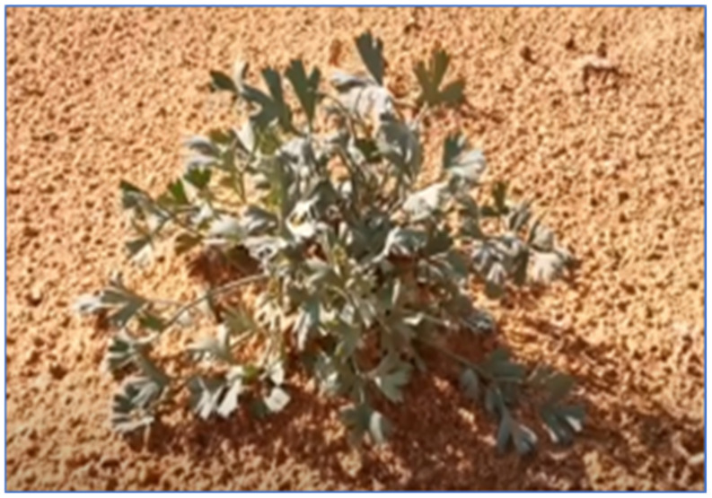
In this proposal, D. flabellifolia (Al-Hazaa; Figure 1) plant species were collected from the Hail region (Al-Mu’ayqilat, 27°16′41.9″ N, 41°22′48.0″ E) in October 2019. A voucher specimen (AN04) was deposited at the herbarium in the Department of Biology (College of Science, University of Hail, Hail, Kingdom of Saudi Arabia). For the experiment, 4 g of air-dried aerial parts were macerated in 100 mL of methanol-80% at room temperature for 72 h. The filtrate was recuperated by lyophilization, and the yield (expressed in percentage) was calculated using the following Equation (1):
| Yield (%) = (W1/100)/W2, | (1) |
where W1 is the weight of extract after the evaporation of solvent and W2 is the dry weight of the sample. The yield of extraction was about 21.56 ± 1.78%.
Figure 1.
D. flabellifolia Boiss. plant species collected from the Hail region.
2.3. Study of the Phytochemical Composition
An Agilent Technologies 1260 Infinity liquid chromatography system (Santa Clara, CA, USA) hyphenated to a 6420 Triple Quad mass spectrometer was used for quantitative analyses. Chromatographic separation was carried out on a Poroshell 120 EC-C18 (100 mm × 4.6 mm I.D., 2.7 μm) column (Santa Clara, CA, USA). The previously validated method was used for the analysis of phenolic compounds by LC-ESI-MS/MS [27]. The mobile phase was made up of solvent A (0.1%, v/v formic acid solution) and solvent B (methanol). The gradient profile was set as follows: 0.00 min 2% B eluent, 3.00 min 2% B eluent, 6.00 min 25% B eluent, 10.00 min 50% B eluent, 14.00 min 95% B eluent, 17.00 min 95% B, and 17.50 min 2% B eluent. The column temperature was maintained at 25 °C. The flow rate was 0.4 mL min−1 and the injection volume was 2.0 μL. The tandem mass spectrometer was interfaced with the LC system via an ESI source. The electrospray source of the MS was operated in negative and positive multiple reaction monitoring (MRM) mode and the interface conditions were as follows: capillary voltage of −3.5 kV, gas temperature of 300 °C, and gas flow of 11 L min−1. The nebulizer pressure was 40 psi. MRM transitions, the optimum collision energies, and retention times for each species are indicated in Supplementary Material S1. In addition, representative LC-ESI-MS/MS chromatograms of phenolic compounds are shown in Supplementary Material S2. Calibration curves and sensitivity properties of the method are also shown in Supplementary Material S3.
In negative and positive multiple reaction monitoring (MRM) mode, the peaks of the analytes were identified by comparing the retention time, together with monitoring ion pairs in an authentic standard solution.
2.4. Screening of the Biological Activities
2.4.1. Antimicrobial Activities
The antimicrobial activity of Al-Haza extracts was tested against twelve ESKAPE bacterial strains including Enterococcus faecium, three Staphylococcus species (S. aureus, S. epidermidis, and S. hominis), Klebsiella pneumoniae, Acinetobacter baumannii, Pseudomonas aeruginosa, two Enterobacter species (E. cloacae and E. faecalis), and Escherichia coli. Four types of Candida species were also tested (C. albicans ATCC 20402, C. tropicalis ATCC 1362. C. guillermondii ATCC 6260, and C. utilis ATCC 9255). The effect of the hydroalcoholic extract from D. flabellifolia was estimated using disc diffusion assay [28] by measuring the diameter of the growth inhibition zone tested on a Mueller Hinton agar plate for bacterial strains and Sabouraud dextrose agar for yeast. Sterile discs were impregnated with three different concentrations of the tested extract (1 mg/disc, 1.5 mg/disc, and 3 mg/disc). After incubation, the mean diameter of the growth inhibition zone (mGIZ) was calculated, and the scheme proposed by Parveen et al. [29] was used to interpret the obtained results (no activity: mGIZ = 0; low activity: mGIZ = 1–6 mm; moderate activity: mGIZ = 7–10 mm; high activity: mGIZ = 11–15 mm; and very high activity: GIZ = 16–20 mm). Ampicillin and Amphotericin B were used as controls.
The microdilution assay was used for the determination of MIC (minimal inhibitory concentration) and MBC/MFC values (minimal bactericidal/fungicidal concentration) as previously described by Khalfaoui et al. [30]. Bacterial and fungal cultures were inoculated into the wells of 96-well microtiter plates in the presence of D. flabellifolia methanolic extract at final concentrations varying from 0.039 mg/mL to 100 mg/mL. To interpret the character of the tested extract, we used the ratios (MBC/MIC ratio and MFC/MIC ratio) described by Gatsing et al. [31] and Moroh et al. [32].
2.4.2. Phytochemistry and Antioxidant Activities Screening
Total phenolic content expressed as milligram (mg) of gallic acid per gram of plant extract (mg GAE/g extract) was estimated by using the Folin–Ciocalteu method as previously described by Kumar et al. [33]. Total flavonoids expressed as mg of quercetin equivalents per gram of plant extract (mg QE/g extract) were determined using the AlCl3 method developed by Benariba et al. [34]. In addition, acidified vanillin method previously described by Broadhurst and Jones [35] was used to estimate the total condensed tannins (expressed mg tannic acid equivalent per gram of plant extract (mg TAE/g extract)).
The ability of the Al-Haza extract against DPPH-H was determined following the same method as Mseddi al. [36]. The method of Koleva et al. [37] for β-Carotene bleaching test and Oyaizu [38] method for the determination of reducing power were used.
2.5. In Silico Study
2.5.1. Molecular Docking
The 3D structures of LC-MS-detected phytochemicals were retrieved in structural data format (sdf) from the PubChem database. The LigPrep module was used to prepare the entire set of phytochemicals, which involved the addition of hydrogen atoms and suitable charges, as well as the correcting of the valences and the protonation and tautomeric states at pH 7.2 ± 2.0 of the molecules using the Epik tool [39,40]. The X-ray crystal structure of S. aureus type IIA topoisomerase, tyrosyl-tRNA synthetases (TyrRS) of S. aureus, Sap1 of C. albicans, and human peroxiredoxin 5 receptor were retrieved from the PDB server with accession codes of 2XCT, 1JIJ, 2QZW, and 1HD2, respectively, as the receptor for the molecular docking study. The Schrodinger Protein Preparation Wizard has been used for protein preparation and energy minimization, in which crystallographic water molecules are removed, then missing hydrogen and/or side chain atoms are added, and the correct charges and protonation states are given to protein residues at pH 7.0 [41,42]. The protein structure was energy minimized using the OPLS3 force field to alleviate steric conflicts in the protein structure. Following that, the prepared protein was considered for grid generation utilizing the “Receptor Grid Generation” panel where active sites in targeted proteins were defined for grid generation by selecting cognate ligands [43]. Glide’s standard precision (SP) mode was used to execute all docking calculations for the prepared ligand molecules, with the default parameters.
2.5.2. Molecular Dynamics (MD) Simulation
MD simulation is regarded as the most important approach for comprehending the nature of biological macromolecules’ underlying structure and function. The MD simulations were conducted utilizing the Schrödinger MD simulation program Desmond, which helps us to understand how a ligand–protein complex binds in simulated physiological conditions [44]. MD simulation analysis was conducted for top score phyto-compound Hyperoside in complex with 1JIJ protein. The simulation system was built through the system builder, with the boundaries set by an orthorhombic shape with a diameter of 10 Å × 10 Å × 10 Å and filled with SPC water molecules.
To neutralize the system’s charges, sodium (+27) and chloride (−12) ions were supplied as counter ions and salt concentration was maintained at 0.15 M. Following the setup of the system builder, the protein–ligand complex system was minimized using the steepest descent method, which was followed by LBFGS algorithms with a maximum of 2000 iterations [45,46]. Further, the system was equilibrated using the NPT ensemble with a Nose–Hoover chain thermostat to evenly distribute the ions and solvent throughout the protein–ligand complex. During the simulation, the temperature was maintained at 300 K. Furthermore, isotropic position scaling was used to control 1bar barostat pressure [47,48,49,50]. A 100 ns simulation was run, with a total of 1000 frames stored in the simulation system for subsequent analysis using the Desmond program’s “Simulation Interactions Diagram” module.
2.6. Statistical Analysis
All experiments were performed in triplicate and average values were calculated using the SPSS 25.0 statistical package for Windows. Duncan’s multiple-range tests for means with a 95% confidence interval (p ≤ 0.05) were used to calculate the differences in means.
3. Results
3.1. Phytochemical Composition of D. Flabellifolia Hydroalcoholic Extract
Using ESI-MS/MS technique, 23 phytocompounds were identified in the hydroalcoholic extract from D. flabellifolia aerial parts (Table 1). This extract was dominated by (mg/g of extract): chlorogenic acid (5980.96 ± 73.12), ferulic acid (180.58 ± 2.77), caffeic acid (70.90 ± 1.75), and sinapic acid (61.74 ± 2.79).
Table 1.
Phytochemical profiling of D. flabellifolia methanol/water extract by using ESI–MS/MS technique.
| No | Identified Compounds | Retention Time (min) |
Abondance (mg/Kg of Extract) |
Chemical Formula | Molecular Weight (g/mol) |
|---|---|---|---|---|---|
| 1 | Gallic acid | 8.891 | 8.78 ± 0.14 | C7H6O5 | 170.12 |
| 2 | Protocatechuic acid | 10.818 | 25.69 ± 0.395 | C7H6O4 | 154.12 |
| 3 | 3,4-Dihydroxyphenylacetic acid | 11.224 | 1.61 ± 0.135 | C8H8O4 | 168.15 |
| 4 | Pyrocatechol | 11.506 | 14.36 ± 0.315 | C6H6O2 | 110.11 |
| 5 | Chlorogenic acid | 11.802 * | 5980.96 ± 73.12 | C16H18O9 | 354.31 |
| 6 | 2,5-Dihydroxybenzoic acid | 12.412 | 59.74 ± 0.945 | C7H6O4 | 154.12 |
| 7 | 4-Hydroxybenzoic acid | 12.439 | 19.25 ± 0.98 | C7H6O3 | 138.12 |
| 8 | (-)-Epicatechin | 12.458 * | 5.15 ± 0.1 | C15H14O6 | 290.27 |
| 9 | Caffeic acid | 12.841 | 70.90 ± 1.75 | C9H8O4 | 180.16 |
| 10 | Syringic acid | 12.963 | 28.92 ± 0.51 | C9H10O5 | 198.17 |
| 11 | 3-Hydroxybenzoic acid | 13.259 | 13.30 ± 0.405 | C7H6O3 | 138.12 |
| 12 | Vanillin | 13.379 | 25.03 ± 0.785 | C8H8O3 | 152.15 |
| 13 | Sinapic acid | 13.992 | 61.74 ± 2.79 | C11H12O5 | 224.21 |
| 14 | p-Coumaric acid | 14.022 | 55.11 ± 0.765 | C9H8O3 | 164.16 |
| 15 | Ferulic acid | 14.120 | 180.58 ± 2.77 | C10H10O4 | 194.18 |
| 16 | Luteolin 7-glucoside | 14.266 | 5.42 ± 0.5 | C21H20O11 | 448.4 |
| 17 | Hyperoside | 14.506 * | 26.55 ± 0.635 | C21H20O12 | 464.4 |
| 18 | Rosmarinic acid | 14.600 | 6.05 ± 0.025 | C18H16O8 | 360.3 |
| 19 | Apigenin 7-glucoside | 14.781 * | 2.52 ± 0.25 | C21H20O10 | 432.4 |
| 20 | 2-Hydroxycinnamic acid | 15.031 | 31.28 ± 0.015 | C9H8O3 | 164.16 |
| 21 | Pinoresinol | 15.118 | 13.13 ± 0.55 | C20H22O6 | 358.4 |
| 22 | Eriodictyol | 15.247 | 2.29 ± 0.005 | C15H12O6 | 288.25 |
| 23 | Quercetin | 15.668 | 2.27 ± 0.075 | C15H10O7 | 302.23 |
*: Compounds identified by positive Ionization mode.
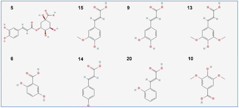
The chemical structures of the main identified phytoconstituents are summarized in Figure 2.
Figure 2.
Chemical structure of the main compounds identified by ESI-MS/MS technique in the hydroalcoholic extract from D. flabellifolia aerial parts. Numbers are the same as listed in Table 1.
3.2. Antioxidant Activities Screening
Table 2 summarizes the results of the quantification of tannins, phenols, and flavonoids. In fact, the hydroalcoholic extract was dominated by phenolic compounds (46.684 ± 0.757 mg GAE/g extract), followed by tannins (6.204 ± 0.401 mg TAE/g extract), and flavonoids (1.816 ± 0.133 mg QE/g extract). The results obtained showed that the tested methanolic extract from D. flabellifolia was able to scavenge the DPPH radical with a low IC50 value (0.014 ± 0.045 mg/mL) as compared to BHT and AA (0.023 ± 3 × 10⁻⁴ mg/mL and 0.022 ± 53 × 10-4 mg/mL, respectively). In addition, the ABTS + radical was scavenged with an IC50 value of about 0.102 ± 0.024 mg/mL of the tested extract. For beta-carotene bleaching inhibiting property, the IC50 value was estimated at 7.80 ± 0.919 mg/mL, significantly different from BHT and AA (p < 0.05).
Table 2.
Antioxidant activities of D. flabellifolia methanol/water extract compared to known drugs.
| Test Systems | D. flabellifolia (Methanol-80%) | Butylated Hydroxytoluene | Ascorbic Acid |
|---|---|---|---|
| DPPH IC50 (mg/mL) | 0.014 ± 0.045 a | 0.023 ± 3 × 10⁻⁴ b | 0.022 ± 5 × 10⁻⁴ b |
| ABTS IC50 (mg/mL) | 0.102 ± 0.024 a | 0.018 ± 4 × 10⁻⁴ b | 0.021 ± 0.001 b |
| β-carotene IC50 (mg/mL) | 7.80 ± 0.919 a | 0.042 ± 3.5 × 10⁻³ b | 0.017 ± 0.001 b |
The letters (a,b) indicate a significant difference between the different antioxidant methods according to the Duncan test (p < 0.05).
3.3. Antimicrobial Activities Screening
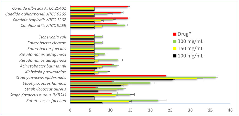
The results of the antimicrobial activity of the hydro-methanolic extract from D. flabellifolia aerial parts showed that the mean diameter of the growth inhibition zone (mGIZ) increased in a concentration-dependent manner (Figure 3). At 3 mg/disc, the mGIZ ranged from 8.00 ± 0.00 mm (E. cloacae) to 36.33 ± 0.58 mm (S. epidermidis). At the same concentration (3 mg/disc), C. utilis ATCC 9255 was the most sensitive strain with mGIZ of about 13.67 ± 0.58 mm. All bacterial strains seem to be more sensitive to the tested extract6 as compared to yeast strains, and the reference drug (ampicillin). Using the scheme proposed by Parveen et al. [29], the tested extract at 3 mg/mL showed high to very high activity against all tested bacterial and fungal strains with mGIZ from 11 to 20 mm.
Figure 3.
Mean diameters of bacterial and fungal growth inhibition zones (mGIZ ± mm) obtained with different concentrations of D. flabellifolia hydro-methanolic extract as compared to standard drugs. *: Ampicillin for bacteria and amphotericin B for Candida strains.
Using the microdilution technique, the minimal inhibitory concentrations values (MICs) ranged from 12.5 to 25 mg/mL for bacterial strains and the minimal bactericidal concentration values (MBCs) ranged from 100 to 200 mg/mL. Using the scheme proposed by Gatsing et al. [31] and Moroh et al. [32], the tested extract showed bacteriostatic activity against almost all tested ESKAPE microorganisms (MBC/MIC ratio higher than 4 and lower than 16), with the exception of E. faecium and E. cloacae (MBC/MIC equal to 4). All these data are summarized in Table 3.
Table 3.
Determination of MICs, MBCs, and MBC/MIC ratio values for ESKAPE strains as compared to known drugs.
| Code | ESKAPE Bacterial Strains |
D. flabellifolia Methanol-80% Extract (mg/mL) |
Ampicillin (mg/mL) |
|||
|---|---|---|---|---|---|---|
| MIC | MBC | MBC/MIC Ratio | MIC | MBC | ||
| 260 | Enterococcus faecium | 25 | 100 | =4; Bactericidal | 0.625 | 5 |
| 445 | Staphylococcus aureus (MRSA) | 12.5 | 100 | 8; Bacteriostatic | 0.625 | 5 |
| 259 | Staphylococcus aureus | 12.5 | 100 | 8; Bacteriostatic | 0.625 | 1.25 |
| 140 BC | Staphylococcus hominis | 12.5 | 100 | 8; Bacteriostatic | 0.625 | 2.5 |
| BC 161 | Staphylococcus epidermidis | 12.5 | 100 | 8; Bacteriostatic | 0.312 | 0.625 |
| 147 | Klebsiella pneumoniae | 25 | 200 | 8; Bacteriostatic | 0.625 | 5 |
| 486 | Acinetobacter baumannii | 25 | 200 | 8; Bacteriostatic | 1.25 | 5 |
| 249 | Pseudomonas aeruginosa | 25 | 200 | 8; Bacteriostatic | 2.5 | 5 |
| 525 | Pseudomonas aeruginosa | 25 | 200 | 8; Bacteriostatic | 2.5 | 5 |
| 268 | Enterobacter faecalis | 12.5 | 100 | 8; Bacteriostatic | 0.312 | 2.5 |
| 235 | Enterobacter cloacae | 50 | 200 | =4; Bacteriostatic | 0.625 | 1.25 |
| 215 | Escherichia coli | 25 | 200 | 8; Bacteriostatic | 1.25 | 5 |
Similarly, the tested D. flabellifolia hydroalcoholic extract was able to inhibit the growth of Candida strains in liquid media at 25 mg/mL (Table 4), while a high concentration of the tested extract was needed to completely kill them (from 100 to 200 mg/mL), with MFC/MIC ratios varying from 4 (C. albicans ATCC 20402; fungicidal action) to 8 (Fungistatic action against C. utilis ATCC 9255, C. tropicalis ATCC 1362, and C. guillermondii ATCC 6260).
Table 4.
Determination of MICs, MFCs, and MFC/MIC ratios for Candida species as compared to Amphotericin B.
| Code | Candida sp. Strains |
D. flabellifolia Methanol-80% Extract (mg/mL) |
Amphotericin B (mg/mL) |
|||
|---|---|---|---|---|---|---|
| MIC | MFC | MFC/MIC Ratio | MIC | MFC | ||
| A1 | Candida utilis ATCC 9255 | 25 | 200 | 8; Fungistatic | 0.78 | 1.56 |
| A8 | Candida tropicalis ATCC 1362 | 25 | 200 | 8; Fungistatic | 0.195 | 0.78 |
| A4 | Candida guillermondii ATCC 6260 | 25 | 200 | 8; Fungistatic | 0.097 | 1.56 |
| A15 | Candida albicans ATCC 20402 | 25 | 100 | =4; Fungicidal | 0.195 | 0.39 |
3.4. Computational Study
3.4.1. Molecular Docking
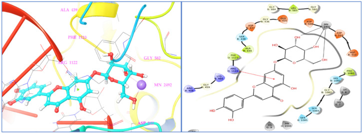
The molecular docking study of identified phyto-compounds was carried out against bacterial fungal and human peroxiredoxin 5 targets. The binding affinities of phyto-compounds are shown in the Supplementary Material (S1). The results of the molecular docking study revealed that phyto-compound luteolin 7-glucoside showed the highest binding affinity toward topoisomerase IIA (2XCT) with a docking score −12.562 Kcal/mol. The phyto-compounds apigenin 7-glucoside, quercetin, (−)-epicatechin, eriodictyol, and hyperoside from D. flabellifolia also showed significant binding affinities against topoisomerase IIA (2XCT) with docking scores of −12.514, −10.06, −9.836, −9.582, and −9.159 Kcal/mol, respectively. The best binding pose of luteolin 7-glucoside inside the active site of topoisomerase IIA and the 2D and 3D representations of the interactions with the amino acids inside the binding pocket are presented in Figure 4.
Figure 4.
Two- and three-dimensional residual interactions network of luteolin 7-glucoside against the active site of S. aureus IIA topoisomerase (PDB ID: 2XCT).
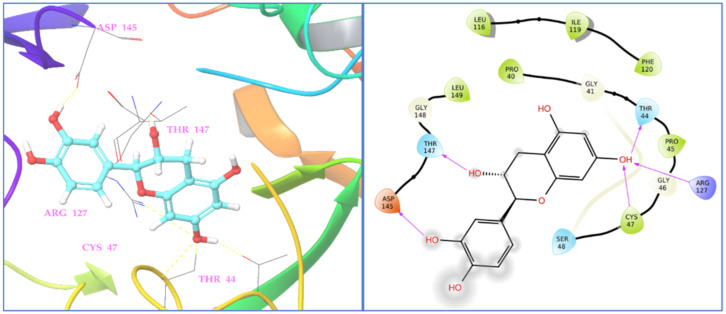
Luteolin 7-glucoside binding interaction reveals that it interacted with Arg1122 and manganese (II) ion via π-cationic and metal coordinates, respectively. Among the assessed phyto-compounds, five compounds, namely, (−)-epicatechin, eriodictyol, 2,5-dihydroxybenzoic acid, chlorogenic acid, luteolin 7-glucoside, and vanillin showed proper and promising antioxidant activity by proper recognition at the binding active site of human peroxiredoxin protein with docking scores of −5.791, −5.255, −5.249, −5.11, −5.06, and −5.02 Kcal/mol, respectively. It was reported that hydrogen bonds and hydrophobic interactions established with the surrounding amino acids are applied to anticipate the binding modes of the novel compounds compared to the antioxidant benzoic acid cognate ligand, at the 1HD2 complex. This active pocket contained conserved amino acid residues such as Thr44, Gly46, Cys47, and Arg127, which play critical roles in docked compound recognition via hydrogen bonding and hydrophobic interactions. The binding interaction shows that two terminal hydroxyl groups connected with asp145 and the147 in the (−)-epicatechin-1HD2 complex, whereas the chromane ring hydroxyl group showed triple hydrogen bonding with Cys47, Thr44, and Arg127 amino acids, suggesting that the identified phyto-compound (−)-epicatechin has antioxidant activity (Figure 5).
Figure 5.
Two- and three-dimensional residual interactions network of (−)-epicatechin in human peroxiredoxin 5 protein (PDB ID: 1HD2).
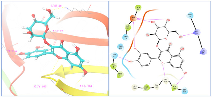
Similarly, in C. albicans Sap1 protein (2QZW), hyperoside showed the highest binding affinity with a docking score of −6.055 Kcal/mol, and phyto-compounds eriodictyol, (−)-epicatechin, and syringic acid showed potential binding affinities against C. albicans Sap1 (2QZW) protein with docking scores of −5.814, −5.461, and −5.042 Kcal/mol, respectively. The residues of Thr6, Asp17, Lys26, Gly103, and Ala104, from Sap1 of C. albicans formed seven significant interactions with the phyto-compound hyperoside with a standard hydrogen binding pattern as shown in Figure 6.
Figure 6.
Two- and three-dimensional residual interactions network of hyperoside against the active site of C. albicans Sap1 (PDB ID: 2QZW).
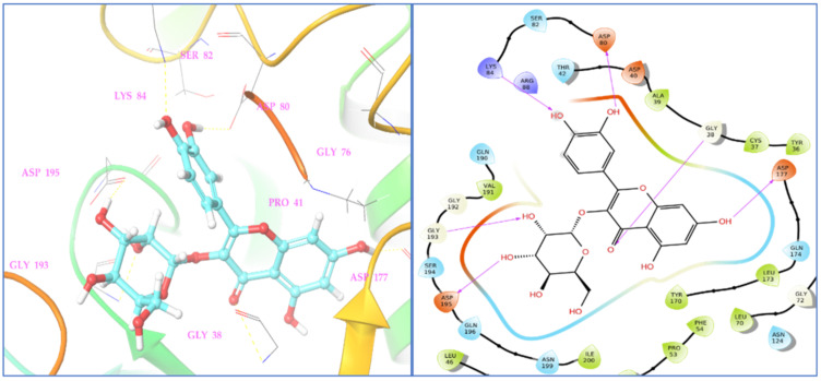
Among assessed phyto-compounds, hyperoside also showed a promising docking score (−8.852 Kcal/mol) in the microbial target TyrRS from S. aureus (1JIJ) in which it interacted with active residues, namely, Asp195, Gly193, Gly38, and Asp177 (Figure 7).
Figure 7.
Two- and three-dimensional residual interactions network of hyperoside against the active site of TyrRS protein (PDB ID: 1JIJ).
Table 5 shows the promising phyto-compounds hydrogen bonding profile in the active site of type IIA topoisomerase, TyrRS, Sap1, and human peroxiredoxin 5 proteins.
Table 5.
Interacting active site residues of receptors with the best phyto-compounds identified in D. flabellifolia hydroalcoholic extract.
| Name of Complex | Interacting Residues |
|---|---|
| Luteolin 7-glucoside-2XCT | Arg1112(5.54 Å): [Arg N of NH2) -Lig (Phenyl ring) *] Mn2492(1.84 Å): [ Mn-Lig (O of OH) **] |
| Mn2492(1.80 Å): [ Mn-Lig (O of OH) **] | |
| Mn2492(1.81 Å): [ Mn-Lig (O of OH) **] | |
| (−)-Epicatechin -1HD2 | Thr44 (1.84 Å): [Thr (O of COO−) -Lig (H of OH) d] |
| Cys47 (2.76 Å): [Cys (H of NH) d-Lig (O of OH)] | |
| Arg127 (2.70 Å): [Arg (H of NH) d-Lig (O of OH)] | |
| Asp145 (1.70 Å): [Asp (O of COO−)-Lig (H of OH) d] | |
| Thr147 (2.20 Å): [Asp (O of COO−)-Lig (H of OH) d] | |
| Hyperoside-2QZW | Thr6 (1.92 Å): [Thr (H of NH) d -Lig (O of OH)] |
| Asp17 (1.74 Å): [Asp (O of COO−)-Lig (H of OH) d] | |
| Asp17 (1.84 Å): [Asp (O of COO−)-Lig (H of OH) d] | |
| Asp17 (1.92 Å): [Asp (O of NHCO−)-Lig (H of OH) d] | |
| Lys26 (1.80 Å): [Lys (H of NH) d -Lig (O of OH)] | |
| Gly103 (2.62 Å): [Gly H of NH) d -Lig (O of C-O-C)] | |
| Ala104 (2.18 Å): [Ala O of COO−) -Lig (H of OH) d] | |
| Hyperoside-1JIJ | Gly38 (2.70 Å): [Gly (H of NH) d -Lig (O of C = O)] |
| Asp80(1.85 Å): [Asp (O of COO−)-Lig (H of OH) d] | |
| Lys84 (1.91 Å): [Lys (H of NH) d -Lig (O of OH)] | |
| Asp177 (1.85 Å): [Asp (H of NH) d -Lig (O of OH)] | |
| Gly193 (2.28 Å): [Gly (H of NH) d -Lig (O of OH)] | |
| Asp195 (1.93 Å): [Asp (O of COO−)-Lig (H of OH) d] |
d Hydrogen bond donor; * π-Cation interaction; ** Metal interaction.
3.4.2. MD Simulation
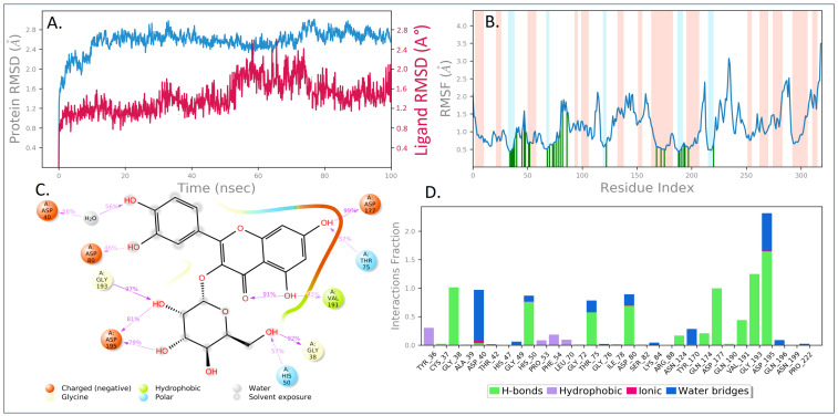
Using molecular dynamics simulation, the docked complex of the phyto-compound hyperoside at the binding site of TyrRS of S. aureus was simulated under biological environments to investigate complex stability and protein flexibility. Hyperoside, a phyto-compound, has shown a high affinity for microbial targets, hence it was chosen for the MD simulation studies. The MD trajectories were used to determine RMSD values, root-mean-square fluctuation (RMSF) values, and protein–ligand interactions. Various MD trajectory data analyses for the hyperoside-1JIJ complex are shown in Figure 8. The RMSD figure showed a stable ligand–protein complex throughout the simulation time, as shown by RMSD values ranging from 1.6 to 2.8 Å for protein Cα atoms in the complex with hyperoside (Figure 8A). RMSD changes remain within 3 Å throughout the simulation period, which is perfectly appropriate for small, globular proteins such as TyrRS. In the case of hyperoside RMSD with respect to protein, it was found that the ligand RMSD ranged from 0.8 to 2.6 Å. Except for a minor fluctuation, the RMSD of hyperoside was found to remain steady throughout the simulation. The maximum ligand RMSD was recorded at 59 and 66 ns, when RMSD reached 2.59 and 2.60 Å, respectively.
Figure 8.
MD simulation analysis of hyperoside in complex with S. aureus tyrosyl-tRNA synthetases (TyrRS) (PDB ID: 1JIJ): (A) time dependent RMSD (protein Cα atoms RMSD is shown in teal blue color while the RMSDs of hyperoside with respect to protein are shown in brown color); (B) protein Cα atoms RMSF; (C) 2D diagram of ligand interactions that occurred more than 30.0% of the simulation time; and (D) protein–ligand contact analysis of throughout the simulation.
Furthermore, the flexibility of the protein system was evaluated during the simulation by computing the RMSF of individual protein amino acid residues. The higher-peaking residues correspond to loop regions (highlighted by white shade) identified by MD trajectories or N- and C-terminal areas. Low RMSF values of binding site residues show that hyperoside binding to TyrRS protein is stable. From Figure 8B, it may be observed that phyto-compound hyperoside interacted with 31 amino acids of TyrRS protein during the simulation time, which were highlighted by green vertical bars, including, Tyr36 (0.425 Å), Cys37 (0.452 Å), Gly38 (0.509 Å), Ala39 (0.534 Å), Asp40 (0.769 Å), Thr42 (1.106 Å), His47 (0.975 Å), Gly49 (1.588 Å), His50 (1.015 Å), Pro53 (0.672 Å), Phe54 (0.712 Å), Leu70 (0.486 Å), Gly72 (0.59 Å), Thr75 (0.628 Å), Gly76 (0.685 Å), Ile78 (0.726 Å), Asp80 (0.788 Å), Ser82 (1.097 Å), Lys84 (1.507 Å), Arg88 (1.551 Å), Asn124 (0.655 Å), Tyr170 (0.538 Å), Gln174 (0.528 Å), Asp177 (0.505 Å), Gln190 (0.434 Å), Val191 (0.46 Å), Gly193 (0.563 Å), Asp195 (0.687 Å), Gln196 (0.604 Å), Asn199 (0.524 Å), and Pro222 (0.662 Å).
The 2D ligand interaction diagram shows the charged negative amino acid Asp establishing a major hydrogen bond with a hydroxyl group of hyperoside. The amino acids His50, Gly38, Val191, and Thr75 interacted with hyperoside for 57%, 92%, 43%, and 57% of simulation time, respectively. Additionally, there are intramolecular hydrogen bonds in hyperoside between the hydrogen atom of hydroxyl and the carbonyl oxygen of chromen-4-one moiety (Figure 8C). The binding interactions between hyperoside and active site amino acid residues inside the binding pocket of TyrRS were computed and represented in Figure 8D. Most of the important interactions of hyperoside with the TyrRS protein determined with MD simulations are hydrogen bonds, polar (amino acids mediated hydrogen bonding) interactions, and hydrophobic interactions.
4. Discussion
In the present study, we report for the first time the identification of bioactive molecules by using the ESI-MS/MS technique in the hydroalcoholic extract from D. flabellifolia aerial parts collected from the Saudi Arabian desert. The main identified phytoconstituents were: protocatechuic acid, chlorogenic acid, 2,5-dihydroxybenzoic acid, 4-hydroxybenzoic acid, caffeic acid, syringic acid, vanillin, sinapic acid, p-coumaric acid, ferulic acid, hyperoside, 2-hydroxycinnamic acid, and pinoresinol.
As far as we know, there are few data available in the literature relating to the chemical compounds from D. flabellifolia extracts. In fact, Talib et al. [14] have demonstrated that D. flabellifolia 95% ethanolic extract collected from Wadi Hassan (Jordan) was a rich source of flavonoids and terpenoids qualitatively estimated by the thin layer chromatography technique. The same team has demonstrated the identification of several flavonoids (flavonols and flavones) by using the HPLC-MS/MS technique in D. flabellifolia ethanolic extract including quercetin, fisetin, kaempferol, luteolin, and apigenin.
Our results have shown that D. flabellifolia hydroalcoholic extract was active against ESKAPE pathogens and Candida species with an increase in the mean diameter of the growth inhibition zone depending on the concentration used. The highest sensitivity for all tested microorganisms was obtained at 3 mg/disc of the tested extract. Using the schemes proposed by Gatsing et al. [31] and Moroh et al. [32], the tested extract exhibited bacteriostatic and fungistatic action against almost all tested microorganisms. No study has hitherto described the antimicrobial activity of D. flabellifolia extracts. While testing Jordanian D. flabellifolia essential oil from dried leaves, Al-Shudiefat et al. [10] reported good activity against C. albicans with a MIC value of about 234 µg/mL; meanwhile, for bacterial strains, the MICs values were about 234 µg/mL against S. aureus, 1870 µg/mL against E. coli, and 1872 µg/mL against P. aeruginosa strain. For D. anethifolia, Elsharkawi and colleagues [11] have demonstrated that the ethyl acetate fraction was active against different Gram-positive bacteria including S. aureus ATCC 25923, S. epidermidis ATCC 49461, Bacillus cereus ATCC 10876, and S. aureus clinical isolate tested at 500 mg/mL. The highest diameter of growth inhibition was recorded against S. epidermidis (14.5 ± 0.5 mm) followed by B. subtilis (14.0 ± 0.00 mm), while no activity was recorded against E. coli ATCC 35218, K. pneumoniae ATCC 700603, K. pneumoniae ATCC 27736, P. aeruginosa, and A. baumannii.
We also reported in this study that D. flabellifolia hydroalcoholic extract was dominated by phenolic compounds, followed by tannins and flavonoids. The same extract showed the ability to scavenge different radicals with low IC50 values (IC50 DPPH radical = 0.014 ± 0.045 mg/mL; IC50 ABTS + radicals = 0.102 ± 0.024 mg/mL; and IC50 for the beta-carotene test = 7.80 ± 0.919 mg/mL). Previously, Mottaghipisheh et al. [13] reported that the IC50 value of the free radical scavenging activity of D. anethifolia ethanolic extract was estimated at 122.02 ppm and 354.37 ppm for the ethyl acetate extract. In addition, Elsharkawi and colleagues [11] revealed the presence of high reduction capacity (EC50: 0.63 ± 0.03 g/L) and ability to scavenge the free radicals of DPPH with an IC50 of 0.38 ± 0.02 g/L in ethyl acetate fraction of D. anethifolia.
Overall, the good antimicrobial activity against all tested Gram-positive/Gram-negative and Candida species, and the antioxidant activity of the tested hydroalcoholic extract can be attributed to the presence of many molecules with potential biological activities (Table 6).
Table 6.
Literature survey showing some biological activities of the identified molecules in D. flabellifolia hydroalcoholic extract.
| Identified Compounds | Biological Activities | References |
|---|---|---|
| Gallic acid | Antioxidant, antimicrobial, anticancer, anti-inflammatory, gastroprotective, cardioprotective, neuroprotective, anti-hyperlipidemia, anti-obesity, and anti-diabetes. | [51,52,53] |
| Protocatechuic acid | Antiviral, anti-oxidation, antibacterial, anti-apoptotic, anti-inflammatory, anti-atherosclerotic, antioxidant, and neuroprotective. | [54,55,56,57,58] |
| 3,4-Dihydroxyphenylacetic acid |
Decrease in the formation of amyloid fibrils and modulator of cell fate in Parkinson’s disease. | [59,60] |
| Pyrocatechol | Antibacterial, antitumor, antioxidant and cytotoxic activities. | [61] |
| Chlorogenic acid | Anti-metastatic, anti-oxidative, nephroprotective, anti-inflammatory, anti-diabetic, anti-hypertensive, hepatoprotective, anti-bacterial, neuroprotective, anti-proliferative, central nervous system stimulator, anti-obesity, cardioprotective, anti-pyretic, anti-viral, anti-angiogenic. | [62] |
| 2,5-Dihydroxybenzoic acid | Anti-inflammatory, anti-oxidant, antibacterial, muscle relaxant, anticarcinogenic, nephroprotective, hepatoprotective, cardioprotective, neuroprotective. | [63,64] |
| 4-Hydroxybenzoic acid | Antibacterial, antiviral, antisickling agent, antialgal, antimutagenic, estrogenic agent, anti-inflammatory, antioxidant. | [65] |
| (−)-Epicatechin | Antiviral (Anti-SARS-CoV-2 virus), gastroprotective, cardioprotective, neuroprotective, hepatoprotective, antioxidant, anti-inflammatory, antidiabetic. | [66,67,68] |
| Caffeic acid | Anti-inflammatory, anticancer, antiviral, antioxidant, antihyperglycemic, antidepressive, antibacterial. | [69,70,71] |
| Syringic acid | Anti-oxidant, anti-microbial, anti-inflammation, antiangiogenic, anti-cancer, anti-diabetic, hepatoprotective, cardioprotective, neuroprotective. | [72] |
| 3-Hydroxybenzoic acid | Antifungal, antimutagenic, antisickling, estrogenic, antimicrobial. | [73] |
| Vanillin | Anticancer, neuroprotective, antihyperglycemic, anti-hyperlipidemic, anti-inflammatory, antimicrobial, antioxidant, antisickling, cardioprotective. | [74,75,76] |
| Sinapic acid | Antioxidant, anti-inflammatory, anti-cancer, antihypertensive, cardioprotective, neuroprotective, renoprotective, hepatoprotective, anti-hyperglycemic, anti-diabetic. | [77,78] |
| p-Coumaric acid | Antioxidant, anti-inflammatory, anti-platelet aggregation, analgesic, anticancer, neuroprotective, anti-necrotic, anti-cholestatic, anti-amoebic. | [79,80] |
| Ferulic acid | Anti-inflammatory, antioxidant, antimicrobial activity, anticancer, and antidiabetic | [81,82,83] |
| Luteolin 7-glucoside | Antioxidant, anti-inflammatory, antiaging, anticancer, vasoprotective | [84,85] |
| Hyperoside | Anti-inflammatory, anti-thrombotic, antidiabetic, anti-viral, anti-fungal, hepato-protective, antioxidant, neuroprotective, antidepressant, cardioprotective, antidiabetic, anticancer, hepatoprotective, Immuno-modulatory activity. | [86,87,88] |
| Rosmarinic acid | Cytoprotective, antioxidative, antibacterial, antiviral, astringent, analgesic, anti-inflammatory, antihyperglycemic, hepatoprotective, immunomodulatory, anticancer, cardioprotective, neuroprotective. | [89,90,91] |
| Apigenin 7-glucoside | Anti-inflammatory, anticandidal, anticancer, antiviral, antibacterial, antioxidant, pro-apoptotic, antimutagenic, antiproliferative, antiallergic, inhibits xenobiotic-metabolizing enzymes. | [92,93,94] |
| 2-Hydroxycinnamic acid | Inhibition of HIV/SARS-CoV S pseudovirus. | [95] |
| Pinoresinol | Neuroprotective, vasorelaxant, hepatoprotective, anti-inflammatory, anticancer. | [96,97,98] |
| Eriodictyol | Cardioprotective, skin protection, antitumor, neuroprotective, antioxidant, antidiabetic, anti-inflammatory, cytoprotective, hepatoprotective, analgesic. | [99,100] |
| Quercetin | Anticancer, antiviral, antiprotozoal, antimicrobial, anti-allergy, anti-inflammatory, cardioprotective, sedative, immunostimulant. | [101,102] |
Molecular docking studies were performed in order to explore binding affinity and interaction of identified phyto-compounds against bacterial, fungal, and human peroxiredoxin 5 receptor. A docking score was used to determine the binding affinities of the phyto-compounds to target proteins. A lower docking score indicates a higher affinity. Luteolin 7-glucoside connected with Arg1122 and manganese (II) ion in the S. aureus IIA topoisomerase target via π -cationic and metal coordinates, respectively. This interaction is comparable to that of the co-crystallized standard drug ciprofloxacin. According to the study, the binding site cavity’s metal interaction had an impact on the substance’s physicochemical properties and antibacterial activity [103,104]. Free radical-scavenging activity properties using the 3-D crystallographic peroxiredoxin 5 (1HD2) were carried out to explore the identified phyto-compounds’ recognition in the active site as potential antioxidants. With a docking score of -5 kcal/mol, five phyto-compounds, namely (−)-epicatechin, eriodictyol, 2,5-dihydroxybenzoic acid, chlorogenic acid, luteolin 7-glucoside, and vanillin, showed promising antioxidant activity by appropriate orientation at the binding active site of human peroxiredoxin protein. In fungal target C. albicans Sap1 and bacterial TyrRS protein, the phyto-compound hyperoside showed the highest binding affinity. SB-219383 and its analogs are a family of bacterial TyrRS inhibitors that are both potent and selective. These inhibitors’ crystal structures have been solved in complex with the TyrRS from S. aureus, the bacterium that causes the majority of hospital-acquired infections, according to crystal structure. SB-219383 and its analogs interacted with charged negative amino acids Asp195 and Asp80, as well as hydrophobic Tyr170, in the active region of TyrRS from S. aureus [105]. Our docking results for hyperoside match those found in the deposited crystal structure, indicating that it has potential antibacterial activity by inhibiting TyrRS S. aureus. Because of its high affinity for microbial targets, hyperoside was selected for MD simulation studies. The RMSD values of the protein Cα atoms were used to calculate the stability of the protein–ligand complex during dynamics analysis. In fact, Cα atoms in proteins’ RMSD is a crucial parameter of the MD simulation trajectory that is used to investigate the backbone deviation of a single frame created in a dynamic environment. The unfolding of the protein molecule results in a high RMSD value, which suggests compactness. The steady fluctuation in the RMSD value across the simulation time indicates that the protein–ligand complex has equilibrated [106,107,108,109,110]. It is anticipated that the lesser RMSD value during the simulation reveals that the protein–ligand complex is more stable. In contrast, the higher RMSD value indicates the protein–ligand complex is less stable [111,112,113]. The overall RMSD revealed that fluctuations in the range of 1.6 Å to 2.8 Å were within the standard range (1–3 Å) of RMSD, indicating that the protein–ligand complex is stable. Throughout the simulation process, the RMSF value shows the mobility and flexibility of each amino acid in the protein. In this plot, the peaks indicate the fluctuation of each amino acid residue over the entire simulation. It implies that higher RMSF values represent higher residue flexibility, whereas lower RMSF values reflect less residue flexibility and better system stability. If the residues in the active site and main chain fluctuated slightly, it indicated that the conformational change was minimal, implying that the reported lead compound was firmly bound within the cavity of the target protein binding pocket [114,115]. In addition, it was observed that the phyto-compound hyperoside interacted with 31 amino acids of the TyrRS protein during the simulation period, which was highlighted by the green vertical bar. All these intercalated residues have low RMSF values, indicating that the hyperoside binding to TyrRS protein is stable.
5. Conclusions
In summary, we report for the first time in this study the identification of various phenolic compounds in the hydroalcoholic extract from D. flabellifolia aerial parts growing wild in Saudi Arabia. The tested extract possessed good antimicrobial activities and exhibited notable potency in scavenging free radicals. Docking studies on the identified phyto-compounds in the bacterial, fungal, and peroxiredoxin 5 receptors were carried out to confirm the in vitro results, and they exhibited satisfactory binding profiles. In-depth protein–ligand interaction stability in the dynamic state was evaluated using 100 ns MD simulation studies, which revealed a significant binding affinity of the discovered hyperoside towards the TyrRS protein, implying anti-bacterial effectiveness via TyrRS enzyme inhibition. The obtained results highlighted the possible use of this plant species as a source of molecules with therapeutic effects to be used as antimicrobial and antioxidant agents.
Supplementary Materials
The following supporting information can be downloaded at: https://www.mdpi.com/article/10.3390/antiox11112174/s1, Supplementary material (S1). ESI–MS/MS Parameters and analytical characteristics for the Analysis of Target Analytes by MRM Negative and Positive Ionization Mode.; Supplementary material (S2). Calibration curves and sensitivity properties of the method; Supplementary material (S3). LC–ESI–MS/MS MRM chromatograms of phenolic compounds. 1–31 represent the chromatograms of gallic acid, protocatechuic acid, 3,4-dihydroxyphenylacetic acid, chlorogenic acid, (−)-epicatechin, caffeic acid, 3-hydroxybenzoic acid, vanillin, verbascoside, taxifolin, p-coumaric acid, luteolin 7-glucoside, hyperoside, rosmarinic acid, apigenin 7-glucoside, 2-hydroxycinnamic acid, eriodictyol, quercetin, luteolin, apigenin, (+)-catechin, pyrocatechol, 2,5-dihydroxybenzoic acid, 4-hydroxybenzoic acid, vanillic acid, syringic acid, sinapic acid, ferulic acid, hesperidin, pinoresinol and kaempferol, respectively; Supplementary material (S4). LC–ESI–MS/MS MRM chromatogram of D. flabellifolia methanol/water extract; Supplementary material (S5). Result of the docking experiment performed between the Target proteins and the identified phyto-compounds.
Author Contributions
Conceptualization, M.S., V.D.F. and A.M.A.A.; methodology, B.T., M.A.A., A.M.A.A., E.N., Y.S.A. and C.S.; software, I.A. and H.P.; formal analysis, B.R., M.A. (Mohd Adnan) and M.A. (Mousa Alreshidi); investigation, A.M.A.A.; resources, A.M.A.A.; writing—original draft preparation, A.M.A.A., I.A., C.S., H.P., B.R. and M.S.; writing—review and editing, A.J.S., E.N., V.D.F., M.A. (Mohd Adnan) and M.A. (Mousa Alreshidi); supervision, M.S.; project administration, M.S.; funding acquisition, M.S. and A.M.A.A. All authors have read and agreed to the published version of the manuscript.
Data Availability Statement
The data presented in this study are available in the article and supplementary materials.
Conflicts of Interest
The authors declare no conflict of interest.
Funding Statement
This research was funded by Scientific Research Deanship at the University of Ha’il, Saudi Arabia through project number GR-22 041.
Footnotes
Publisher’s Note: MDPI stays neutral with regard to jurisdictional claims in published maps and institutional affiliations.
References
- 1.Sayed-Ahmad B., Thierry T., Saad Z., Hijazi A., Merah O. The Apiaceae: Ethnomedicinal family as source for industrial uses. Ind. Crop. Prod. 2017;109:661–671. doi: 10.1016/j.indcrop.2017.09.027. [DOI] [Google Scholar]
- 2.Amiri M.S., Joharchi M.R. Ethnobotanical knowledge of Apiaceae family in Iran: A review. Avicenna J. Phytomed. 2016;6:621–635. [PMC free article] [PubMed] [Google Scholar]
- 3.Sousa R.M.O.F., Cunha A.C., Fernandes-Ferreira M. The potential of Apiaceae species as sources of singular phytochemicals and plant-based pesticides. Phytochemistry. 2021;187:112714. doi: 10.1016/j.phytochem.2021.112714. [DOI] [PubMed] [Google Scholar]
- 4.Thiviya P., Gamage A., Piumali D., Merah O., Madhujith T. Apiaceae as an Important Source of Antioxidants and Their Applications. Cosmetics. 2021;8:111. doi: 10.3390/cosmetics8040111. [DOI] [Google Scholar]
- 5.Aati H., El-Gamal A., Shaheen H., Kayser O. Traditional use of ethnomedicinal native plants in the Kingdom of Saudi Arabia. J. Ethnobiol. Ethnomed. 2019;15:2. doi: 10.1186/s13002-018-0263-2. [DOI] [PMC free article] [PubMed] [Google Scholar]
- 6.Ullah R., Alqahtani A.S., Noman O.M.A., Alqahtani A.M., Ibenmoussa S., Bourhia M. A review on ethno-medicinal plants used in traditional medicine in the Kingdom of Saudi Arabia. Saudi J. Biol. Sci. 2020;27:2706–2718. doi: 10.1016/j.sjbs.2020.06.020. [DOI] [PMC free article] [PubMed] [Google Scholar]
- 7.Mottaghipisheh J., Boveiri Dehsheikh A., Mahmoodi Sourestani M., Kiss T., Hohmann J., Csupor D. Ducrosia spp., Rare Plants with Promising Phytochemical and Pharmacological Characteristics: An Updated Review. Pharmaceuticals. 2020;13:175. doi: 10.3390/ph13080175. [DOI] [PMC free article] [PubMed] [Google Scholar]
- 8.Chaudhary S. Flora of the Kingdom of Saudi Arabia (Vascular Plants) National Agriculture and Water Research Center, National Herbarium, Ministry of Agriculture and Water Press; Riyadh, Saudi Arabia: 2001. [Google Scholar]
- 9.Al-Hassan H. Wild Plants of the Northern Region of the Kingdom of Saudi Arabia. Ministry of Agriculture Press; Riyadh, Saudi Arabia: 2006. [Google Scholar]
- 10.Al-Shudiefat M., Al-Khalidi K., Abaza I., Afifi F.U. Chemical Composition Analysis and Antimicrobial Screening of the Essential Oil of a Rare Plant from Jordan: Ducrosia flabellifolia. Anal. Lett. 2014;47:422–432. doi: 10.1080/00032719.2013.841176. [DOI] [Google Scholar]
- 11.Elsharkawy E.R., Abdallah E.M., Shiboob M.H., Alghanem S. Phytochemical, Antioxidant and Antibacterial Potential of Ducrosia anethifolia in Northern Border Region of Saudi Arabia. Int. J. Pharm. Res. 2019;31:1–8. doi: 10.9734/jpri/2019/v31i630361. [DOI] [Google Scholar]
- 12.Shahabipour S., Firuzi O., Asadollahi M., Faghihmirzaei E., Javidnia K. Essential oil composition and cytotoxic activity of Ducrosia anethifolia and Ducrosia flabellifolia from Iran. J. Essent. Oil Res. 2014;25:160–163. doi: 10.1080/10412905.2013.773656. [DOI] [Google Scholar]
- 13.Mottaghipisheh J., Maghsoudlou M.T., Valizadeh J., Arjomandi R. Antioxidant Activity and Chemical Composition of the Essential oil of Ducrosia anethifolia (DC.) Boiss. from Neyriz. J. Med. Plants By Prod. 2014;2:215–218. [Google Scholar]
- 14.Talib W.H., Reem A., Issa R.A., Feryal Kherissat F., Mahasneh A.M. Jordanian Ducrosia flabellifolia Inhibits Proliferation of Breast Cancer Cells by Inducing Apoptosis. Br. J. Med. Med. Res. 2013;3:771–783. doi: 10.9734/BJMMR/2013/3077. [DOI] [Google Scholar]
- 15.Kherissata F., Al-Esawib D. Checklist of Wadi Hassan flora, Northeastern Badia, Jordan. Plant Divers. 2019;41:166–173. doi: 10.1016/j.pld.2019.05.001. [DOI] [PMC free article] [PubMed] [Google Scholar]
- 16.Oueslati M.H., Bouajila J., Belkacem M.A., Harrath A.H., Alwasel S.H., Ben Jannet H. Cytotoxicity of new secondary metabolites, fatty acids and tocols composition of seeds of Ducrosia anethifolia (DC.) Boiss. Nat. Prod. Res. 2019;33:708–714. doi: 10.1080/14786419.2017.1408101. [DOI] [PubMed] [Google Scholar]
- 17.Javidnia K., Miri R., Assadollahi M., Gholami M., Ghaderi M. Screening of selected plants growing in Iran for antimicrobial activity. Iran J. Sci. Technol. Trans. A. 2009;33:329–333. [Google Scholar]
- 18.Nawash O., Shudiefat M., Al-Tabini R., Al-Khalidi K. Ethnobotanical study of medicinal plants commonly used by local bedouins in the badia region of Jordan. J. Ethnopharmacol. 2013;148:921–925. doi: 10.1016/j.jep.2013.05.044. [DOI] [PubMed] [Google Scholar]
- 19.Mottaghipisheh J., Nové M., Spengler G., Kúsz N., Hohmann J., Csupora D. Antiproliferative and cytotoxic activities of furocoumarins of Ducrosia anethifolia. Pharm. Biol. 2018;56:658–664. doi: 10.1080/13880209.2018.1548625. [DOI] [PMC free article] [PubMed] [Google Scholar]
- 20.Haghi G., Safaei A., Safari J. Extraction and determination of the main components of the essential oil of Ducrosia anethifolia by GC and GC/MS. Iran. J. Pharm. Res. 2004;3((Suppl. 2)):275. [Google Scholar]
- 21.Nouri M., Khaki A., Fathi Azar F., Rashidi M. The protective effects of carrot seed extraction spermatogenesis and cauda epididymal sperm reservesin gentamicin treated rats. Yakhteh Med. J. 2009;11:327–332. [Google Scholar]
- 22.Zargari A. Medicinal Plants. Tehran University Publications; Tehran, Iran: 1994. [Google Scholar]
- 23.Hajhashemi V., Rabbani M., Ghanadi A., Davari E. Evaluation of antianxiety and sedative effects of essential oil of Ducrosia anethifolia in mice. Clinics. 2010;65:1037–1042. doi: 10.1590/S1807-59322010001000020. [DOI] [PMC free article] [PubMed] [Google Scholar]
- 24.Assadipour A., Sharififar F., Robati M., Samzadeh V., Esmaeilpour K. Composition and antioxidant effect of the essential oils of the flowers and fruits of Ducrosia assadii Alava., a unique endemic plant from Iran. J. Biol. Sci. 2013;13:288–292. doi: 10.3923/jbs.2013.288.292. [DOI] [Google Scholar]
- 25.Mozaffarian V. Flora of Iran: Umbelliferae. Publication of Research Institute of Forests and Rangelands; Tehran, Iran: 2007. [Google Scholar]
- 26.de Kraker M.E., Stewardson A.J., Harbarth S. Will 10 Million People Die a Year due to Antimicrobial Resistance by 2050? PLoS Med. 2016;13:e1002184. doi: 10.1371/journal.pmed.1002184. [DOI] [PMC free article] [PubMed] [Google Scholar]
- 27.Cittan M., Çelik A. Development and validation of an analytical methodology based on Liquid Chromatography–Electrospray Tandem Mass Spectrometry for the simultaneous determination of phenolic compounds in olive leaf extract. J. Chromatogr. Sci. 2018;56:336–343. doi: 10.1093/chromsci/bmy003. [DOI] [PubMed] [Google Scholar]
- 28.Ghannay S., Aouadi K., Kadri A., Snoussi M. GC-MS Profiling, Vibriocidal, Antioxidant, Antibiofilm, and Anti-Quorum Sensing Properties of Carum carvi L. Essential Oil: In Vitro and In Silico Approaches. Plants. 2022;11:1072. doi: 10.3390/plants11081072. [DOI] [PMC free article] [PubMed] [Google Scholar]
- 29.Parveen M., Ghalib R.M., Khanam Z., Mehdi S.H., Ali M. A Novel antimicrobial agent from the leaves of Peltophorum vogelianum (Benth.) Nat. Prod. Res. 2010;24:1268–1273. doi: 10.1080/14786410903387688. [DOI] [PubMed] [Google Scholar]
- 30.Khalfaoui A., Noumi E., Belaabed S., Aouadi K., Lamjed B., Adnan M., Defant A., Kadri A., Snoussi M., Khan M.A., et al. LC-ESI/MS-Phytochemical Profiling with Antioxidant, Antibacterial, Antifungal, Antiviral and In Silico Pharmacological Properties of Algerian Asphodelus tenuifolius (Cav.) Organic Extracts. Antioxidants. 2021;10:628. doi: 10.3390/antiox10040628. [DOI] [PMC free article] [PubMed] [Google Scholar]
- 31.Gatsing D., Tchakoute V., Ngamga D., Kuiate J.R., Tamokou J.D.D., Nji-Nkah B.F., Tchouanguep F.M., Fodouop S.P.C. In vitro antibacterial activity of Crinum purpurascens Herb. leaf extract against the Salmonella species causing typhoid fever and its toxicological evaluation. Iran J. Med. Sci. 2009;34:126–136. [Google Scholar]
- 32.Moroh J.L., Bahi C., Dje K., Loukou Y.G., Guide Guina F. Etude de l’activité antibactérienne de l’extrait acétatique de Morinda morindoides (Baker) Milne-Redheat (Rubiaceae) sur la croissance in vitro des souches d’Escherichia coli. Bull. Soc. R Sci. Liege. 2008;77:44–61. [Google Scholar]
- 33.Kumar M., Gupta V., Kumari P., Reddy C., Jha B. Assessment of nutrient composition and antioxidant potential of Caulerpaceae seaweeds. J. Food Compos. Anal. 2011;24:270–278. doi: 10.1016/j.jfca.2010.07.007. [DOI] [Google Scholar]
- 34.Benariba N., Djaziri R., Bellakhdar W., Belkacem N., Kadiata M., Malaisse W. Phytochemical screening and free radical scavenging activity of Citrullus colocynthis seeds extract. Asian Pac. J. Trop. Biomed. 2011;3:35–40. doi: 10.1016/S2221-1691(13)60020-9. [DOI] [PMC free article] [PubMed] [Google Scholar]
- 35.Broadhurst R.B., Jones W.T. Analysis of condensed tannins using acidified vanillin. J. Sci. Food Agric. 1998;29:788–794. doi: 10.1002/jsfa.2740290908. [DOI] [Google Scholar]
- 36.Mseddi K., Alimi F., Noumi E., Veettil V.N., Deshpande S., Adnan M., Hamdi A., Elkahoui S., Alghamdi A., Kadri A., et al. Thymus musilii Velen. as a promising source of potent bioactive compounds with its pharmacological properties: In vitro and in silico analysis. Arab. J. Chem. 2020;13:6782–6801. doi: 10.1016/j.arabjc.2020.06.032. [DOI] [Google Scholar]
- 37.Koleva I.I., van Beek T.A., Linssen J.P., de Groot A., Evstatieva L.N. Screening of plant extracts for antioxidant activity: A comparative study on three testing methods. Phytochem. Anal. 2002;13:8–17. doi: 10.1002/pca.611. [DOI] [PubMed] [Google Scholar]
- 38.Oyaizu M. Studies on product of browning reaction prepared from glucoseamine. Jpn J. Nutr. 1986;44:307–315. doi: 10.5264/eiyogakuzashi.44.307. [DOI] [Google Scholar]
- 39.Patel H., Ahmad I., Jadhav H., Pawara R., Lokwani D., Surana S. Investigating the Impact of Different Acrylamide (Electrophilic Warhead) on Osimertinib’s Pharmacological Spectrum by Molecular Mechanic and Quantum Mechanic Approach. Comb. Chem. High Throughput Screen. 2021;25:149–166. doi: 10.2174/1386207323666201204125524. [DOI] [PubMed] [Google Scholar]
- 40.Malani A., Makwana A., Monapara J., Ahmad I., Patel H., Desai N. Synthesis, molecular docking, DFT study, and in vitro antimicrobial activity of some 4-(biphenyl-4-yl)-1,4-dihydropyridine and 4-(biphenyl-4-yl)pyridine derivatives. J. Biochem. Mol. Toxicol. 2021;35:e22903. doi: 10.1002/jbt.22903. [DOI] [PubMed] [Google Scholar]
- 41.Pawara R., Ahmad I., Surana S., Patel H. Computational identification of 2,4-disubstituted amino-pyrimidines as L858R/T790M-EGFR double mutant inhibitors using pharmacophore mapping, molecular docking, binding free energy calculation, DFT study and molecular dynamic simulation. Silico Pharmacol. 2021;6:54. doi: 10.1007/s40203-021-00113-x. [DOI] [PMC free article] [PubMed] [Google Scholar]
- 42.Patel H., Ansari A., Pawara R., Ansari I., Jadhav H., Surana S. Design and synthesis of novel 2,4-disubstituted aminopyrimidines: Reversible non-covalent T790M EGFR inhibitors. J. Recept. Signal Transduct. 2018;38:393–412. doi: 10.1080/10799893.2018.1557207. [DOI] [PubMed] [Google Scholar]
- 43.Bharadwaj K.K., Ahmad I., Pati S., Ghosh A., Sarkar T., Rabha B., Patel H., Baishya D., Edinur H.A., Abdul Kari Z., et al. Potent Bioactive Compounds from Seaweed Waste to Combat Cancer Through Bioinformatics Investigation. Front. Nutr. 2022;9:889276. doi: 10.3389/fnut.2022.889276. [DOI] [PMC free article] [PubMed] [Google Scholar]
- 44.Bowers K.J., Chow E., Xu H., Dror R.O., Eastwood M.P., Gregersen B.A., Klepeis J.L., Kolossvary I., Moraes M.A., Sacerdoti F.D., et al. Scalable Algorithms for Molecular Dynamics Simulations on Commodity Clusters; Proceedings of the ACM/IEEE Conference on Supercomputing (SC06); Tampa, FL, USA. 11–17 November 2006. [Google Scholar]
- 45.Girase R., Ahmad I., Pawara R., Patel H. Optimizing cardio, hepato and phospholipidosis toxicity of the Bedaquiline by chemoinformatics and molecular modelling approach. SAR and QSAR in environmental research, 1–21. SAR QSAR Environ. Res. 2022;33:215–235. doi: 10.1080/1062936X.2022.2041724. Advance online publication. [DOI] [PubMed] [Google Scholar]
- 46.Ahmad I., Kumar D., Patel H. Computational investigation of phytochemicals from Withania somnifera (Indian ginseng/ashwagandha) as plausible inhibitors of GluN2B-containing NMDA receptors. J. Biomol. Struct. Dyn. 2021;10:1–13. doi: 10.1080/07391102.2021.1926325. [DOI] [PubMed] [Google Scholar]
- 47.Jorgensen W.L., Maxwell D.S., Tirado-Rives J. Development and testing of the OPLS all atom force field on conformational energetics and properties of organic liquids. J. Am. Chem. Soc. 1996;118:11225–11236. doi: 10.1021/ja9621760. [DOI] [Google Scholar]
- 48.Pawara R., Ahmad I., Nayak D., Wagh S., Wadkar A., Ansari A., Belamkar S., Surana S., Nath Kundu C., Patil C., et al. Novel, selective acrylamide linked quinazolines for the treatment of double mutant EGFR-L858R/T790M Non-Small-Cell lung cancer (NSCLC) Bioorg. Chem. 2021;115:105234. doi: 10.1016/j.bioorg.2021.105234. [DOI] [PubMed] [Google Scholar]
- 49.Kalibaeva G., Ferrario M., Ciccotti G. Constant pressure-constant temperature molecular dynamics: A correct constrained NPT ensemble using the molecular virial. Mol. Phys. 2003;101:765–778. doi: 10.1080/0026897021000044025. [DOI] [Google Scholar]
- 50.Martyna G.J. Remarks on Constant-temperature molecular dynamics with momentum conservation. Phys. Rev. E. 1994;50:3234–3236. doi: 10.1103/PhysRevE.50.3234. [DOI] [PubMed] [Google Scholar]
- 51.Yang K., Zhang L., Liao P., Xiao Z., Zhang F., Sindaye D., Xin Z., Tan C., Deng J., Yin Y., et al. Impact of Gallic Acid on Gut Health: Focus on the Gut Microbiome, Immune Response, and Mechanisms of Action. Front. Immunol. 2020;16:580208. doi: 10.3389/fimmu.2020.580208. [DOI] [PMC free article] [PubMed] [Google Scholar]
- 52.Xu Y., Tang G., Zhang C., Wang N., Feng Y. Gallic Acid and Diabetes Mellitus: Its Association with Oxidative Stress. Molecules. 2021;26:7115. doi: 10.3390/molecules26237115. [DOI] [PMC free article] [PubMed] [Google Scholar]
- 53.Kahkeshani N., Farzaei F., Fotouhi M., Alavi S.S., Bahramsoltani R., Naseri R., Momtaz S., Abbasabadi Z., Rahimi R., Farzaei M.H., et al. Pharmacological effects of gallic acid in health and diseases: A mechanistic review. Iran J. Basic Med. Sci. 2019;22:225–237. doi: 10.22038/ijbms.2019.32806.7897. [DOI] [PMC free article] [PubMed] [Google Scholar]
- 54.Lee S.H., Choi B.Y., Lee S.H., Kho A.R., Jeong J.H., Hong D.K., Suh S.W. Administration of Protocatechuic Acid Reduces Traumatic Brain Injury-Induced Neuronal Death. Int. J. Mol. Sci. 2017;18:2510. doi: 10.3390/ijms18122510. [DOI] [PMC free article] [PubMed] [Google Scholar]
- 55.Khan A.K., Rashid R., Fatima N., Mahmood S., Mir S., Khan S., Jabeen N., Murtaza G. Pharmacological activities of protocatechuic acid. Acta Pol. Pharm. 2015;72:643–650. [PubMed] [Google Scholar]
- 56.Semaming Y., Pannengpetch P., Chattipakorn S.C., Chattipakorn N. Pharmacological properties of protocatechuic Acid and its potential roles as complementary medicine. Evid. Based Complement. Altern. Med. 2015;2015:593902. doi: 10.1155/2015/593902. [DOI] [PMC free article] [PubMed] [Google Scholar]
- 57.Kakkar S., Bais S. A review on protocatechuic acid and its pharmacological potential. ISRN Pharmacol. 2014;2014:952943. doi: 10.1155/2014/952943. [DOI] [PMC free article] [PubMed] [Google Scholar]
- 58.Wang Q., Ren X., Wu J., Li H., Yang L., Zhang Y., Wang X., Li Z. Protocatechuic acid protects mice from influenza A virus infection. Eur. J. Clin. Microbiol. Infect. Dis. 2022;41:589–596. doi: 10.1007/s10096-022-04401-y. [DOI] [PMC free article] [PubMed] [Google Scholar]
- 59.Fongaro B., Cappelletto E., Sosic A., Spolaore B., Polverino de Laureto P. 3,4-Dihydroxyphenylethanol and 3,4-dihydroxyphenylacetic acid affect the aggregation process of E46K variant of α-synuclein at different extent: Insights into the interplay between protein dynamics and catechol effect. Protein Sci. 2022;31:e4356. doi: 10.1002/pro.4356. [DOI] [PMC free article] [PubMed] [Google Scholar]
- 60.Nunes C., Almeida L., Laranjinha J. 3,4-Dihydroxyphenylacetic acid (DOPAC) modulates the toxicity induced by nitric oxide in PC-12 cells via mitochondrial dysfunctioning. Neurotoxicology. 2008;29:998–1007. doi: 10.1016/j.neuro.2008.07.003. [DOI] [PubMed] [Google Scholar]
- 61.Smolyaninov I.V., Burmistrova D.A., Arsenyev M.V., Polovinkina M.A., Pomortseva N.P., Fukin G.K., Poddel’sky A.I., Berberova N.T. Synthesis and Antioxidant Activity of New Catechol Thioethers with the Methylene Linker. Molecules. 2022;27:3169. doi: 10.3390/molecules27103169. [DOI] [PMC free article] [PubMed] [Google Scholar]
- 62.Nwafor E.O., Lu P., Zhang Y., Liu R., Peng H., Xing B., Liu Y., Li Z., Zhang K., Zhang Y., et al. Chlorogenic acid: Potential source of natural drugs for the therapeutics of fibrosis and cancer. Transl. Oncol. 2022;15:101294. doi: 10.1016/j.tranon.2021.101294. [DOI] [PMC free article] [PubMed] [Google Scholar]
- 63.Mardani-Ghahfarokhi A., Farhoosh R. Antioxidant activity and mechanism of inhibitory action of gentisic and α-resorcylic acids. Sci. Rep. 2020;10:19487. doi: 10.1038/s41598-020-76620-2. [DOI] [PMC free article] [PubMed] [Google Scholar]
- 64.Abedi F., Razavi B.M., Hosseinzadeh H. A review on gentisic acid as a plant derived phenolic acid and metabolite of aspirin: Comprehensive pharmacology, toxicology, and some pharmaceutical aspects. Phytother. Res. 2020;34:729–741. doi: 10.1002/ptr.6573. [DOI] [PubMed] [Google Scholar]
- 65.Manuja R., Sachdeva S., Jain A., Chaudhary J. A Comprehensive Review on Biological Activities of P-Hydroxy Benzoic Acid and Its Derivatives. Int. J. Pharm. Sci. Rev. Res. 2013;22:109–115. [Google Scholar]
- 66.Al-Shuhaib M.B.S., Hashim H.O., Al-Shuhaib J.M.B. Epicatechin is a promising novel inhibitor of SARS-CoV-2 entry by disrupting interactions between angiotensin-converting enzyme type 2 and the viral receptor binding domain: A computational/simulation study. Comput. Biol. Med. 2022;141:105155. doi: 10.1016/j.compbiomed.2021.105155. [DOI] [PMC free article] [PubMed] [Google Scholar]
- 67.Rozza A.L., Hiruma-Lima C.A., Tanimoto A., Pellizzon C.H. Morphologic and Pharmacological Investigations in the Epicatechin Gastroprotective Effect. Evid. Based Complement. Altern. Med. 2012;2012:1–8. doi: 10.1155/2012/708156. [DOI] [PMC free article] [PubMed] [Google Scholar]
- 68.Bernatova I. Biological activities of (−)-epicatechin and (−)-epicatechin-containing foods: Focus on cardiovascular and neuropsychological health. Biotechnol. Adv. 2018;36:666–681. doi: 10.1016/j.biotechadv.2018.01.009. [DOI] [PubMed] [Google Scholar]
- 69.Cizmarova B., Hubkova B., Bolerazska B., Marekova M., Birkova A. Caffeic acid: A brief overview of its presence, metabolism, and bioactivity. Bioact. Compd. Health Dis. 2020;3:74–81. [Google Scholar]
- 70.Jiang R.W., Lau K.M., Hon P.M., Mak T.C., Woo K.S., Fung K.P. Chemistry and biological activities of caffeic acid derivatives from Salvia miltiorrhiza. Curr. Med. Chem. 2005;12:237–246. doi: 10.2174/0929867053363397. [DOI] [PubMed] [Google Scholar]
- 71.Espíndola K.M.M., Ferreira R.G., Narvaez L.E.M., Silva Rosario A.C.R., da Silva A.H.M., Silva A.G.B., Vieira A.P.O., Monteiro M.C. Chemical and Pharmacological Aspects of Caffeic Acid and Its Activity in Hepatocarcinoma. Front. Oncol. 2019;9:541. doi: 10.3389/fonc.2019.00541. [DOI] [PMC free article] [PubMed] [Google Scholar]
- 72.Srinivasulu C., Ramgopal M., Ramanjaneyulu G., Anuradha C.M., Suresh Kumar C. Syringic acid (SA)—A Review of Its Occurrence, Biosynthesis, Pharmacological and Industrial Importance. Biomed. Pharm. 2018;108:547–557. doi: 10.1016/j.biopha.2018.09.069. [DOI] [PubMed] [Google Scholar]
- 73.Khadem S., Marles R.J. Monocyclic phenolic acids; hydroxy- and polyhydroxybenzoic acids: Occurrence and recent bioactivity studies. Molecules. 2010;15:7985–8005. doi: 10.3390/molecules15117985. [DOI] [PMC free article] [PubMed] [Google Scholar]
- 74.Arya S.S., Rookes J.E., Cahill D.M., Lenka S.K. Vanillin: A review on the therapeutic prospects of a popular flavouring molecule. Adv. Tradit. Med. 2021;21:1–17. doi: 10.1007/s13596-020-00531-w. [DOI] [Google Scholar]
- 75.Bezerra-Filho C.S.M., Barboza J.N., Souza M.T.S., Sabry P., Ismail N.S.M., de Sousa D.P. Therapeutic Potential of Vanillin and its Main Metabolites to Regulate the Inflammatory Response and Oxidative Stress. Mini Rev. Med. Chem. 2019;19:1681–1693. doi: 10.2174/1389557519666190312164355. [DOI] [PubMed] [Google Scholar]
- 76.Olatunde A., Mohammed A., Ibrahim M.A., Tajuddeen N., Shuaibu M.N. Vanillin: A food additive with multiple biological activities. Eur. J. Med. Chem. 2022;5:100055. doi: 10.1016/j.ejmcr.2022.100055. [DOI] [Google Scholar]
- 77.Pandi A., Kalappan V.M. Pharmacological and therapeutic applications of Sinapic acid-an updated review. Mol. Biol. Rep. 2021;48:3733–3745. doi: 10.1007/s11033-021-06367-0. [DOI] [PubMed] [Google Scholar]
- 78.Chen C. Sinapic Acid and Its Derivatives as Medicine in Oxidative Stress-Induced Diseases and Aging. Oxid. Med. Cell. Longev. 2016;2016:3571614. doi: 10.1155/2016/3571614. [DOI] [PMC free article] [PubMed] [Google Scholar]
- 79.Luceri C., Giannini L., Lodovici M., Antonucci E., Abbate R., Masini E., Dolara P. p-Coumaric acid, a common dietary phenol, inhibits platelet activity in vitro and in vivo. Br. J. Nutr. 2007;97:458–463. doi: 10.1017/S0007114507657882. [DOI] [PubMed] [Google Scholar]
- 80.Aldaba-Muruato L.R., Ventura-Juárez J., Perez-Hernandez A.M., Hernández-Morales A., Muñoz-Ortega M.H., Martínez-Hernández S.L., Alvarado-Sánchez B., Macías-Pérez J.R. Therapeutic perspectives of p-coumaric acid: Anti-necrotic, anti-cholestatic and anti-amoebic activities. World Acad. Sci. J. 2021;3:47. doi: 10.3892/wasj.2021.118. [DOI] [Google Scholar]
- 81.Zduńska K., Dana A., Kolodziejczak A., Rotsztejn H. Antioxidant Properties of Ferulic Acid and Its Possible Application. Ski. Pharmacol. Physiol. 2018;31:332–336. doi: 10.1159/000491755. [DOI] [PubMed] [Google Scholar]
- 82.Mancuso C., Santangelo R. Ferulic acid: Pharmacological and toxicological aspects. Food Chem. Toxicol. 2014;65:185–195. doi: 10.1016/j.fct.2013.12.024. [DOI] [PubMed] [Google Scholar]
- 83.Li D., Rui Y.X., Guo S.D., Luan F., Liu R., Zeng N. Ferulic acid: A review of its pharmacology, pharmacokinetics and derivatives. Life Sci. 2021;284:119921. doi: 10.1016/j.lfs.2021.119921. [DOI] [PubMed] [Google Scholar]
- 84.Song Y.S., Park C.M. Luteolin and luteolin-7-O-glucoside strengthen antioxidative potential through the modulation of Nrf2/MAPK mediated HO-1 signaling cascade in RAW 264.7 cells. Food Chem. Toxicol. 2014;65:70–75. doi: 10.1016/j.fct.2013.12.017. [DOI] [PubMed] [Google Scholar]
- 85.Caporali S., De Stefano A., Calabrese C., Giovannelli A., Pieri M., Savini I., Tesauro M., Bernardini S., Minieri M., Terrinoni A. Anti-Inflammatory and Active Biological Properties of the Plant-Derived Bioactive Compounds Luteolin and Luteolin 7-Glucoside. Nutrients. 2022;14:1155. doi: 10.3390/nu14061155. [DOI] [PMC free article] [PubMed] [Google Scholar]
- 86.Li Q., Song F., Zhu M., Wangm Q., Han Y., Ling Y., Qiao L., Zhong N., Zhang L. Hyperoside: A review of pharmacological effects. F1000Res. 2022;11:635. doi: 10.12688/f1000research.122341.1. [DOI] [Google Scholar]
- 87.Kim S.J., Um J.Y., Lee J.Y. Anti-inflammatory activity of hyperoside through the suppression of nuclear factor-κB activation in mouse peritoneal macrophages. Am. J. Chin. Med. 2011;39:171–181. doi: 10.1142/S0192415X11008737. [DOI] [PubMed] [Google Scholar]
- 88.Raza A., Xu X., Sun H., Tang J., and Zhen O. Pharmacological activities and pharmacokinetic study of hyperoside: A short review. Trop. J. Pharm. Res. 2017;16:483–489. doi: 10.4314/tjpr.v16i2.30. [DOI] [Google Scholar]
- 89.Nunes S., Madureira A.R., Campos D., Sarmento B., Gomes A.M., Pintado M., Reis F. Therapeutic and nutraceutical potential of rosmarinic acid-Cytoprotective properties and pharmacokinetic profile. Crit. Rev. Food Sci. Nutr. 2017;57:1799–1806. doi: 10.1080/10408398.2015.1006768. [DOI] [PubMed] [Google Scholar]
- 90.Hitl M., Kladar N., Gavarić N., Božin B. Rosmarinic Acid-Human Pharmacokinetics and Health Benefits. Planta Med. 2021;87:273–282. doi: 10.1055/a-1301-8648. [DOI] [PubMed] [Google Scholar]
- 91.Noor S., Mohammad T., Rub M.A., Raza A., Azum N., Yadav D.K., Hassan M.I., Asiri A.M. Biomedical features and therapeutic potential of rosmarinic acid. Arch. Pharm. Res. 2022;45:205–228. doi: 10.1007/s12272-022-01378-2. [DOI] [PMC free article] [PubMed] [Google Scholar]
- 92.Smiljkovic M., Stanisavljevic D., Stojkovic D., Petrovic I., Marjanovic Vicentic J., Popovic J., Golic Grdadolnik S., Markovic D., Sankovic-Babice S., Glamoclija J., et al. Apigenin-7-O-glucoside versus apigenin: Insight into the modes of anticandidal and cytotoxic actions. EXCLI J. 2017;16:795–807. doi: 10.17179/excli2017-300. [DOI] [PMC free article] [PubMed] [Google Scholar]
- 93.Liu M.M., Ma R.H., Ni Z.J., Thakur K., Cespedes-Acuña C.L., Jiang L., Wei Z.J. Apigenin 7-O-glucoside promotes cell apoptosis through the PTEN/PI3K/AKT pathway and inhibits cell migration in cervical cancer HeLa cells. Food Chem. Toxicol. 2020;146:111843. doi: 10.1016/j.fct.2020.111843. [DOI] [PubMed] [Google Scholar]
- 94.Hanske L., Loh G., Sczesny S., Blaut M., Braune A. The Bioavailability of Apigenin-7-Glucoside Is Influenced by Human Intestinal Microbiota in Rats. J. Nutr. 2009;139:1095–1102. doi: 10.3945/jn.108.102814. [DOI] [PubMed] [Google Scholar]
- 95.Zhuang M., Jiang H., Suzuki Y., Li X., Xiao P., Tanaka T., Ling H., Yang B., Saitoh H., Zhang L., et al. Procyanidins and butanol extract of Cinnamomi Cortex inhibit SARS-CoV infection. Antivir. Res. 2009;82:73–81. doi: 10.1016/j.antiviral.2009.02.001. [DOI] [PMC free article] [PubMed] [Google Scholar]
- 96.Pu Y., Cai Y., Zhang Q., Hou T., Zhang T., Zhang T., Wang B. Comparison of Pinoresinol and its Diglucoside on their ADME Properties and Vasorelaxant Effects on Phenylephrine-Induced Model. Front. Pharmacol. 2021;12:695530. doi: 10.3389/fphar.2021.695530. [DOI] [PMC free article] [PubMed] [Google Scholar]
- 97.Lei S., Wu S., Wang G., Li B., Liu B., Lei X. Pinoresinol diglucoside attenuates neuroinflammation, apoptosis and oxidative stress in a mice model with Alzheimer’s disease. Neuroreport. 2021;32:259–267. doi: 10.1097/WNR.0000000000001583. [DOI] [PubMed] [Google Scholar]
- 98.Kim H.Y., Kim J.K., Choi J.H., Jung J.Y., Oh W.Y., Kim D.C., Lee H.S., Kim Y.S., Kang S.S., Lee S.H., et al. Hepatoprotective effect of pinoresinol on carbon tetrachloride-induced hepatic damage in mice. J. Pharmacol. Sci. 2010;112:105–112. doi: 10.1254/jphs.09234FP. [DOI] [PubMed] [Google Scholar]
- 99.Islam A., Islam M.S., Rahman M.K., Uddin M.N., Akanda M.R. The pharmacological and biological roles of eriodictyol. Arch. Pharm. Res. 2020;43:582–592. doi: 10.1007/s12272-020-01243-0. [DOI] [PubMed] [Google Scholar]
- 100.Deng Z., Sabba Hassan H., Rafiq M., Li H., He Y., Cai Y., Kang X., Liu Z., Yan T. Pharmacological Activity of Eriodictyol: The Major Natural Polyphenolic Flavanone. Evid. Based Complement. Altern. Med. 2020;2020:1–11. doi: 10.1155/2020/6681352. [DOI] [PMC free article] [PubMed] [Google Scholar]
- 101.Yang D., Wang T., Long M., Li P. Quercetin: Its Main Pharmacological Activity and Potential Application in Clinical Medicine. Oxid. Med. Cell. Longev. 2020;2020:8825387. doi: 10.1155/2020/8825387. [DOI] [PMC free article] [PubMed] [Google Scholar]
- 102.Batiha G.E., Beshbishy A.M., Ikram M., Mulla Z.S., El-Hack M.E.A., Taha A.E., Algammal A.M., Elewa Y.H.A. The Pharmacological Activity, Biochemical Properties, and Pharmacokinetics of the Major Natural Polyphenolic Flavonoid: Quercetin. Foods. 2020;9:374. doi: 10.3390/foods9030374. [DOI] [PMC free article] [PubMed] [Google Scholar]
- 103.Bax B.D., Chan P.F., Eggleston D.S., Fosberry A., Gentry D.R., Gorrec F., Giordano I., Hann M.M., Hennessy A., Hibbs M., et al. Type IIA topoisomerase inhibition by a new class of antibacterial agents. Nature. 2010;466:935–940. doi: 10.1038/nature09197. [DOI] [PubMed] [Google Scholar]
- 104.Declercq J.P., Evrard C., Clippe A., Stricht D.V., Bernard A., Knoops B. Crystal structure of human peroxiredoxin 5, a novel type of mammalian peroxiredoxin at 1.5 A resolution. J. Mol. Biol. 2001;311:751–759. doi: 10.1006/jmbi.2001.4853. [DOI] [PubMed] [Google Scholar]
- 105.Qiu X., Janson C.A., Smith W.W., Green S.M., McDevitt P., Johanson K., Carter P., Hibbs M., Lewis C., Chalker A., et al. Crystal structure of Staphylococcus aureus tyrosyl-tRNA synthetase in complex with a class of potent and specific inhibitors. Protein Sci. A Publ. Protein Soc. 2001;10:2008–2016. doi: 10.1110/ps.18001. [DOI] [PMC free article] [PubMed] [Google Scholar]
- 106.Acar Çevik U., Celik I., Işık A., Ahmad I., Patel H., Özkay Y., Kaplancıklı Z.A. Design, synthesis, molecular modeling, DFT, ADME and biological evaluation studies of some new 1,3,4-oxadiazole linked benzimidazoles as anticancer agents and aromatase inhibitors. J. Biomol. Struct. Dyn. 2022:1–15. doi: 10.1080/07391102.2022.2025906. [DOI] [PubMed] [Google Scholar]
- 107.Boulaamane Y., Ahmad I., Patel H., Das N., Britel M.R., Maurady A. Structural exploration of selected C6 and C7-substituted coumarin isomers as selective MAO-B inhibitors. J. Biomol. Struct. Dyn. 2022:1–15. doi: 10.1080/07391102.2022.2033643. [DOI] [PubMed] [Google Scholar]
- 108.Ghosh S., Das S., Ahmad I., Patel H. In silico validation of anti-viral drugs obtained from marine sources as a potential target against SARS-CoV-2 Mpro. J. Indian Chem. Soc. 2021;98:100272. doi: 10.1016/j.jics.2021.100272. [DOI] [Google Scholar]
- 109.Lee H.Y., Cho D.Y., Ahmad I., Patel H.M., Kim M.J., Jung J.G., Jeong E.H., Haque M.A., Cho K.M. Mining of a novel esterase (est3S) gene from a cow rumen metagenomic library with organosphosphorus insecticides degrading capability: Catalytic insights by site directed mutations, docking, and molecular dynamic simulations. Int. J. Biol. Macromol. 2021;190:441–455. doi: 10.1016/j.ijbiomac.2021.08.224. [DOI] [PubMed] [Google Scholar]
- 110.Pawara R., Ahmad I., Nayak D., Belamkar S., Surana S., Kundu C.N., Patil C., Patel H. Design and Synthesis of the Novel, Selective WZ4002 analogue as EGFR-L858R/T790M Tyrosine Kinase Inhibitors for Targeted Drug Therapy in Non-Small-Cell Lung Cancer (NSCLC) J. Mol. Struct. 2022;1254:132313. doi: 10.1016/j.molstruc.2021.132313. [DOI] [Google Scholar]
- 111.Chaudhari B., Patel H., Thakar S., Ahmad I., Bansode D. Optimizing the Sunitinib for cardiotoxicity and thyro-toxicity by scaffold hopping approach. Silico Pharmacol. 2022;10:10. doi: 10.1007/s40203-022-00125-1. [DOI] [PMC free article] [PubMed] [Google Scholar]
- 112.Farhan M.M., Guma M.A., Rabeea M.A., Ahmad I., Patel H. Synthesizes, Characterization, Molecular docking and in vitro Bioactivity study of new compounds containing Triple Beta Lactam Rings. J. Mol. Struct. 2022;1269:133781. doi: 10.1016/j.molstruc.2022.133781. [DOI] [Google Scholar]
- 113.Tople M.S., Patel N.B., Patel P.P., Kumar A., Ahmad P.I., Patel H. An in silico-in vitro antimalarial and antimicrobial investigation of newer 7-Chloroquinoline based Schiff-bases. J. Mol. Struct. 2022;1271:134016. doi: 10.1016/j.molstruc.2022.134016. [DOI] [Google Scholar]
- 114.Paul R.K., Ahmad I., Patel H., Kumar V., Raza K. Phytochemicals from Amberboa ramosa as potential DPP-IV inhibitors for the management of Type-II Diabetes Mellitus: Inferences from In-silico Investigations. J. Mol. Struct. 2022;1271:134045. doi: 10.1016/j.molstruc.2022.134045. [DOI] [Google Scholar]
- 115.Ben Hammouda M., Ahmad I., Hamdi A., Dbeibia A., Patel H., Bouali N., Hamadou W.S., Hosni K., Ghannay S., Alminderej F., et al. Design, synthesis, biological evaluation and in silico studies of novel 1,2,3-triazole linked benzoxazine-2,4-dione conjugates as potent antimicrobial, antioxidant and anti-inflammatory agents. Arab. J. Chem. 2022;15:104226. doi: 10.1016/j.arabjc.2022.104226. [DOI] [Google Scholar]
Associated Data
This section collects any data citations, data availability statements, or supplementary materials included in this article.
Supplementary Materials
Data Availability Statement
The data presented in this study are available in the article and supplementary materials.


