Abstract
Interleukin 6 (IL-6) belongs to a broad class of cytokines involved in the regulation of various homeostatic and pathological processes. These activities range from regulating embryonic development, wound healing and ageing, inflammation, and immunity, including COVID-19. In this review, we summarise the role of IL-6 signalling pathways in cancer biology, with particular emphasis on cancer cell invasiveness and metastasis formation. Targeting principal components of IL-6 signalling (e.g., IL-6Rs, gp130, STAT3, NF-κB) is an intensively studied approach in preclinical cancer research. It is of significant translational potential; numerous studies strongly imply the remarkable potential of IL-6 signalling inhibitors, especially in metastasis suppression.
Keywords: IL-6, cancer, metastasis
1. Introduction
Interleukin 6 (IL-6) belongs to a broad class of small proteins involved in the regulation of various homeostatic and pathological processes, including embryonic development, wound healing and ageing, inflammation, and immunity, including COVID-19 [1].
Specifically, IL-6 is classified to be a part of the IL-6 family group of cytokines whose receptor complexes associate with either two (IL-6 and IL-11) or one (the rest of the cytokines) glycoprotein 130 (gp130) subunits [2]. Additional cytokines belonging to the family include IL-6 itself, IL-11, ciliary neurotrophic factor, cardiotrophin 1, cardiotrophin-like cytokine, leukaemia inhibitory factor, oncostatin M, and IL-27 [3,4].
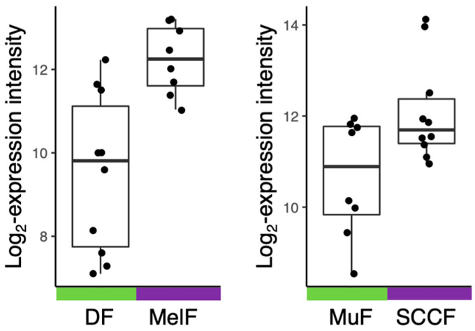
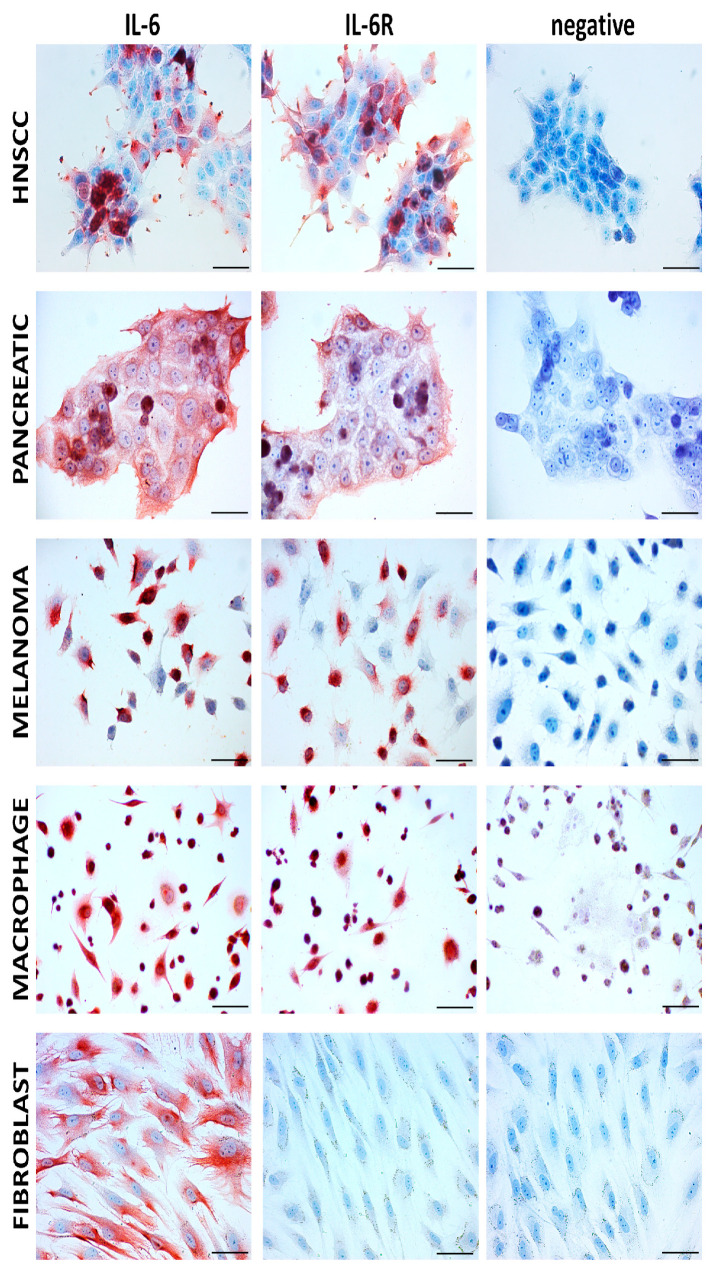
IL-6 was initially identified in 1986 by Hirano et al. as a pro-inflammatory cytokine produced by immune cells and since then has been implicated in a wide variety of pathologies ranging from chronic inflammatory conditions to cancer [5]. Currently, it is understood from numerous reviews that IL-6 production is not limited exclusively to immune cells but can also be synthesised by parenchymal cells of the skin, intestinal tract, smooth muscle, lung tissue, or stroll cells such as mesenchymal cells and fibroblasts [3,6]. Release of IL-6 from these stromal cells, such as fibroblasts, is depicted in Figure 1 based on Novák et al. [7] and the author’s unpublished data. Pathologically, IL-6 is produced by tumour stromal cells, immune cells, trafficking to the cancerous lesion, or the cancer cells themselves (Figure 2). Sources of IL-6 do not have to be limited to the tumour microenvironment (TME) but can also be produced by hematopoietic stem and progenitor cells (HPSC), epithelial cells, or muscle tissue, all contributing to the rigorous inter-cellular cross-talk, vital for the advancement of the disease [8,9]. This aberrant activation of downstream IL-6 signalling pathways is associated clinically and experimental with poor outcomes in oncological patients or cancer models [8,10]. Therefore, what is of particular interest in the area of cancer research are the effects of IL-6 on both stromal and parenchymal cells in promoting the invasiveness of the tumour and its ensuing metastasis.
Figure 1.
Expression of the IL6 gene in stromal elements of melanoma and head and neck squamous cell carcinoma (HNSCC). Normal dermal fibroblasts (DF, (left panel)) and normal mucous fibroblasts (MuF, (right panel)) express lower quantities of IL-6 mRNA than cancer-associated fibroblasts isolated from melanoma (MelF, (left panel)) or HNSCC (SCCF, (right panel)).
Figure 2.
Immunocytochemical analysis of IL-6 and IL-6 receptor in cell lines representing the malignant (several types) and the stromal component of tumours using immunoperoxidase reaction; positive staining is visualised by red AEC (3-amino-9-ethylcarbazole) substrate deposition. HNSCC–head and neck squamous cell carcinoma–cell line FaDu (CVCL_1218), pancreatic ductal carcinoma cell line PaTu n (CVCL_1846), melanoma cell line BLM (CVCL_7035). Macrophages were obtained by the standard protocol using the THP-1 monocytic cell line (CVCL_0006). Fibroblasts represented here are primary human isolates of dermal origin. Negative control was performed using isotype control. Gill’s haematoxylin (blue) was used for counterstaining. The bar represents 100 μm.
Given that, IL-6 is of particular interest in cancer research. In this review, we aimed to summarise the effects of IL-6 on both stromal and parenchymal cells, particularly on promoting tumour invasiveness and thus ensuing metastasis.
2. IL-6 Signalling and Downstream Effects
IL-6 signalling can follow either the classical or the trans-signalling pathway. Whereas the classical pathway is vital in the acute-phase immune response, regeneration, and haematopoiesis, the trans pathway allows cells not expressing the IL-6 receptor (IL-6R) to become responsive to this signal and initiate downstream signalling of IL-6 [3,10,11]. This allows such cells to engage in response to IL-6 stimuli and to become active participants rather than bystanders.
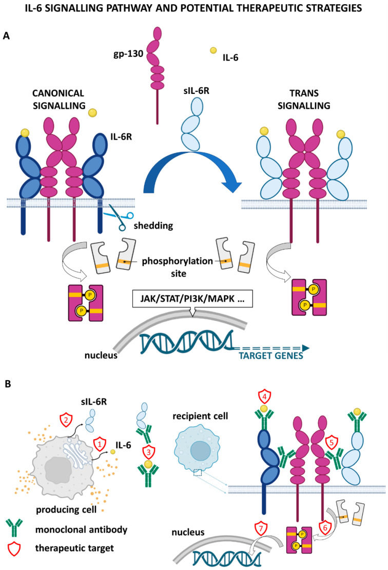
The classical IL-6 signalling pathway is initiated by IL-6 binding to a membrane-bound specific receptor, IL-6R. The ligand/receptor complex then associates with membrane-spanning gp130, resulting in the formation of a trimeric complex. Consequently, by further dimerisation, a heterohexameric signal-transducing receptor complex arises [5,11,12,13]. While IL-6R is limited in its expression to neutrophils, hepatocytes, monocytes/macrophages, and some lymphocytes, gp130 is ubiquitously expressed in most cell types [4,5,11,12]. However, cells that express gp130 alone are unable to bind IL-6 and are, therefore, not responsive to its effects. This highlights the relevance of the alternative trans pathway [2].
The trans-signalling pathway was discovered as a consequence of the detection of soluble IL-6R (sIL-6R) in human serum and urine samples [13]. The presence of sIL-6R and other cytokine receptors in body fluids is a general phenomenon that occurs under physiological conditions [13]. It was later confirmed that sIL-6R was markedly increased in several inflammatory diseases, such as chronic inflammatory bowel disease (in the serum) and rheumatoid arthritis (in the serum and synovial fluid). Of note, sIL-6R can promote tumorigenesis in cancers linked to long-standing inflammation, such as colitis-associated cancer [4,10,14,15,16,17,18]. sIL-6R is generated by cleavage via metalloproteases ADAM10 and ADAM17 and is shed from the membrane. It can bind circulating IL-6 and then form the necessary trimeric complex with gp130. The complex formation is followed by dimerisation and activation of downstream signalling [10,12,14]. It is of interest that, to a lesser extent, sIL-6R can also be generated via alternative splicing of pre-mRNA [19].
The trans pathway is critical in the context of cancer because it influences tumour and surrounding stromal cells that do not express IL-6R, thus modifying the activity and recruitment of cells into the TME [5,11]. For example, direct stimulation of tumour cells via IL-6 can induce increased proliferation and invasiveness. Paracrine or autocrine IL-6 signalling prompts stromal and immune cells to secrete additional signalling molecules such as VEGF for angiogenesis or pro-inflammatory cytokine IL-1β [10]. Thus, IL-6-initiated signalling gains higher complexity and involves multifaceted mechanisms of action crucial for shaping the course of cancer progression.
Regardless of the mechanism of IL-6/IL-6R/gp130 complex formation, it uniformly leads to the recruitment of Janus kinases (JAKs). JAKs, in turn, provide protein docking sites for additional pro-proliferative, pro-survival signalling pathways such as JAK/signal transducer and activator of transcription (STAT), PI3K/AKT, or for the RAS/RAF/MEK/MAPK pathways [5,10,12]. However, there are notable differences in downstream effects. In the classical pathway activation, the effects are associated with regenerative and anti-inflammatory results, as shown in several studies in murine models post partial hepatectomy [11,20]. On the other hand, it is the pro-inflammatory effect of the trans pathway activation that is implicated in the TME [10,11].
In order to emphasise the relevance of IL-6 downstream signalling and its role in cancer, we will primarily focus on the effects of the JAK/STAT3 signal transduction pathway, which has the ability to module tumour cell proliferation, survival, invasion, and metastasis, and thus is strongly associated with the progression of malignant disease. Following heterohexameric signal-transducing receptor complex formation, kinases JAK1, JAK2, and tyrosine kinase 2 (TYK2) associate with gp130. These kinases undergo activation via reciprocal transphosphorylation, thus allowing phosphorylation of tyrosine residues in the cytoplasmic region of gp130. Phosphorylated gp130 will now be able to interact with STAT3. Due to the proximity of STAT3 to activated JAKs, STAT3 is also activated via phosphorylation. Activated STAT3 forms a homodimer and acts as a transcription factor. In the nucleus, activated STAT3 targets regulatory sequences of genes encoding pro-proliferation factors such as cyclin-D1 and cMYC, pro-survival factors such as Bcl-XL and Bcl-2, and pro-angiogenic factors such as VEGF [10,11,14,21]. Increased STAT3 signalling and upregulated levels of cyclin-D1 and cMYC expedite progression through the cell cycle, while pro-survival factors also suppress apoptosis in cancer cells [22]. Furthermore, IL-6 downstream effects also modulate the activity of neutrophils, natural killer cells, or T cells, resulting in a decreased immune response to the neoplasm, despite the apparent trafficking of these immune cells to the lesion. This mechanism allows for the development of an immune tolerance [10,23]. IL-6 simultaneously upregulates T regulatory cells and myeloid-derived suppressor cells. Their activation further contributes to the remarkably immunosuppressed TME, resulting in a severely impaired anti-tumour immune response.
Thus, it becomes increasingly apparent how versatile IL-6 signalling is. In cancer, via stimulation of proliferation, survival, angiogenesis, or evasion of immune detection, it potentiates and propagates pro-cancerogenic signals within the TME. Further discussion will concentrate on the effects of IL-6 in the invasion-metastasis cascade across a range of cancer types, showing the applicability of targeting both IL-6 signalling and tumour cell migration as a therapeutic goal in cancer treatment.
3. IL-6 Signalling in Promoting Tumorigenesis, Invasiveness, and Metastasis in Cancer
IL-6 has been identified as a cytokine abundantly present in the TME of various tumour types, including head and neck squamous cell carcinoma (HNSCC) [24,25,26,27], pancreatic cancer [28,29], non-small-cell lung cancer [30], breast cancer [14,31], ovarian cancer [19,32], and melanoma [33,34]. In addition to being relevant in the course of tumorigenesis, IL-6 also facilitates the series of events that must occur as a prerequisite for the formation of a secondary tumour, a metastasis.
Investigation of the isolated mechanisms contributing to cancer progression in individual tissues and cancer types is extremely valuable. However, a unifying hallmark across nearly all types of solid tumours that also accounts for the highest mortality is the process of metastatic spread. As such, it merits in-depth investigation [31,35]. Metastasis formation is understood as a series of events beginning with (i) localised migration and invasion of the surrounding extracellular matrix (ECM), (ii) intravasation into nearby vessels, (iii) survival in extreme conditions in the bloodstream or lymphatic vessels, (iv) extravasation into the parenchyma of the tissue, and finally (v) modification of the activity of tumour cells to allow them to thrive in a new environment [36,37,38]. Just as metastasis formation is a common process occurring in a variety of types of cancer, aberrant IL-6 signalling provides another unifying motif that supports tumour growth and metastasis. Therefore, it provides IL-6 with a position as a promising therapeutic target. To demonstrate just how consequential this cytokine is, the metastatic cascade will be discussed in light of IL-6 signalling in selected cancer types.
4. IL-6 Contributes to the Formation of the Pre-Metastatic Niche
Early observations in breast cancer led Stephen Paget to coin his “seed” (cancer cells) and “soil” (host tissue) hypothesis of metastasis. In these pioneer times, Paget scrutinised why and how some organs were affected by metastases while others remained unscathed [39]. Only more recently, sufficient data were gathered to shed light on this process mediated by tumour-secreted factors, extracellular vesicles (EV), and bone marrow-derived cells (BMDCs). All these tightly orchestrated mechanisms prepare the preselected pre-metastatic niche (PMN) for the arrival of cancer cells [9,40,41,42]. The sequential preparation of the PMN involves changes such as vascular leakiness, allowing CSC extravasation and activation of stromal cells and restructuring of the extracellular matrix (ECM), facilitating recruitment of cell types such as BMDCs for PMN formation [41,42]. The most important cellular component of the PMN is BMDCs, especially immune cells, all derived from haematopoietic progenitor and stem cells (HPSCs) [9,42]. HPSCs can respond to conditions such as injury and inflammation or to tumour-secreted stimuli, promoting their differentiation [9,42]. Perhaps the most significant population of differentiated HPSCs are myeloid-derived suppressor cells (MDSCs). MDSCs maintain a chronic pro-inflammatory and yet immunosuppressed environment within the PMN [9,43]. It is unclear how and why the MDSCs migrate to the PMN. However, data show that a number of soluble factors, including IL-6, may regulate MDSC recruitment, activation, and differentiation within the PMN [Talmadge history].
Magidey-Klein et al. used paired breast cancer or melanoma cell lines, one with a high frequency of metastasis (met-high) and one with a low frequency of metastasis (met-low), to study the role of IL-6 in HPSC differentiation, metastasis, and its involvement in the generation of the PMN. Cells were transplanted into mouse models with comparable success and presented similar tumour growth between the met-high and met-low groups. However, lung metastases were significantly more frequent in the high-met group [9]. When analysing the cellular composition of the bone marrow, mice with tumours were also observed to have a higher percentage of LSK cells (considered as HPSCs) than control mice. Specifically, the monocyte dendritic progenitor (MDP) population was elevated in both cancer types when compared to other progenitor cell types. Overall, the results showed that met-high tumours induce myeloid-biased differentiation of HPSCs, which correlates with tumour aggressiveness and metastatic potential. IL-6 was identified as a mediator for the cross-talk between bone marrow and cancer cells, and the levels of IL-6 correlated with MDP growth and increased incidence of metastasis. In met-low melanoma cells, overexpression of IL-6 was a sufficient signal to educate MDPs to induce metastasis and metastatic switch. MDPs further differentiated into M2 pro-inflammatory and immunosuppressive macrophages, localised at the metastatic sites. Magidey-Klein and co-workers put forward a new role of tumour-derived IL-6 in driving the differentiation of HPSCs toward pro-metastatic MDPs. This shows the importance of IL-6 not only in the context of TME but also in its potential to orchestrate the bone marrow niche, which is vital for the eventual formation of the PMN.
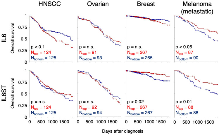
The influence of mRNA expression of the IL-6 signalling components on patient survival is, however, not direct. While in HNSCC, low expression of IL6 mRNA improves survival of the patients with marginal statistical significance (Figure 3, XENA [44]), in ovarian and breast cancers, there is no difference in survival of the patients with high IL6 expression. In metastatic melanoma, low expression of IL6 shortens the patient survival, paradoxically. High expression of IL6ST, the gene coding for gp130, significantly improves the survival of patients suffering from breast cancer and metastatic melanoma (Figure 3). No such correlation was observed for IL6R mRNA expression.
Figure 3.
Correlation of patient survival with mRNA expression of IL-6 pathway components. Overall survival of patients suffering from HNSCC, ovarian cancer, breast cancer, or metastatic melanoma is evaluated for patients with different levels of IL6 mRNA expression (top) and IL6ST mRNA expression (bottom). Survival of the patients with the highest gene expression (4th quartile, Ntop patients) was compared with the survival of the patients with the lowest expression (1st quartile, Nbottom) using Kaplan–Meier curves and the log-rank test. The analysis was performed within the Xena platform [44].
5. Metastasis Repression by Targeting IL-6 Signalling
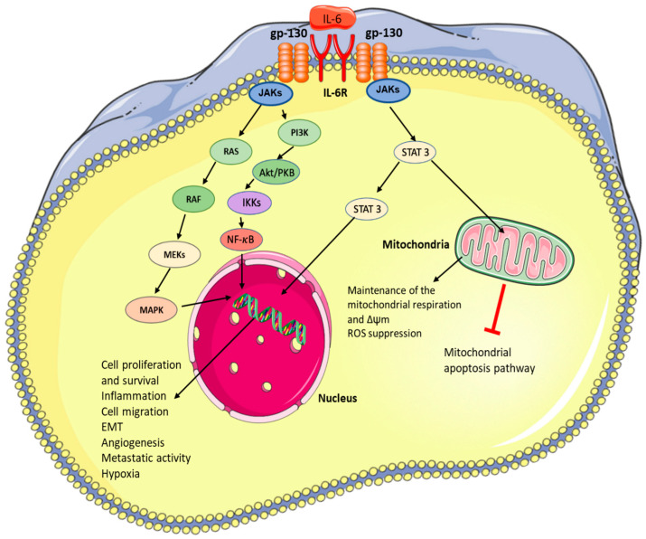
As a result of the ubiquitousness of IL-6 pro-cancerogenic signalling, the molecule presents itself as a therapeutic target that will “pack a punch”. IL-6 signalling is associated with increased invasiveness, aggressiveness, and incidence of metastasis across many tumours (Figure 4) [3,8,45,46].
Figure 4.
Influence of IL-6 signalling on cancer development and metastasis formation. The figure was created using Servier Medical Art available at http://smart.servier.com/ (accessed on 15 August 2022).
Recent data also implicate this protein in preparation for the PMN, which is another essential step prior to cancer cell dissemination. Therefore, since IL-6 plays a central role in the invasion-metastasis cascade, which is the leading cause of cancer-related deaths worldwide, it is an absolutely obligatory avenue for novel pharmacological interventions [3,8,45,46]. Inhibition of IL-6 and its signalling pathways is an intensively studied therapeutic approach in cancer treatment. Strategies explore general inhibition of the IL-6 signalling axis (IL-6/6IL-6R/gp130), including downstream signalling proteins such as STAT3, NF-κB, and HIF-1α. The most frequently used/studied strategies for the inhibition of IL-6 signalling are shown in Figure 5, and examples of possible therapeutic agents for targeting IL-6 and its pathway are summarised in Table 1.
Figure 5.
Therapeutic strategies for the inhibition of IL-6 signalling.(A) intracellular signaling, (B)intercellular signaling. (1) cytokine synthesis/release blockade, (2) sIL-6R shedding blockade, (3) cytokine/soluble receptor neutralizing, (4) cytokine binding to receptor prevention, (5) heterotetrameric complex formation blockade, (6) signal transducer kinase activity inhibition, (7) downstream signalling blockade/gene transcription blockade.
Table 1.
Examples studied agents for the inhibition of IL-6 signalling with an emphasis on their clinical applicability.
| Name (Trade Name) | Target: Function | Status | Ref. |
|---|---|---|---|
| Human monoclonal antibody | |||
| Tocilizumab (RoActemra) | IL-6R: receptor inhibition | Clinically used for rheumatoid arthritis | [47] |
| Case report: Reducing IL-6-mediated cachexia | [48] | ||
| Sarilumab (Kevzara) | IL-6R: receptor inhibition | Clinically used for rheumatoid arthritis | [49] |
| Under clinical trial (EMPOWER NCT04333706): triple-negative breast cancer (stage I-III, high-risk residual diseases) combination with Capecitabine | |||
| Siltuximab | IL-6: neutralization | approved for CAR-T | [50] |
| Low molecular inhibitor | |||
| Bazedoxifene (Conbriza) | Estrogen receptor modulator | Clinically used in the treatment of osteoporosis | [51] |
| gp130: inhibitor | In vivo | [52] | |
| CD40 receptor: inhibitor | In vitro | [53] | |
| Tofacitinib (Xeljanz) | JAK1/3 inhibitor | Clinically used in the treatment of moderate-severe ulcerative colitis | [54] |
| JAK pathway | Clinical trial: developed malignancies lung, breast, gastric cancer, and lymphoma; rate of malignancies by 6-month intervals of tofacitinib exposure indicates rates remained stable over time | [55] | |
| Ruxolitinib (Jakafi) | JAK1/2 inhibitor | Clinically used in the treatment of steroid refractory graft-versus-host disease | [56] |
| JAK pathway | Clinical trials: inadequately controlled polycythaemia; decrease in thromboembolic events | [57] | |
| Momelotinib | ACVR1/ALK2, JAK1 and JAK2, inhibitor | FDA accepts for the treatment of the myelofibrosis | |
| Momelotinib | Clinical trials: myelofibrosis; higher overall and leukaemia-free survival. | [58] | |
| Madindoline A and B | gp130: inhibitor | In vitro | [59,60] |
| ERBF | IL-6R: blocking interaction IL-6R with IL-6, or gp130 | In vivo | [61,62,63] |
| Stattic | STAT3: inhibition of activation | In vivo | [64] |
| OPB-31121 | STAT3: inhibition of activation | Clinical trials: advanced colon and rectal tumours; tumour shrinkage, bad pharmacokinetic (very low blood concentration) | [65,66] |
| Galiellalactone | STAT3: inhibition of DNA binding | prostatectomy samples: reduction IL-6 induced AR signalling | [67] |
| In vivo | [68] | ||
| GPB730 | STAT3: inhibition of DNA binding | In vivo | [69] |
| OPB-51602 | STAT3: activation of aggregation | Clinical trials: refractory haematological malignancies; no clear therapeutic response was observed | [70] |
| Ixazomib | NF-κB: inhibition of ubiquitin-proteasome pathway leading to loss of NF-κB activity | Clinical trials: Relapsed or Refractory Cutaneous or Peripheral T-cell Lymphomas; reduction in NF-κB activation and subsequently GATA-3 expression in the biopsy specimens | [71] |
| Theofyline (Elixophyllin, Elixophylline, Pulmophylline, Quibron-T, Theo-24, Theolair, Uniphyl) | phosphodiesterase inhibitor, adenosine receptor blocker, and histone deacetylase activator | Clinically used in chronic obstructive pulmonary disease and asthma | [72,73] |
| NF-κB: inhibition of activation | In vitro | [74] | |
| Rapamycin (Sirolimus, Rapamur) | mTOR: inhibitor | Clinically used immunosuppressive therapy | [75] |
| Clinical trials: acute myelogenous leukaemia; no effects on the composite complete remission rate | [76] | ||
| IL-6, TNF-α and IL-1β: decrease cytokine level | In vivo | [77] | |
| Zotarolimus | IL-1β, TNF-α, IL-6 and NF-κB: decrease cytokine level and NF-KB activity | In vivo | [78] |
|
NSAIDs (e.g., celecoxib, aspirin, ibuprofen, naproxen, meloxicam) |
cyclooxygenase inhibitors | in a broad spectrum of conditions; Analgetic, antipyretics, in rheumatic diseases | [79] |
| Celecoxib | IL-6: decrease expression by COX-2 inhibition | Clinical trials: former-smokers; bronchoscopy samples (reduction IL-6 and Ki-67 expression) | [80] |
| Food supplements | |||
| Curcumin | IL-6: decrease expression | Clinical trials: patients with solid tumour; decrease in plasma level of IL-6, TNF-a, TGF-b, substance P, hs-CRP, CGRP and MCP-1, increase patient quality life | [81] |
| STAT3: decrease activity | In vivo | [82] | |
| NF-κB: activity and expression | Clinical trials: advanced pancreatic cancer; peripheral blood mononuclear cells (decrease in expression of NF-κB, STAT-3 and COX-2) decrease in serum cytokine levels (IL-6, IL-8, IL-10, and IL-1 receptor antagonists) | [83] | |
| HIF-1α: decrease expression and activity | In vitro and In vivo | [84,85] | |
| Epigallocatechin-3-gallate | NF-κB: decrease expression | Clinical trial: subjects with a high risk of colorectal cancer; lover expression of the NF-κB and DNMT1 | [86] |
| STAT3: inhibition of activation | Molecular assay and In vitro | [87] | |
Currently, the clinical and/or research applications of IL-6R targeting antibodies are utilised in a variety of fields. Tocilizumab, an IL-6R antibody, is clinically used in the treatment of various autoimmune diseases, such as rheumatoid arthritis, which is associated with pathologically hyper-activated IL-6 signalling [47]. Despite the practical applications in the field of rheumatology, the therapeutic use of such biological treatments in oncology still requires optimisation. However, there are some encouraging data supporting this concept.
In the case of recurrent ovarian carcinoma, tocilizumab decreased STAT3 activation/phosphorylation in patient immune cells (e.g., myeloid cells, CD4+ T and CD8+ T only at a high dose), most probably due to the suppression of IL-6R signalling [88]. It suggests that tocilizumab could suppress IL-6-induced immunosuppression (e.g., induction of macrophage M2 phenotype and Treg attraction) [89,90]. However, patients with acute leukaemia or myelodysplasia did not show any improvements in long-term survival on tocilizumab treatment [91]. A combination of Sarilumab (antibody targeting soluble and membrane IL-6R; FDA-approved for rheumatoid arthritis) and Capecitabine is currently tested in a clinical trial (EMPOWER; NCT04333706) in triple-negative breast cancer patients (stage I-III, high-risk residual disease). Moreover, Nguyen et al. reported that siltuximab (IL-6R antibody) could repress the Wnt/β-catenin pathway [92].
An alternative strategy employs small molecular inhibitors of IL-6/gp130 signalling rather than IL-6R inhibitors [3]. This approach also shows potential for a beneficial anti-metastatic effect. Bazedoxifene (repurposed selective oestrogen receptor modulator) displays a strong inhibitory effect on gp130 (receptor kinase of IL-6R). In the case of cervical cancer cells (SiHa, HeLa, CaSki), bazedoxifene treatment leads to a decrease in cell migration and invasion and additionally decreases Siha tumour burden in mouse models [52]. Its effect is associated with a reduction of IL-6-induced GP130, STAT3 and ERK1/2 phosphorylation. Possible agents for inhibition of IL-6 signalling could also be based on the structural motif of madindolines (gp130 inhibition) [93] and bufadienolide (blocking interaction of IL-6R with IL-6 or gp130) [61,62].
STAT3 plays a key role in IL-6 signalling and carcinogenesis. STAT3 (respective STAT3α isoform) [94,95] is involved in the EMT and self-renewal of cancer stem cells, promoting metastasis and invasion. Its downstream effects are crucial for the formation of an immunosuppressive TME [10]. Specifically, in the context of TME, active STAT3 signalling induces repression of neutrophils, natural killer (NK) cells, effector T cells, and dendritic cells (DCs) and activates regulatory T (Treg) cells and MDSC populations. This immunosuppressed landscape is thought to contribute to the weakened ability of the immune system to respond to developing cancer. Despite STAT3′s critical contribution to forming a cancer-supportive TME, no drug targeting STAT3 itself has been clinically approved for this purpose until now. Nevertheless, several promising compounds are being intensively studied [96,97]. For example, Stattic (low molecular direct inhibitor of Src homology 2 domain) significantly represses STAT3 activation and expression in a murine orthotopic xenograft model of HNSCC. This effect is associated with a reduction of STAT3-mediated HIF-1α expression and tumour progression [64]. Similarly, the application of OPB-31121 (oral STAT3 inhibitor) repressed constitutive and IL-6-induced JAK/STAT3 signalling in gastric cancer lines [98]. In this cohort of patients with advanced colon and rectal tumours, OPB-31121 treatment was associated with tumour shrinkage. The application was safe and relatively well tolerated [66]. Nevertheless, its pharmacokinetics displays significant variability, and the mean blood Cmax (1.19–12.9 ng/mL) was much lower than in this case of the mouse tumour xenograft model [65,66].
In the past, when targeting particular signalling cascades, it has proven to be both important and effective to target the pathway at all levels, e.g., receptor, second messenger, and subsequent signalling proteins. Therefore, another therapeutic opportunity is emerging at the level of gene expression since IL-6 cascade-activated STAT3 acts as a transcription factor. By inhibiting STAT3′s DNA binding ability, its capacity to act as a transcription factor would be eliminated. While galiellalactone inhibits STAT3 signalling by binding to its DNA-binding region, STAT3 phosphorylation/activation is not repressed [67,99]. In prostate cancer cell lines (LNCap DuCaP and VCaP), galiellalactone strongly repressed IL-6-induced activity of the androgen receptor (AR; e.g., PSA expression) [67]. In these primary tissue slice cultures from radical prostatectomy samples, reduced expression of AR-controlled genes (e.g., PSA, TMPRSS2 and FKBP5) was also observed. AR plays one of the key roles in the development of prostate cancer, and inhibition of its signalling has been a main therapeutic option to manage locally advanced and metastatic prostate cancer in clinics [100].
While cytosolic STAT3 is an obvious choice as a therapeutic target, it was shown that the dysfunction of mitochondrial STAT3 can create sufficient stress for the cancer cells to induce apoptosis. Therefore, a combination of mitochondria-targeting drugs (e.g., arsenic trioxide, tamoxifen, hydrocortisone and others) [101] with IL-6 signalling inhibitors could be a base for another attractive alternative. Mitochondrial STAT3 plays an important role in the control of cellular respiration in the mitochondria (e.g., complexes I and II of the electron transport chain) [102,103] and can strongly support the oncogenic process [104,105]. Over the last years, it was reported that the phenotype of metastatic cancer is associated with active mitochondrial oxidative phosphorylation. This plays a central role in the generation of ROS, cell death, survival, and metastasis [106,107]. In detail, metastatic cells’ survival in the blood and their homing to the metastatic site may depend on the mitochondrial oxidative phosphorylation [108,109]. The redox balance is regulated by very high glucose uptake, and the stimulated citrate cycle enhances mitochondrial membrane potential. Cancer cells can also capture glycolytic lactate produced by fibroblast, tumour, and stromal cells [110,111]. The obtained lactate is converted to pyruvate, and in the mitochondria, it provides electrons for the mitochondrial electron transport chains and energy for ATP production. This process is called the “reverse Warburg” effect.
Targeting STAT3 could be an effective way to repress the mitochondrial function in cancer cells. OPB-51602 (an SH2 domain-targeting STAT3 inhibitor) stimulates the formation of proteotoxic STAT3 aggregates and resulting mitochondrial dysfunction [112]. OPB-51602-related cytotoxicity was induced by glucose starvation and increased reliance of prostate cancer cells DU 145 on mitochondrial function. Similarly, a decrease in IL-6-induced STAT3 mitochondrial localisation leads to mitochondrial oxidative stress, loss of mitochondrial membrane potential, and subsequent apoptosis of cancer cells [113].
Repression of NF-κB activity could also be a promising approach to the inhibition of IL-6 signalling. NF-κB proteins are a group of oncogenic factors that, in turn, control the expression of pro-inflammatory signalling proteins, such as IL-6. There is increasing evidence suggesting that NF-κB targeting could also increase tumour sensitivity to the therapy (e.g., chemo- and radiotherapy) and delay/repress the loss of therapeutic effectiveness. Chemotherapeutics (e.g., paclitaxel, 5-fluorouracil or doxorubicin) can stimulate strongly increased production of cytokines such as IL-1β, IL-6, IL-8, CSF2, and CCL2 [106,114]. IL-6 activates NF-κB (via STAT3/AKT pathway) [115,116], which subsequently promotes the production and secretion of more cytokines [114].
In TME, IL-6 signalling can significantly enhance the hypoxic phenotype [117]. Chronic inflammation stimulates cycling hypoxia as a result of limited oxygen diffusion and increases consumption of oxygen by filtrating immune and hyperproliferating cancer cells, leading to the lack of oxygen in TME [118,119,120]. A hypoxic phenotype is associated with EMT transition and resistance to therapy, especially immunotherapy. For example, macrophage-derived IL-6 promotes EMT in primary hepatocellular carcinoma cells under hypoxic versus oxygenated conditions [117]. Notably, this EMT can be strongly repressed by tocilizumab. The increased levels of intracellular ROS produced during hypoxia lead to the stabilisation of HIF-1α and NF-κB. HIF-1α induces an immunosuppressive tumour microenvironment by recruiting Tregs, MDSCs and macrophages. The NF-κB signalling pathway can also support the production of inflammatory factors and the recruitment of inflammatory immune cells [119]. Finally, higher levels/activity of infiltrated inflammatory cells result in a repeated hypoxia cycle. It suggests that targeting the intratumoral inflammatory mechanism by targeting IL-6 signalling could repress the hypoxic phenotype. Nevertheless, hypoxia can stimulate the activity of IL-6 signalling and thereby potentially decrease the effectiveness of this therapeutic strategy.
6. Inhibition of IL-6 Signalling in Combination Therapy
It is well known that oncological diseases display significant heterogeneity and interindividual variability. Therefore, general oncogenic signalling pathways may be at least partially substitutable targets, and their simultaneous modulation can synergically abrogate tumorigenesis. For example, both IL-6 and IL-8 can activate STAT3 signalling via JAK 2 and significantly increase cell migration when the signalling occurs concurrently, as opposed to stimulation by IL-6 or IL-8 alone [121]. A possible approach to this corroborative effect could be their simultaneous dual targeting. Simultaneous inhibition of IL-6 and IL-8 receptors via Tocilizumab and Reparixin (inhibitor of C-X-C motif chemokine receptor 1) significantly decreased the expression of matrix metalloprotease in mouse MDA-MB-231 breast cancer model models and decreased the incidence of liver and lung metastasis [122]. Similarly, a combination of bazedoxifene and SCH527123 (inhibitor of C-X-C motif chemokine receptors; Il-8 receptors) synergically repressed STAT3 and Akt phosphorylation in ovarian cancer cells (OVCAR3, SKOV3, and CAOV3) and in mice bearing CAOV3 tumours when compared to agents inhibiting just IL-6 or IL-8 [123]. According to the proposed model, in this case, the effect of combination therapy on tumour growth was sometimes smaller compared to the application of single agents. In this connection, it is interesting to note that bazedoxifene could repress TNF-α activation of CD40 receptors and subsequent activation of the NF-κB, STAT3, and PI3K/AKT/mTOR signalling [53]. Moreover, in the tumour microenvironment, TNF-α is one of the activators of IL-8 expression [44].
Similarly, simultaneous activation of NF-κB and HIF-1α can synergically enhance tumour development and metastasis formation [119]. Nevertheless, their effects could be strongly suppressed by the co-application of low-toxic, multi-targeting natural compounds such as curcuminoids and flavonoids with potent anti-metastatic effects [124]. For example, curcumin is a direct inhibitor of NF-κB signalling, and its application can also repress the hypoxic phenotype by targeting HSP90 and mTOR (HIF-1α stabilisation and expression) [84,125,126]. Furthermore, flavonoids inhibit various signalling pathways associated with cell migration and metastatic activity, such as MAPK, AKT, mTOR, STAT3, and/or NF-κB pathways [127]. Moreover, both types of agents display low toxicity, and their application is favourable for patients.
A promising strategy could also be based on a dual inhibitor for simultaneous targeting of mitochondrial metabolism and IL-6 signalling pathway. In a recent study, it was reported that bis-pentamethinium salts could inhibit the gp130 protein and disturb mitochondrial respiration [128]. Nevertheless, their mitochondrial uptake is too fast, and thereby their effect on IL-6 may be limited. However, their structural motif can be used as an appropriate starting point in the design of these novel dual inhibitors. It is interesting to note that suitably designed pentamethimium salts display very strong inhibition activity against dihydroorotate dehydrogenase [129], which catalyses the mitochondrial step of de novo pyrimidine synthesis (conversion of dihydroorotate to orotate) [130]. Because the enzyme prosthetic group flavin mononucleotide serves as an acceptor of a dihydroorotate electron, its inhibition can cause disturbance of mitochondrial respiration [131]. In prostate cancer cells, pentamethinium application led to the imbalance of the mitochondrial metabolism, which is strongly associated with the repression of cell migration and invasiveness [129].
Clinically used inhibitors of IL-6 signalling, such as IL-6R antibody, could be promising tools to assist classical anticancer treatment (e.g., chemotherapy and radiotherapy). For example, in the mouse model of mucoepidermoid carcinoma, tocilizumab repression of STAT3 and AKT phosphorylation caused a significant decrease in tumour growth, drug resistance, and increased overall survival [132]. Although in vitro, the tocilizumab application did not display any cytotoxicity in mucoepidermoid carcinoma cells. It decreased the subpopulation of cancer stem cells (ALDHhighCD44high) and in vivo repressed paclitaxel-related induction of this cancer stem cell phenotype. Tocilizumab could also be a prospective agent in combination therapy for treating radiotherapy patients. Matsuoka et al. reported that higher IL-6 levels could be observed in squamous cell carcinoma cells and tissue samples from squamous cell carcinoma patients [133]. In squamous cell carcinoma cells, IL-6 supports cell survival via STAT3 and nuclear factor erythroid 2-related factor 2 signalling. Based on that, it was hypothesised that tocilizumab application could strongly enhance the radiosensitivity of the tested cells. Moreover, in order to increase the efficacy of treatment, therapeutic strategies can also simultaneously target various levels of IL-6 signalling. Dual application of tocilizumab and stattic significantly repressed IL-6-induced expression of vimentin and VEGF and downregulation of E-cadherin in DU-145 prostate cancer [134]. This phenomenon was associated with a substantial decrease in cell viability, colony formation, and migratory and invasive capacity against single-target inhibition.
The above-mentioned facts strongly suggest that targeting IL-6 signalling could greatly enhance the commonly used anticancer modalities. Nevertheless, numerous clinical trials are requested for the validation of this hypothesis and the optimisation of possible therapeutic strategies.
7. IL-6 Signalling in Selected Cancer Types
7.1. Head and Neck Squamous Cell Carcinoma
HNSCC is the most common cancer type of the head and neck region. Epidemiologically, HNSCC is one of the top ten most common cancer types worldwide. It affects the epithelium of the oral cavity, pharynx, and larynx [135]. It is most commonly associated with the use of tobacco products, alcohol consumption, poor oral hygiene or infection, namely by human papillomaviruses (HPV) [135,136]. Population-based screening for HNSCC has proven ineffective, as most patients do not present with pre-malignant symptoms [135]. While 5-year survival rates for patients with early-stage HNSCC are good, around 80%, this figure rapidly drops with cancer spread to lymph nodes down to 40%. Further, the survival with metastatic spread falls to 20% only [137]. IL-6 has been shown to be one of the molecules whose levels correlate with HNSCC progression and patient survival [24].
In HNSCC, a specialised subpopulation of cells, cancer stem cells (CSC), localise to a perivascular niche, and this convenient proximity to an organ’s vasculature most likely allows subsequent migration and intravasation into blood vessels [25,138]. CSC have unlimited potential for proliferation and self-renewal, thus being able to perpetuate the growth of HNSCC [25,136]. The tumorigenic potential of CSC correlated with IL-6 levels, as confirmed in both mice transplanted with HNSCC and in tissue sections from HNSCC patients [25]. Kim and colleagues generated CRISPR/Cas9 IL-6 knockout endothelial cells, which were co-implanted with UM-SCC-22B cells to form xenograft tumours and then implanted into mouse models. Slowed tumour growth was observed with IL-6 knockout endothelial cells when compared to the control, suggesting that secreted endothelial IL-6 advanced the migratory phenotype of the cancer cells. Furthermore, cancer stem cell migration in vitro was also reduced when treated with antiIL-6 antibodies or tocilizumab, and cultures had a smaller fraction of cancer stem cells, a key piece of data helping to further describe the contribution of cancer stem cells and IL-6 in tumorigenesis and metastatic spread [138,139]. Additionally, Wang et al. found increased expression of mRNA of IL-6 and IL-6R in human tumour samples when compared to the physiological oral mucosa, with higher expression also being associated with larger tumours and more advanced histological grade [140]. Overall, data have shown that IL-6 prepares cancer stem cells, likely via a chemotactic mechanism, for the epithelial-to-mesenchymal transition (EMT), essential for the next step of the invasion-metastasis cascade [25,138].
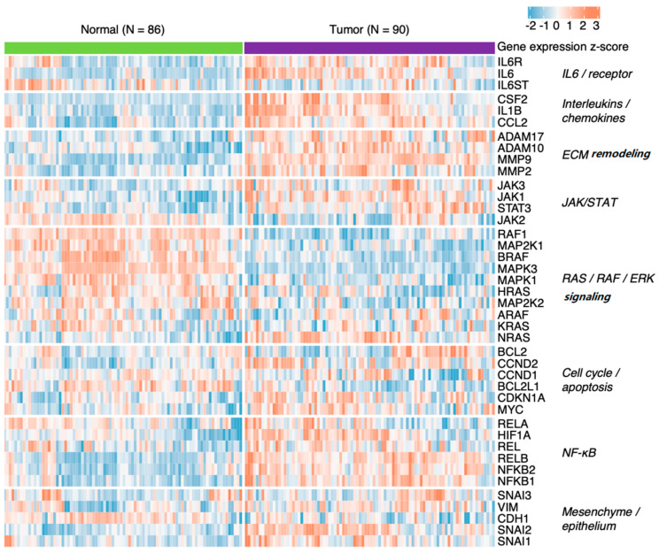
Novotný et al. (2020) [141] suggested compensatory deregulation of the genes coding for cyclins D1 and D2 in HNSCC. Moreover, analysis of their publicly available data (ArrayExpress accession E-MTAB-8588) using the DESeq2 Bioconductor package [142] showed deregulation of IL-6 pathway components, including some of its downstream targets (Figure 6).
Figure 6.
Deregulation of IL-6 signalling and its downstream targets in HNSCC tumours compared to matched samples of adjacent healthy mucosa (see Novotný et al., 2020 [141] for details).
7.2. Ovarian Cancer
Ovarian cancer is the most lethal of female genital cancers because of a lack of early clinical symptoms in the patient and a lack of effective screening methods. As a result, the malignancy is usually discovered at an advanced stage, worsening the survival rate of patients. IL-6 is prevalent in the TME of ovarian cancer and, via complex signalling and response by both cancer and stromal cells, is able to promote proliferation, angiogenesis, and migration while inhibiting apoptosis [19,32,143]. The overactivation of IL-6 pathways, particularly activation of STAT3, has been implicated in the aggressiveness of ovarian cancer [144]. Activation of STAT3 by IL-6 allows expression of cell cycle-promoting proteins such as cyclin D1, D2, and c-MYC and downregulation of cyclin-dependent kinase inhibitor p21, facilitating entry into the cell cycle, thus enhancing the growth of the tumour [32,145]. Saini and co-workers observed that activated STAT3 is highly expressed in ascites-derived ovarian cancer cells (ADOCC). When transplanted into the ovarian bursa in mice, ADOCC proceeded rapidly to generate large tumours as well as extensive metastases to the liver and peritoneum. STAT3-knocked-down ADOCC failed to form metastases and resulted in slower tumour growth [144]. Using the SKOV-3 cell line and treatment with ascites fluid from three patients with advanced serous ovarian carcinoma, Kim et al. showed an increase in cellular migration and invasion in response to treatment. The effect was only seen in SKOV-3 ovarian cancer cells and not in immortalised ovarian surface epithelial cells [26]. Additionally, the ascites-treated SKOV-3 cells showed a mesenchymal phenotype with decreased levels of E-cadherin (epithelial marker) and increased levels of Snail and vimentin (mesenchymal markers) [26]. Subsequent analysis showed increased levels of IL-6 in ovarian cancer patient ascites, and treatment using anti-IL-6 antibodies showed decreased invasion and migration, decreased mesenchymal phenotype, and decreased activation of the JAK/STAT3 downstream signalling [26]. Increased invasiveness of IL-6-expressing ovarian cancer cells was also demonstrated by Wang et al. Invasiveness was evaluated based on cell proliferation, ability to invade Matrigel-coated Transwell chambers, and expression of matrix metalloproteinases 2 and 9. In A2780 (cells not expressing IL-6), highly invasive behaviour was observed after overexpression of IL-6. Compared to A2780 untransfected controls, the authors observed better anchorage-independent growth and enhanced cell migration. The effect was abrogated when IL-6-expressing SKOV-3 cells transfected with an antisense IL-6 plasmid showed decreased invasive abilities [146].
7.3. Breast Cancer
Breast cancer is the second leading cause of cancer-related deaths in women [147]. It was estimated that every one in eight women would develop breast cancer in their lifetime. In breast cancer, several studies have shown that patients with breast cancer have increased serum levels of IL-6. Moreover, there is evidence that IL-6 levels correlate with worse survival rates in patients, especially in those with metastatic breast cancer [14,148]. Early research in vitro demonstrated IL-6-dependent motility of human ductal carcinoma cells. Morphologically, the carcinoma cells initially had an epithelioid morphology, but upon exposure to IL-6, promptly adopted a stellate or fusiform shape. This was associated with increased motility of the cells and loss of their cell-cell junctions [149]. Recently, it was shown that IL-6-dependent repression of E-cadherin expression and weakening of adherens junctions correlates with invasiveness and metastatic potential by promoting an EMT phenotype both in in vitro experiments and mouse models [150]. Chang et al. demonstrated in mouse models that both IL-6 and downstream activated transcription factor STAT3 were present at the leading edge of breast tumours, suggesting a link between the presence of IL-6 and the invasive behaviour of the tumour itself. Indeed, Chang and colleagues proposed a feed-forward mechanism. In the suggested model, paracrine IL-6 signalling from tumour cells also activated p-STAT3 and IL-6 expression in stromal components–namely endothelial cells, CAFs (cancer-associated fibroblasts), and myeloid cells. This further functionally enhanced the IL-6/JAK/STAT3 signalling axis within the TME. This feed-forward mechanism dictated the ability of the tumour to proliferate, establish vascular supply, regulate the degree of inflammation, and determine the metastatic potential of the tumour [31]. Additional studies further proved the pivotal role that IL-6 plays in the aggressiveness of breast cancer by stimulating a stem-like phenotype in MCF-7 mammospheres, characteristic of basal-like breast carcinoma, and IL-1β-dependent expression of IL-6, which increased stemness, invasiveness, and survival in MCF-7 cells [151,152].
7.4. Melanoma and Cutaneous Squamous Cell Carcinoma
The incidence of melanoma has been rising around the world, despite the incidence of other cancers decreasing [153]. Melanoma is an aggressive malignant disease of multifactorial aetiology. Melanoma is often primarily resistant to various oncological therapeutical modalities, making treatment difficult. Until recently, melanoma was nearly incurable in the case of metastatic disease [154].
In a spheroid-based model using A2058 human melanoma cells, Jobe et al. showed that in cultures with conditioned media (CM) from A2058 + CAF, CAF, or normal primary fibroblasts, invasiveness increased the most in spheroids cultured in CM from the A2508 + CAF condition [34]. Upon analysing levels of IL-6, it was found that the levels in CM were increased especially in cultures of CAF or fibroblasts, suggesting that these cells are the dominant producers of IL-6 within the TME. However, the most marked increase in IL-6 was from the co-culture of A2508 or BLM cells together with CAF [34]. When anti-IL-6 antibodies were added to the cell culture, the invasive effects were significantly diminished [34]. Fibroblasts co-cultured with invasive human melanoma cell lines showed increased expression of chemokines and cytokines such as IL-1β, IL-8, and IL-6. IL-1β is thought to promote invasiveness by inducing the expression of pro-inflammatory signalling molecules such as IL-6, and subsequent siRNA silencing of IL-1β attenuated the invasiveness of the cells [33].
Weber and co-workers recently observed in a murine melanoma model that IL-6 induced CCR5 expression and thus induced potent immunosuppressive activity of MDSC in the TME [155,156]. Increasingly in the last decade, immune checkpoint blockade therapy redefined the therapeutic options to treat advanced melanoma. However, it is a great success in clinical oncology; acquired resistance and treatment-related toxicities are widespread. Hailemichael et al. suggested recently that the combination of IL-6 blockade and dual inhibition of CTLA-4 and PD-1 can overcome these issues [157]. This identifies another critical mechanism in melanoma immune response avoidance and makes IL-6 a promising target for melanoma immunotherapy.
Depner and colleagues were able to identify a possible mechanism by which IL-6 activation of stromal fibroblasts contributes to the metastatic potential of human squamous cell carcinoma (SCC) [158]. In in vitro organotypic co-cultures with human fibroblasts and a human skin carcinogenesis model (HaCaT-ras A-5RT1 cell line), IL-6 was found to activate fibroblasts and encourage progression to the tumour-associated fibroblast phenotype, activating expression of metalloproteinase-2, thus promoting the invasive capabilities [158]. In addition to paracrine signalling, exosomes produced by melanoma cells stimulated the production of IL-6 by CAFs, which improved in vitro migration of melanoma cells from the heterogeneous spheroids containing melanoma cells and CAFs in 3D collagen gels [159,160].
Taken together, these experiments demonstrate the significance of the cross-talk between stromal and tumour cells in cancers of the skin and elucidate the mechanisms by which IL-6 is able to promote invasiveness in cancers of the skin.
8. Conclusions
The IL-6 signalling pathway plays a significant role in cancer biology, particularly in its involvement in metastasis formation. Targeting its principal components (e.g., IL-6Rs, gp130, STAT3, NF-κB) is an intensively studied approach that is of translation potential in patients, as it can affect the course of treatment. Should targeting IL-6 be insufficient, it can be used as a complementary treatment along with chemo- and radiotherapy. Nevertheless, IL-6 signalling is not an isolated phenomenon. It must be observed as an important part of a complex system, and unlocking its full potential will very likely require either targeting IL-6 signalling at several levels or in combination with inhibition of other signalling pathways. However, numerous well-conducted studies strongly imply the remarkable potential of IL-6 signalling inhibitors, especially in metastasis suppression.
Acknowledgments
We acknowledge bioinformatics support by Jiří Novotný and ELIXIR CZ (MEYS LM2018131). We thank S. Takacova for a careful language revision of our manuscript.
Author Contributions
Writing—original draft preparation, M.R., L.L. and. A.V.; Writing—review and editing, Z.K., M.S., M.K. and D.R.; Conceptualization and supervision, M.J., K.S.J. and J.B. All authors have read and agreed to the published version of the manuscript.
Institutional Review Board Statement
Not applicable.
Informed Consent Statement
Not applicable.
Data Availability Statement
Not applicable.
Conflicts of Interest
The authors declare no conflict of interest.
Funding Statement
This work was funded by Operational Programme Research, Development and Education, within the projects: Centre for Tumour Ecology—Research of the Cancer Microenvironment Supporting Cancer Growth and Spread (reg. No. CZ.02.1.01/0.0/0.0/16_019/0000785), project National Institute for Cancer Research (Programme EXCELES, ID Project No. LX22NPO5102)—funded by the European Union—Next Generation EU. This work was also supported by the Ministry of Education, Youth and Sports, grant No. LM2018133 (EATRIS-CZ), and by the Ministry of Health of the Czech Republic (grants Nos. NU21-08-00407 and NU22-D-136).
Footnotes
Publisher’s Note: MDPI stays neutral with regard to jurisdictional claims in published maps and institutional affiliations.
References
- 1.Gál P., Brábek J., Holub M., Jakubek M., Šedo A., Lacina L., Strnadová K., Dubový P., Hornychová H., Ryška A., et al. Autoimmunity, cancer and COVID-19 abnormally activate wound healing pathways: Critical role of inflammation. Histochem. Cell Biol. 2022;158:415–434. doi: 10.1007/s00418-022-02140-x. [DOI] [PMC free article] [PubMed] [Google Scholar]
- 2.Rose-John S. IL-6 trans-signaling via the soluble IL-6 receptor: Importance for the pro-inflammatory activities of IL-6. Int. J. Biol. Sci. 2012;8:1237–1247. doi: 10.7150/ijbs.4989. [DOI] [PMC free article] [PubMed] [Google Scholar]
- 3.Brábek J., Jakubek M., Vellieux F., Novotný J., Kolář M., Lacina L., Szabo P., Strnadová K., Rösel D., Dvořánková B., et al. Interleukin-6: Molecule in the Intersection of Cancer, Ageing and COVID-19. Int. J. Mol. Sci. 2020;21:7937. doi: 10.3390/ijms21217937. [DOI] [PMC free article] [PubMed] [Google Scholar]
- 4.Rose-John S., Scheller J., Elson G., Jones S.A. Interleukin-6 biology is coordinated by membrane-bound and soluble receptors: Role in inflammation and cancer. J. Leukoc. Biol. 2006;80:227–236. doi: 10.1189/jlb.1105674. [DOI] [PubMed] [Google Scholar]
- 5.Hirano T., Yasukawa K., Harada H., Taga T., Watanabe Y., Matsuda T., Kashiwamura S., Nakajima K., Koyama K., Iwamatsu A., et al. Complementary DNA for a novel human interleukin (BSF-2) that induces B lymphocytes to produce immunoglobulin. Nature. 1986;324:73–76. doi: 10.1038/324073a0. [DOI] [PubMed] [Google Scholar]
- 6.Talmadge J.E., Fidler I.J. AACR centennial series: The biology of cancer metastasis: Historical perspective. Cancer Res. 2010;70:5649–5669. doi: 10.1158/0008-5472.CAN-10-1040. [DOI] [PMC free article] [PubMed] [Google Scholar]
- 7.Novák Š., Kolář M., Szabó A., Vernerová Z., Lacina L., Strnad H., Šáchová J., Hradilová M., Havránek J., Španko M., et al. Desmoplastic Crosstalk in Pancreatic Ductal Adenocarcinoma Is Reflected by Different Responses of Panc-1, MIAPaCa-2, PaTu-8902, and CAPAN-2 Cell Lines to Cancer-associated/Normal Fibroblasts. Cancer Genom. Proteom. 2021;18:221–243. doi: 10.21873/cgp.20254. [DOI] [PMC free article] [PubMed] [Google Scholar]
- 8.Španko M., Strnadová K., Pavlíček A.J., Szabo P., Kodet O., Valach J., Dvořánková B., Smetana K., Lacina L. IL-6 in the Ecosystem of Head and Neck Cancer: Possible Therapeutic Perspectives. Int. J. Mol. Sci. 2021;22:11027. doi: 10.3390/ijms222011027. [DOI] [PMC free article] [PubMed] [Google Scholar]
- 9.Magidey-Klein K., Cooper T.J., Kveler K., Normand R., Zhang T., Timaner M., Raviv Z., James B.P., Gazit R., Ronai Z.A., et al. IL-6 contributes to metastatic switch via the differentiation of monocytic-dendritic progenitors into prometastatic immune cells. J. Immunother. Cancer. 2021;9:e002856. doi: 10.1136/jitc-2021-002856. [DOI] [PMC free article] [PubMed] [Google Scholar]
- 10.Johnson D.E., O’Keefe R.A., Grandis J.R. Targeting the IL-6/JAK/STAT3 signalling axis in cancer. Nat. Rev. Clin. Oncol. 2018;15:234–248. doi: 10.1038/nrclinonc.2018.8. [DOI] [PMC free article] [PubMed] [Google Scholar]
- 11.Scheller J., Chalaris A., Schmidt-Arras D., Rose-John S. The pro- and anti-inflammatory properties of the cytokine interleukin-6. Biochim. Biophys. Acta. 2011;1813:878–888. doi: 10.1016/j.bbamcr.2011.01.034. [DOI] [PubMed] [Google Scholar]
- 12.Lacina L., Brábek J., Král V., Kodet O., Smetana K., Jr. Interleukin-6: A molecule with complex biological impact in cancer. Histol. Histopathol. 2019;34:125–136. doi: 10.14670/hh-18-033. [DOI] [PubMed] [Google Scholar]
- 13.Novick D., Engelmann H., Wallach D., Rubinstein M. Soluble cytokine receptors are present in normal human urine. J. Exp. Med. 1989;170:1409–1414. doi: 10.1084/jem.170.4.1409. [DOI] [PMC free article] [PubMed] [Google Scholar]
- 14.Manore S.G., Doheny D.L., Wong G.L., Lo H.W. IL-6/JAK/STAT3 Signaling in Breast Cancer Metastasis: Biology and Treatment. Front. Oncol. 2022;12:866014. doi: 10.3389/fonc.2022.866014. [DOI] [PMC free article] [PubMed] [Google Scholar]
- 15.Böttcher J.P., Schanz O., Garbers C., Zaremba A., Hegenbarth S., Kurts C., Beyer M., Schultze J.L., Kastenmüller W., Rose-John S., et al. IL-6 trans-signaling-dependent rapid development of cytotoxic CD8+ T cell function. Cell Rep. 2014;8:1318–1327. doi: 10.1016/j.celrep.2014.07.008. [DOI] [PubMed] [Google Scholar]
- 16.Chalaris A., Garbers C., Rabe B., Rose-John S., Scheller J. The soluble Interleukin 6 receptor: Generation and role in inflammation and cancer. Eur. J. Cell Biol. 2011;90:484–494. doi: 10.1016/j.ejcb.2010.10.007. [DOI] [PubMed] [Google Scholar]
- 17.Mantovani A., Allavena P., Sica A., Balkwill F. Cancer-related inflammation. Nature. 2008;454:436–444. doi: 10.1038/nature07205. [DOI] [PubMed] [Google Scholar]
- 18.McLoughlin R.M., Jenkins B.J., Grail D., Williams A.S., Fielding C.A., Parker C.R., Ernst M., Topley N., Jones S.A. IL-6 trans-signaling via STAT3 directs T cell infiltration in acute inflammation. Proc. Natl. Acad. Sci. USA. 2005;102:9589–9594. doi: 10.1073/pnas.0501794102. [DOI] [PMC free article] [PubMed] [Google Scholar]
- 19.Szulc-Kielbik I., Kielbik M., Nowak M., Klink M. The implication of IL-6 in the invasiveness and chemoresistance of ovarian cancer cells. Systematic review of its potential role as a biomarker in ovarian cancer patients. Biochim. Biophys. Acta Rev. Cancer. 2021;1876:188639. doi: 10.1016/j.bbcan.2021.188639. [DOI] [PubMed] [Google Scholar]
- 20.Cressman D.E., Greenbaum L.E., DeAngelis R.A., Ciliberto G., Furth E.E., Poli V., Taub R. Liver failure and defective hepatocyte regeneration in interleukin-6-deficient mice. Science. 1996;274:1379–1383. doi: 10.1126/science.274.5291.1379. [DOI] [PubMed] [Google Scholar]
- 21.Yu H., Pardoll D., Jove R. STATs in cancer inflammation and immunity: A leading role for STAT3. Nat. Rev. Cancer. 2009;9:798–809. doi: 10.1038/nrc2734. [DOI] [PMC free article] [PubMed] [Google Scholar]
- 22.Kamran M.Z., Patil P., Gude R.P. Role of STAT3 in cancer metastasis and translational advances. BioMed Res. Int. 2013;2013:421821. doi: 10.1155/2013/421821. [DOI] [PMC free article] [PubMed] [Google Scholar]
- 23.Yu H., Kortylewski M., Pardoll D. Crosstalk between cancer and immune cells: Role of STAT3 in the tumour microenvironment. Nat. Rev. Immunol. 2007;7:41–51. doi: 10.1038/nri1995. [DOI] [PubMed] [Google Scholar]
- 24.Stanam A., Love-Homan L., Joseph T.S., Espinosa-Cotton M., Simons A.L. Upregulated interleukin-6 expression contributes to erlotinib resistance in head and neck squamous cell carcinoma. Mol. Oncol. 2015;9:1371–1383. doi: 10.1016/j.molonc.2015.03.008. [DOI] [PMC free article] [PubMed] [Google Scholar]
- 25.Krishnamurthy S., Warner K.A., Dong Z., Imai A., Nör C., Ward B.B., Helman J.I., Taichman R.S., Bellile E.L., McCauley L.K., et al. Endothelial interleukin-6 defines the tumorigenic potential of primary human cancer stem cells. Stem Cells. 2014;32:2845–2857. doi: 10.1002/stem.1793. [DOI] [PMC free article] [PubMed] [Google Scholar]
- 26.Kim S., Gwak H., Kim H.S., Kim B., Dhanasekaran D.N., Song Y.S. Malignant ascites enhances migratory and invasive properties of ovarian cancer cells with membrane bound IL-6R in vitro. Oncotarget. 2016;7:83148–83159. doi: 10.18632/oncotarget.13074. [DOI] [PMC free article] [PubMed] [Google Scholar]
- 27.Gasche J.A., Hoffmann J., Boland C.R., Goel A. Interleukin-6 promotes tumorigenesis by altering DNA methylation in oral cancer cells. Int. J. Cancer. 2011;129:1053–1063. doi: 10.1002/ijc.25764. [DOI] [PMC free article] [PubMed] [Google Scholar]
- 28.Mace T.A., Ameen Z., Collins A., Wojcik S., Mair M., Young G.S., Fuchs J.R., Eubank T.D., Frankel W.L., Bekaii-Saab T., et al. Pancreatic cancer-associated stellate cells promote differentiation of myeloid-derived suppressor cells in a STAT3-dependent manner. Cancer Res. 2013;73:3007–3018. doi: 10.1158/0008-5472.CAN-12-4601. [DOI] [PMC free article] [PubMed] [Google Scholar]
- 29.Lesina M., Kurkowski M.U., Ludes K., Rose-John S., Treiber M., Klöppel G., Yoshimura A., Reindl W., Sipos B., Akira S., et al. Stat3/Socs3 activation by IL-6 transsignaling promotes progression of pancreatic intraepithelial neoplasia and development of pancreatic cancer. Cancer Cell. 2011;19:456–469. doi: 10.1016/j.ccr.2011.03.009. [DOI] [PubMed] [Google Scholar]
- 30.Abulaiti A., Shintani Y., Funaki S., Nakagiri T., Inoue M., Sawabata N., Minami M., Okumura M. Interaction between non-small-cell lung cancer cells and fibroblasts via enhancement of TGF-β signaling by IL-6. Lung Cancer. 2013;82:204–213. doi: 10.1016/j.lungcan.2013.08.008. [DOI] [PubMed] [Google Scholar]
- 31.Chang Q., Bournazou E., Sansone P., Berishaj M., Gao S.P., Daly L., Wels J., Theilen T., Granitto S., Zhang X., et al. The IL-6/JAK/Stat3 feed-forward loop drives tumorigenesis and metastasis. Neoplasia. 2013;15:848–862. doi: 10.1593/neo.13706. [DOI] [PMC free article] [PubMed] [Google Scholar]
- 32.Browning L., Patel M.R., Horvath E.B., Tawara K., Jorcyk C.L. IL-6 and ovarian cancer: Inflammatory cytokines in promotion of metastasis. Cancer Manag. Res. 2018;10:6685–6693. doi: 10.2147/CMAR.S179189. [DOI] [PMC free article] [PubMed] [Google Scholar]
- 33.Li L., Dragulev B., Zigrino P., Mauch C., Fox J.W. The invasive potential of human melanoma cell lines correlates with their ability to alter fibroblast gene expression in vitro and the stromal microenvironment in vivo. Int. J. Cancer. 2009;125:1796–1804. doi: 10.1002/ijc.24463. [DOI] [PubMed] [Google Scholar]
- 34.Jobe N.P., Rösel D., Dvořánková B., Kodet O., Lacina L., Mateu R., Smetana K., Brábek J. Simultaneous blocking of IL-6 and IL-8 is sufficient to fully inhibit CAF-induced human melanoma cell invasiveness. Histochem. Cell Biol. 2016;146:205–217. doi: 10.1007/s00418-016-1433-8. [DOI] [PubMed] [Google Scholar]
- 35.Gandalovičová A., Rosel D., Fernandes M., Veselý P., Heneberg P., Čermák V., Petruželka L., Kumar S., Sanz-Moreno V., Brábek J. Migrastatics-Anti-metastatic and Anti-invasion Drugs: Promises and Challenges. Trends Cancer. 2017;3:391–406. doi: 10.1016/j.trecan.2017.04.008. [DOI] [PMC free article] [PubMed] [Google Scholar]
- 36.Hanahan D., Weinberg R.A. Hallmarks of cancer: The next generation. Cell. 2011;144:646–674. doi: 10.1016/j.cell.2011.02.013. [DOI] [PubMed] [Google Scholar]
- 37.Meirson T., Gil-Henn H., Samson A.O. Invasion and metastasis: The elusive hallmark of cancer. Oncogene. 2020;39:2024–2026. doi: 10.1038/s41388-019-1110-1. [DOI] [PubMed] [Google Scholar]
- 38.van Zijl F., Krupitza G., Mikulits W. Initial steps of metastasis: Cell invasion and endothelial transmigration. Mutat. Res. 2011;728:23–34. doi: 10.1016/j.mrrev.2011.05.002. [DOI] [PMC free article] [PubMed] [Google Scholar]
- 39.Paget S. THE DISTRIBUTION OF SECONDARY GROWTHS IN CANCER OF THE BREAST. Lancet. 1889;133:571–573. doi: 10.1016/S0140-6736(00)49915-0. [DOI] [PubMed] [Google Scholar]
- 40.Wang Y., Ding Y., Guo N., Wang S. MDSCs: Key Criminals of Tumor Pre-metastatic Niche Formation. Front. Immunol. 2019;10:172. doi: 10.3389/fimmu.2019.00172. [DOI] [PMC free article] [PubMed] [Google Scholar]
- 41.Kaplan R.N., Riba R.D., Zacharoulis S., Bramley A.H., Vincent L., Costa C., MacDonald D.D., Jin D.K., Shido K., Kerns S.A., et al. VEGFR1-positive haematopoietic bone marrow progenitors initiate the pre-metastatic niche. Nature. 2005;438:820–827. doi: 10.1038/nature04186. [DOI] [PMC free article] [PubMed] [Google Scholar]
- 42.Peinado H., Zhang H., Matei I.R., Costa-Silva B., Hoshino A., Rodrigues G., Psaila B., Kaplan R.N., Bromberg J.F., Kang Y., et al. Pre-metastatic niches: Organ-specific homes for metastases. Nat. Rev. Cancer. 2017;17:302–317. doi: 10.1038/nrc.2017.6. [DOI] [PubMed] [Google Scholar]
- 43.Giles A.J., Reid C.M., Evans J.D., Murgai M., Vicioso Y., Highfill S.L., Kasai M., Vahdat L., Mackall C.L., Lyden D., et al. Activation of Hematopoietic Stem/Progenitor Cells Promotes Immunosuppression Within the Pre-metastatic Niche. Cancer Res. 2016;76:1335–1347. doi: 10.1158/0008-5472.CAN-15-0204. [DOI] [PMC free article] [PubMed] [Google Scholar]
- 44.Goldman M.J., Craft B., Hastie M., Repečka K., McDade F., Kamath A., Banerjee A., Luo Y., Rogers D., Brooks A.N., et al. Visualizing and interpreting cancer genomics data via the Xena platform. Nat. Biotechnol. 2020;38:675–678. doi: 10.1038/s41587-020-0546-8. [DOI] [PMC free article] [PubMed] [Google Scholar]
- 45.Han Z.J., Li Y.B., Yang L.X., Cheng H.J., Liu X., Chen H. Roles of the CXCL8-CXCR1/2 Axis in the Tumor Microenvironment and Immunotherapy. Molecules. 2021;27:137. doi: 10.3390/molecules27010137. [DOI] [PMC free article] [PubMed] [Google Scholar]
- 46.Matsushima K., Yang D., Oppenheim J.J. Interleukin-8: An evolving chemokine. Cytokine. 2022;153:155828. doi: 10.1016/j.cyto.2022.155828. [DOI] [PubMed] [Google Scholar]
- 47.Biggioggero M., Crotti C., Becciolini A., Favalli E.G. Tocilizumab in the treatment of rheumatoid arthritis: An evidence-based review and patient selection. Drug Des. Devel. 2019;13:57–70. doi: 10.2147/DDDT.S150580. [DOI] [PMC free article] [PubMed] [Google Scholar]
- 48.Ando K., Takahashi F., Motojima S., Nakashima K., Kaneko N., Hoshi K., Takahashi K. Possible Role for Tocilizumab, an Anti–Interleukin-6 Receptor Antibody, in Treating Cancer Cachexia. J. Clin. Oncol. 2013;31:e69–e72. doi: 10.1200/JCO.2012.44.2020. [DOI] [PubMed] [Google Scholar]
- 49.KEVZARA (Sarilumab) [Prescribing Information] 2017. [(accessed on 20 October 2022)]. Available online: https://www.ema.europa.eu/en/documents/product-information/kevzara-epar-product-information_en.pdf.
- 50.Ferreros P., Trapero I. Interleukin Inhibitors in Cytokine Release Syndrome and Neurotoxicity Secondary to CAR-T Therapy. Diseases. 2022;10:41. doi: 10.3390/diseases10030041. [DOI] [PMC free article] [PubMed] [Google Scholar]
- 51.Zafar E., Maqbool M.F., Iqbal A., Maryam A., Shakir H.A., Irfan M., Khan M., Li Y., Ma T. A comprehensive review on anticancer mechanism of bazedoxifene. Biotechnol. Appl. Biochem. 2022;69:767–782. doi: 10.1002/bab.2150. [DOI] [PubMed] [Google Scholar]
- 52.Kim L., Park S.A., Park H., Kim H., Heo T.H. Bazedoxifene, a GP130 Inhibitor, Modulates EMT Signaling and Exhibits Antitumor Effects in HPV-Positive Cervical Cancer. Int. J. Mol. Sci. 2021;22:8693. doi: 10.3390/ijms22168693. [DOI] [PMC free article] [PubMed] [Google Scholar]
- 53.Song W., Lv Y., Tang Z., Nie F., Huang P., Pei Q., Guo R. Bazedoxifene Plays a Protective Role against Inflammatory Injury of Endothelial Cells by Targeting CD40. Cardiovasc. Ther. 2020;2020:1795853. doi: 10.1155/2020/1795853. [DOI] [PMC free article] [PubMed] [Google Scholar]
- 54.Taneja V., El-Dallal M., Haq Z., Tripathi K., Systrom H.K., Wang L.F., Said H., Bain P.A., Zhou Y., Feuerstein J.D. Effectiveness and Safety of Tofacitinib for Ulcerative Colitis: Systematic Review and Meta-analysis. J. Clin. Gastroenterol. 2022;56:e323–e333. doi: 10.1097/MCG.0000000000001608. [DOI] [PubMed] [Google Scholar]
- 55.Curtis J.R., Lee E.B., Kaplan I.V., Kwok K., Geier J., Benda B., Soma K., Wang L., Riese R. Tofacitinib, an oral Janus kinase inhibitor: Analysis of malignancies across the rheumatoid arthritis clinical development programme. Ann. Rheum. Dis. 2016;75:831–841. doi: 10.1136/annrheumdis-2014-205847. [DOI] [PMC free article] [PubMed] [Google Scholar]
- 56.Fan S., Huo W.X., Yang Y., Shen M.Z., Mo X.D. Efficacy and safety of ruxolitinib in steroid-refractory graft-versus-host disease: A meta-analysis. Front. Immunol. 2022;13:954268. doi: 10.3389/fimmu.2022.954268. [DOI] [PMC free article] [PubMed] [Google Scholar]
- 57.Passamonti F., Palandri F., Saydam G., Callum J., Devos T., Guglielmelli P., Vannucchi A.M., Zor E., Zuurman M., Gilotti G., et al. Ruxolitinib versus best available therapy in inadequately controlled polycythaemia vera without splenomegaly (RESPONSE-2): 5-year follow up of a randomised, phase 3b study. Lancet Haematol. 2022;9:e480–e492. doi: 10.1016/S2352-3026(22)00102-8. [DOI] [PubMed] [Google Scholar]
- 58.Mesa R., Harrison C., Oh S.T., Gerds A.T., Gupta V., Catalano J., Cervantes F., Devos T., Hus M., Kiladjian J.J., et al. Overall survival in the SIMPLIFY-1 and SIMPLIFY-2 phase 3 trials of momelotinib in patients with myelofibrosis. Leukemia. 2022;36:2261–2268. doi: 10.1038/s41375-022-01637-7. [DOI] [PMC free article] [PubMed] [Google Scholar]
- 59.Hayashi M., Kim Y.P., Takamatsu S., Enomoto A., Shinose M., Takahashi Y., Tanaka H., Komiyama K., Omura S. Madindoline, a novel inhibitor of IL-6 activity from Streptomyces sp. K93-0711. I. Taxonomy, fermentation, isolation and biological activities. J. Antibiot. 1996;49:1091–1095. doi: 10.7164/antibiotics.49.1091. [DOI] [PubMed] [Google Scholar]
- 60.Hayashi M., Rho M.C., Enomoto A., Fukami A., Kim Y.P., Kikuchi Y., Sunazuka T., Hirose T., Komiyama K., Omura S. Suppression of bone resorption by madindoline A, a novel nonpeptide antagonist to gp130. Proc. Natl. Acad. Sci. USA. 2002;99:14728–14733. doi: 10.1073/pnas.232562799. [DOI] [PMC free article] [PubMed] [Google Scholar]
- 61.Kino T., Boos T.L., Sulima A., Siegel E.M., Gold P.W., Rice K.C., Chrousos G.P. 3-O-Formyl-20R,21-epoxyresibufogenin suppresses IL-6-type cytokine actions by targeting the glycoprotein 130 subunit: Potential clinical implications. J. Allergy Clin. Immunol. 2007;120:437–444. doi: 10.1016/j.jaci.2007.03.018. [DOI] [PubMed] [Google Scholar]
- 62.Hayashi M., Rho M.C., Fukami A., Enomoto A., Nonaka S., Sekiguchi Y., Yanagisawa T., Yamashita A., Nogawa T., Kamano Y., et al. Biological activity of a novel nonpeptide antagonist to the interleukin-6 receptor 20S,21-epoxy-resibufogenin-3-formate. J. Pharm. Exp. 2002;303:104–109. doi: 10.1124/jpet.102.036137. [DOI] [PubMed] [Google Scholar]
- 63.Enomoto A., Rho M.C., Komiyama K., Hayashi M. Inhibitory effects of bufadienolides on interleukin-6 in MH-60 cells. J. Nat. Prod. 2004;67:2070–2072. doi: 10.1021/np049950e. [DOI] [PubMed] [Google Scholar]
- 64.Adachi M., Cui C., Dodge C.T., Bhayani M.K., Lai S.Y. Targeting STAT3 inhibits growth and enhances radiosensitivity in head and neck squamous cell carcinoma. Oral Oncol. 2012;48:1220–1226. doi: 10.1016/j.oraloncology.2012.06.006. [DOI] [PMC free article] [PubMed] [Google Scholar]
- 65.Bendell J.C., Hong D.S., Burris H.A., 3rd, Naing A., Jones S.F., Falchook G., Bricmont P., Elekes A., Rock E.P., Kurzrock R. Phase 1, open-label, dose-escalation, and pharmacokinetic study of STAT3 inhibitor OPB-31121 in subjects with advanced solid tumors. Cancer Chemother. Pharm. 2014;74:125–130. doi: 10.1007/s00280-014-2480-2. [DOI] [PubMed] [Google Scholar]
- 66.Oh D.Y., Lee S.H., Han S.W., Kim M.J., Kim T.M., Kim T.Y., Heo D.S., Yuasa M., Yanagihara Y., Bang Y.J. Phase I Study of OPB-31121, an Oral STAT3 Inhibitor, in Patients with Advanced Solid Tumors. Cancer Res. Treat. 2015;47:607–615. doi: 10.4143/crt.2014.249. [DOI] [PMC free article] [PubMed] [Google Scholar]
- 67.Handle F., Puhr M., Schaefer G., Lorito N., Hoefer J., Gruber M., Guggenberger F., Santer F.R., Marques R.B., van Weerden W.M., et al. The STAT3 Inhibitor Galiellalactone Reduces IL6-Mediated AR Activity in Benign and Malignant Prostate Models. Mol. Cancer Ther. 2018;17:2722–2731. doi: 10.1158/1535-7163.MCT-18-0508. [DOI] [PubMed] [Google Scholar]
- 68.Escobar Z., Bjartell A., Canesin G., Evans-Axelsson S., Sterner O., Hellsten R., Johansson M.H. Preclinical Characterization of 3β-(N-Acetyl l-cysteine methyl ester)-2aβ,3-dihydrogaliellalactone (GPA512), a Prodrug of a Direct STAT3 Inhibitor for the Treatment of Prostate Cancer. J. Med. Chem. 2016;59:4551–4562. doi: 10.1021/acs.jmedchem.5b01814. [DOI] [PubMed] [Google Scholar]
- 69.Witt K., Evans-Axelsson S., Lundqvist A., Johansson M., Bjartell A., Hellsten R. Inhibition of STAT3 augments antitumor efficacy of anti-CTLA-4 treatment against prostate cancer. Cancer Immunol. Immunother. 2021;70:3155–3166. doi: 10.1007/s00262-021-02915-6. [DOI] [PMC free article] [PubMed] [Google Scholar]
- 70.Ogura M., Uchida T., Terui Y., Hayakawa F., Kobayashi Y., Taniwaki M., Takamatsu Y., Naoe T., Tobinai K., Munakata W., et al. Phase I study of OPB-51602, an oral inhibitor of signal transducer and activator of transcription 3, in patients with relapsed/refractory hematological malignancies. Cancer Sci. 2015;106:896–901. doi: 10.1111/cas.12683. [DOI] [PMC free article] [PubMed] [Google Scholar]
- 71.Boonstra P.S., Polk A., Brown N., Hristov A.C., Bailey N.G., Kaminski M.S., Phillips T., Devata S., Mayer T., Wilcox R.A. A single center phase II study of ixazomib in patients with relapsed or refractory cutaneous or peripheral T-cell lymphomas. Am. J. Hematol. 2017;92:1287–1294. doi: 10.1002/ajh.24895. [DOI] [PMC free article] [PubMed] [Google Scholar]
- 72.Shuai T., Zhang C., Zhang M., Wang Y., Xiong H., Huang Q., Liu J. Low-dose theophylline in addition to ICS therapy in COPD patients: A systematic review and meta-analysis. PLoS ONE. 2021;16:e0251348. doi: 10.1371/journal.pone.0251348. [DOI] [PMC free article] [PubMed] [Google Scholar]
- 73.Montaño L.M., Sommer B., Gomez-Verjan J.C., Morales-Paoli G.S., Ramírez-Salinas G.L., Solís-Chagoyán H., Sanchez-Florentino Z.A., Calixto E., Pérez-Figueroa G.E., Carter R., et al. Theophylline: Old Drug in a New Light, Application in COVID-19 through Computational Studies. Int. J. Mol. Sci. 2022;23:4167. doi: 10.3390/ijms23084167. [DOI] [PMC free article] [PubMed] [Google Scholar]
- 74.Ichiyama T., Hasegawa S., Matsubara T., Hayashi T., Furukawa S. Theophylline inhibits NF-κB activation and IκBα degradation in human pulmonary epithelial cells. Naunyn-Schmiedeberg’s Arch. Pharmacol. 2001;364:558–561. doi: 10.1007/s00210-001-0494-x. [DOI] [PubMed] [Google Scholar]
- 75.Viana S.D., Reis F., Alves R. Therapeutic Use of mTOR Inhibitors in Renal Diseases: Advances, Drawbacks, and Challenges. Oxid. Med. Cell. Longev. 2018;2018:3693625. doi: 10.1155/2018/3693625. [DOI] [PMC free article] [PubMed] [Google Scholar]
- 76.Liesveld J.L., Baran A., Azadniv M., Misch H., Nedrow K., Becker M., Loh K.P., O’Dwyer K.M., Mendler J.H. A phase II study of sequential decitabine and rapamycin in acute myelogenous leukemia. Leuk. Res. 2022;112:106749. doi: 10.1016/j.leukres.2021.106749. [DOI] [PubMed] [Google Scholar]
- 77.Zhang X., Jiang N., Li J., Zhang D., Lv X. Rapamycin alleviates proinflammatory cytokines and nociceptive behavior induced by chemotherapeutic paclitaxel. Neurol. Res. 2019;41:52–59. doi: 10.1080/01616412.2018.1531199. [DOI] [PubMed] [Google Scholar]
- 78.Wu C.-F., Wu C.-Y., Chiou R.Y.Y., Yang W.-C., Lin C.-F., Wang C.-M., Hou P.-H., Lin T.-C., Kuo C.-Y., Chang G.-R. The Anti-Cancer Effects of a Zotarolimus and 5-Fluorouracil Combination Treatment on A549 Cell-Derived Tumors in BALB/c Nude Mice. Int. J. Mol. Sci. 2021;22:4562. doi: 10.3390/ijms22094562. [DOI] [PMC free article] [PubMed] [Google Scholar]
- 79.Chen J.S., Alfajaro M.M., Chow R.D., Wei J., Filler R.B., Eisenbarth S.C., Wilen C.B. Non-steroidal anti-inflammatory drugs dampen the cytokine and antibody response to SARS-CoV-2 infection. J. Virol. 2021;95:e00014-21. doi: 10.1128/JVI.00014-21. [DOI] [PMC free article] [PubMed] [Google Scholar]
- 80.Mao J.T., Roth M.D., Fishbein M.C., Aberle D.R., Zhang Z.F., Rao J.Y., Tashkin D.P., Goodglick L., Holmes E.C., Cameron R.B., et al. Lung cancer chemoprevention with celecoxib in former smokers. Cancer Prev. Res. 2011;4:984–993. doi: 10.1158/1940-6207.CAPR-11-0078. [DOI] [PMC free article] [PubMed] [Google Scholar]
- 81.Panahi Y., Saadat A., Beiraghdar F., Sahebkar A. Adjuvant Therapy with Bioavailability-Boosted Curcuminoids Suppresses Systemic Inflammation and Improves Quality of Life in Patients with Solid Tumors: A Randomized Double-Blind Placebo-Controlled Trial. Phytother. Res. 2014;28:1461–1467. doi: 10.1002/ptr.5149. [DOI] [PubMed] [Google Scholar]
- 82.Liao F., Liu L., Luo E., Hu J. Curcumin enhances anti-tumor immune response in tongue squamous cell carcinoma. Arch. Oral Biol. 2018;92:32–37. doi: 10.1016/j.archoralbio.2018.04.015. [DOI] [PubMed] [Google Scholar]
- 83.Dhillon N., Aggarwal B.B., Newman R.A., Wolff R.A., Kunnumakkara A.B., Abbruzzese J.L., Ng C.S., Badmaev V., Kurzrock R. Phase II trial of curcumin in patients with advanced pancreatic cancer. Clin. Cancer Res. 2008;14:4491–4499. doi: 10.1158/1078-0432.CCR-08-0024. [DOI] [PubMed] [Google Scholar]
- 84.Du Y., Long Q., Zhang L., Shi Y., Liu X., Li X., Guan B., Tian Y., Wang X., Li L., et al. Curcumin inhibits cancer-associated fibroblast-driven prostate cancer invasion through MAOA/mTOR/HIF-1α signaling. Int. J. Oncol. 2015;47:2064–2072. doi: 10.3892/ijo.2015.3202. [DOI] [PMC free article] [PubMed] [Google Scholar]
- 85.Choi H., Chun Y.-S., Kim S.-W., Kim M.-S., Park J.-W. Curcumin Inhibits Hypoxia-Inducible Factor-1 by Degrading Aryl Hydrocarbon Receptor Nuclear Translocator: A Mechanism of Tumor Growth Inhibition. Mol. Pharmacol. 2006;70:1664–1671. doi: 10.1124/mol.106.025817. [DOI] [PubMed] [Google Scholar]
- 86.Hu Y., McIntosh G.H., Le Leu R.K., Somashekar R., Meng X.Q., Gopalsamy G., Bambaca L., McKinnon R.A., Young G.P. Supplementation with Brazil nuts and green tea extract regulates targeted biomarkers related to colorectal cancer risk in humans. Br. J. Nutr. 2016;116:1901–1911. doi: 10.1017/S0007114516003937. [DOI] [PubMed] [Google Scholar]
- 87.Wang Y., Ren X., Deng C., Yang L., Yan E., Guo T., Li Y., Xu M.X. Mechanism of the inhibition of the STAT3 signaling pathway by EGCG. Oncol. Rep. 2013;30:2691–2696. doi: 10.3892/or.2013.2743. [DOI] [PubMed] [Google Scholar]
- 88.Dijkgraaf E.M., Santegoets S.J., Reyners A.K., Goedemans R., Wouters M.C., Kenter G.G., van Erkel A.R., van Poelgeest M.I., Nijman H.W., van der Hoeven J.J., et al. A phase I trial combining carboplatin/doxorubicin with tocilizumab, an anti-IL-6R monoclonal antibody, and interferon-α2b in patients with recurrent epithelial ovarian cancer. Ann. Oncol. 2015;26:2141–2149. doi: 10.1093/annonc/mdv309. [DOI] [PubMed] [Google Scholar]
- 89.Dijkgraaf E.M., Heusinkveld M., Tummers B., Vogelpoel L.T., Goedemans R., Jha V., Nortier J.W., Welters M.J., Kroep J.R., van der Burg S.H. Chemotherapy alters monocyte differentiation to favor generation of cancer-supporting M2 macrophages in the tumor microenvironment. Cancer Res. 2013;73:2480–2492. doi: 10.1158/0008-5472.CAN-12-3542. [DOI] [PubMed] [Google Scholar]
- 90.Preston C.C., Maurer M.J., Oberg A.L., Visscher D.W., Kalli K.R., Hartmann L.C., Goode E.L., Knutson K.L. The ratios of CD8+ T cells to CD4+CD25+ FOXP3+ and FOXP3- T cells correlate with poor clinical outcome in human serous ovarian cancer. PLoS ONE. 2013;8:e80063. doi: 10.1371/journal.pone.0080063. [DOI] [PMC free article] [PubMed] [Google Scholar]
- 91.Kennedy G.A., Tey S.K., Buizen L., Varelias A., Gartlan K.H., Curley C., Olver S.D., Chang K., Butler J.P., Misra A., et al. A phase 3 double-blind study of the addition of tocilizumab vs placebo to cyclosporin/methotrexate GVHD prophylaxis. Blood. 2021;137:1970–1979. doi: 10.1182/blood.2020009050. [DOI] [PubMed] [Google Scholar]
- 92.Nguyen M.L.T., Bui K.C., Scholta T., Xing J., Bhuria V., Sipos B., Wilkens L., Nguyen Linh T., Velavan T.P., Bozko P., et al. Targeting interleukin 6 signaling by monoclonal antibody siltuximab on cholangiocarcinoma. J. Gastroenterol. Hepatol. 2021;36:1334–1345. doi: 10.1111/jgh.15307. [DOI] [PubMed] [Google Scholar]
- 93.Saleh A.Z., Greenman K.L., Billings S., Van Vranken D.L., Krolewski J.J. Binding of madindoline A to the extracellular domain of gp130. Biochemistry. 2005;44:10822–10827. doi: 10.1021/bi050439+. [DOI] [PubMed] [Google Scholar]
- 94.Aigner P., Just V., Stoiber D. STAT3 isoforms: Alternative fates in cancer? Cytokine. 2019;118:27–34. doi: 10.1016/j.cyto.2018.07.014. [DOI] [PubMed] [Google Scholar]
- 95.Zhang H.X., Yang P.L., Li E.M., Xu L.Y. STAT3beta, a distinct isoform from STAT3. Int. J. Biochem. Cell Biol. 2019;110:130–139. doi: 10.1016/j.biocel.2019.02.006. [DOI] [PubMed] [Google Scholar]
- 96.Lavecchia A., Di Giovanni C., Cerchia C. Novel inhibitors of signal transducer and activator of transcription 3 signaling pathway: An update on the recent patent literature. Expert Opin. Ther. Pat. 2014;25:1305–1317. doi: 10.1517/13543776.2014.877443. [DOI] [PubMed] [Google Scholar]
- 97.Rah B., Rather R.A., Bhat G.R., Baba A.B., Mushtaq I., Farooq M., Yousuf T., Dar S.B., Parveen S., Hassan R., et al. JAK/STAT Signaling: Molecular Targets, Therapeutic Opportunities, and Limitations of Targeted Inhibitions in Solid Malignancies. Front. Pharmacol. 2022;13:821344. doi: 10.3389/fphar.2022.821344. [DOI] [PMC free article] [PubMed] [Google Scholar]
- 98.Kim M.-J., Nam H.-J., Kim H.-P., Han S.-W., Im S.-A., Kim T.-Y., Oh D.-Y., Bang Y.-J. OPB-31121, a novel small molecular inhibitor, disrupts the JAK2/STAT3 pathway and exhibits an antitumor activity in gastric cancer cells. Cancer Lett. 2013;335:145–152. doi: 10.1016/j.canlet.2013.02.010. [DOI] [PubMed] [Google Scholar]
- 99.Hellsten R., Johansson M., Dahlman A., Dizeyi N., Sterner O., Bjartell A. Galiellalactone is a novel therapeutic candidate against hormone-refractory prostate cancer expressing activated Stat3. Prostate. 2008;68:269–280. doi: 10.1002/pros.20699. [DOI] [PubMed] [Google Scholar]
- 100.Gao L., Han B., Dong X. The Androgen Receptor and Its Crosstalk With the Src Kinase During Castrate-Resistant Prostate Cancer Progression. Front. Oncol. 2022;12:905398. doi: 10.3389/fonc.2022.905398. [DOI] [PMC free article] [PubMed] [Google Scholar]
- 101.Fialova J.L., Raudenska M., Jakubek M., Kejik Z., Martasek P., Babula P., Matkowski A., Filipensky P., Masarik M. Novel Mitochondria-targeted Drugs for Cancer Therapy. Mini Rev. Med. Chem. 2021;21:816–832. doi: 10.2174/1389557520666201118153242. [DOI] [PubMed] [Google Scholar]
- 102.Wegrzyn J., Potla R., Chwae Y.J., Sepuri N.B., Zhang Q., Koeck T., Derecka M., Szczepanek K., Szelag M., Gornicka A., et al. Function of mitochondrial Stat3 in cellular respiration. Science. 2009;323:793–797. doi: 10.1126/science.1164551. [DOI] [PMC free article] [PubMed] [Google Scholar]
- 103.Mantel C., Messina-Graham S., Moh A., Cooper S., Hangoc G., Fu X.Y., Broxmeyer H.E. Mouse hematopoietic cell-targeted STAT3 deletion: Stem/progenitor cell defects, mitochondrial dysfunction, ROS overproduction, and a rapid aging-like phenotype. Blood. 2012;120:2589–2599. doi: 10.1182/blood-2012-01-404004. [DOI] [PMC free article] [PubMed] [Google Scholar]
- 104.Gough D.J., Corlett A., Schlessinger K., Wegrzyn J., Larner A.C., Levy D.E. Mitochondrial STAT3 supports Ras-dependent oncogenic transformation. Science. 2009;324:1713–1716. doi: 10.1126/science.1171721. [DOI] [PMC free article] [PubMed] [Google Scholar]
- 105.Zhang Q., Raje V., Yakovlev V.A., Yacoub A., Szczepanek K., Meier J., Derecka M., Chen Q., Hu Y., Sisler J., et al. Mitochondrial localized Stat3 promotes breast cancer growth via phosphorylation of serine 727. J. Biol. Chem. 2013;288:31280–31288. doi: 10.1074/jbc.M113.505057. [DOI] [PMC free article] [PubMed] [Google Scholar]
- 106.Denisenko T.V., Gorbunova A.S., Zhivotovsky B. Mitochondrial Involvement in Migration, Invasion and Metastasis. Front. Cell Dev. Biol. 2019;7:355. doi: 10.3389/fcell.2019.00355. [DOI] [PMC free article] [PubMed] [Google Scholar]
- 107.Passaniti A., Kim M.S., Polster B.M., Shapiro P. Targeting mitochondrial metabolism for metastatic cancer therapy. Mol. Carcinog. 2022;61:827–838. doi: 10.1002/mc.23436. [DOI] [PMC free article] [PubMed] [Google Scholar]
- 108.Caino M.C., Altieri D.C. Molecular Pathways: Mitochondrial Reprogramming in Tumor Progression and Therapy. Clin. Cancer Res. 2016;22:540–545. doi: 10.1158/1078-0432.CCR-15-0460. [DOI] [PMC free article] [PubMed] [Google Scholar]
- 109.Viale A., Corti D., Draetta G.F. Tumors and Mitochondrial Respiration: A Neglected Connection. Cancer Res. 2015;75:3687–3691. doi: 10.1158/0008-5472.CAN-15-0491. [DOI] [PubMed] [Google Scholar]
- 110.Jia D., Park J.H., Jung K.H., Levine H., Kaipparettu B.A. Elucidating the Metabolic Plasticity of Cancer: Mitochondrial Reprogramming and Hybrid Metabolic States. Cells. 2018;7:21. doi: 10.3390/cells7030021. [DOI] [PMC free article] [PubMed] [Google Scholar]
- 111.Patra S., Elahi N., Armorer A., Arunachalam S., Omala J., Hamid I., Ashton A.W., Joyce D., Jiao X., Pestell R.G. Mechanisms Governing Metabolic Heterogeneity in Breast Cancer and Other Tumors. Front. Oncol. 2021;11:700629. doi: 10.3389/fonc.2021.700629. [DOI] [PMC free article] [PubMed] [Google Scholar]
- 112.Genini D., Brambilla L., Laurini E., Merulla J., Civenni G., Pandit S., D’Antuono R., Perez L., Levy D.E., Pricl S., et al. Mitochondrial dysfunction induced by a SH2 domain-targeting STAT3 inhibitor leads to metabolic synthetic lethality in cancer cells. Proc. Natl. Acad. Sci. USA. 2017;114:E4924–E4933. doi: 10.1073/pnas.1615730114. [DOI] [PMC free article] [PubMed] [Google Scholar]
- 113.Mackenzie G.G., Huang L., Alston N., Ouyang N., Vrankova K., Mattheolabakis G., Constantinides P.P., Rigas B. Targeting mitochondrial STAT3 with the novel phospho-valproic acid (MDC-1112) inhibits pancreatic cancer growth in mice. PLoS ONE. 2013;8:e61532. doi: 10.1371/journal.pone.0061532. [DOI] [PMC free article] [PubMed] [Google Scholar]
- 114.Jia D., Li L., Andrew S., Allan D., Li X., Lee J., Ji G., Yao Z., Gadde S., Figeys D., et al. An autocrine inflammatory forward-feedback loop after chemotherapy withdrawal facilitates the repopulation of drug-resistant breast cancer cells. Cell Death Dis. 2017;8:e2932. doi: 10.1038/cddis.2017.319. [DOI] [PMC free article] [PubMed] [Google Scholar]
- 115.Filimon A., Preda I.A., Boloca A.F., Negroiu G. Interleukin-8 in Melanoma Pathogenesis, Prognosis and Therapy-An Integrated View into Other Neoplasms and Chemokine Networks. Cells. 2021;11:120. doi: 10.3390/cells11010120. [DOI] [PMC free article] [PubMed] [Google Scholar]
- 116.Xia T., Li J., Ren X., Liu C., Sun C. Research progress of phenolic compounds regulating IL-6 to exert antitumor effects. Phytother. Res. 2021;35:6720–6734. doi: 10.1002/ptr.7258. [DOI] [PubMed] [Google Scholar]
- 117.Jiang J., Wang G.Z., Wang Y., Huang H.Z., Li W.T., Qu X.D. Hypoxia-induced HMGB1 expression of HCC promotes tumor invasiveness and metastasis via regulating macrophage-derived IL-6. Exp. Cell Res. 2018;367:81–88. doi: 10.1016/j.yexcr.2018.03.025. [DOI] [PubMed] [Google Scholar]
- 118.Harris A.J., Thompson A.R., Whyte M.K., Walmsley S.R. HIF-mediated innate immune responses: Cell signaling and therapeutic implications. Hypoxia. 2014;2:47–58. doi: 10.2147/hp.S50269. [DOI] [PMC free article] [PubMed] [Google Scholar]
- 119.Korbecki J., Simińska D., Gąssowska-Dobrowolska M., Listos J., Gutowska I., Chlubek D., Baranowska-Bosiacka I. Chronic and Cycling Hypoxia: Drivers of Cancer Chronic Inflammation through HIF-1 and NF-κB Activation: A Review of the Molecular Mechanisms. Int. J. Mol. Sci. 2021;22:10701. doi: 10.3390/ijms221910701. [DOI] [PMC free article] [PubMed] [Google Scholar]
- 120.Malkov M.I., Lee C.T., Taylor C.T. Regulation of the Hypoxia-Inducible Factor (HIF) by Pro-Inflammatory Cytokines. Cells. 2021;10:2340. doi: 10.3390/cells10092340. [DOI] [PMC free article] [PubMed] [Google Scholar]
- 121.Jayatilaka H., Tyle P., Chen J.J., Kwak M., Ju J., Kim H.J., Lee J.S.H., Wu P.H., Gilkes D.M., Fan R., et al. Synergistic IL-6 and IL-8 paracrine signalling pathway infers a strategy to inhibit tumour cell migration. Nat. Commun. 2017;8:15584. doi: 10.1038/ncomms15584. [DOI] [PMC free article] [PubMed] [Google Scholar]
- 122.Jayatilaka H., Umanzor F.G., Shah V., Meirson T., Russo G., Starich B., Tyle P., Lee J.S.H., Khatau S., Gil-Henn H., et al. Tumor cell density regulates matrix metalloproteinases for enhanced migration. Oncotarget. 2018;9:32556–32569. doi: 10.18632/oncotarget.25863. [DOI] [PMC free article] [PubMed] [Google Scholar]
- 123.Zhang R., Roque D.M., Reader J., Lin J. Combined inhibition of IL-6 and IL-8 pathways suppresses ovarian cancer cell viability and migration and tumor growth. Int. J. Oncol. 2022;60:5340. doi: 10.3892/ijo.2022.5340. [DOI] [PMC free article] [PubMed] [Google Scholar]
- 124.Kejík Z., Kaplánek R., Dytrych P., Masařík M., Veselá K., Abramenko N., Hoskovec D., Vašáková M., Králová J., Martásek P., et al. Circulating Tumour Cells (CTCs) in NSCLC: From Prognosis to Therapy Design. Pharmaceutics. 2021;13:1879. doi: 10.3390/pharmaceutics13111879. [DOI] [PMC free article] [PubMed] [Google Scholar]
- 125.Rashid M., Zadeh L.R., Baradaran B., Molavi O., Ghesmati Z., Sabzichi M., Ramezani F. Up-down regulation of HIF-1α in cancer progression. Gene. 2021;798:145796. doi: 10.1016/j.gene.2021.145796. [DOI] [PubMed] [Google Scholar]
- 126.Li M., Lin L., Guo T., Wu Y., Lin J., Liu Y., Yang K., Hu C. Curcumin Administered in Combination with Glu-GNPs Induces Radiosensitivity in Transplanted Tumor MDA-MB-231-luc Cells in Nude Mice. BioMed Res. Int. 2021;2021:9262453. doi: 10.1155/2021/9262453. [DOI] [PMC free article] [PubMed] [Google Scholar]
- 127.Kubatka P., Mazurakova A., Samec M., Koklesova L., Zhai K., Al-Ishaq R., Kajo K., Biringer K., Vybohova D., Brockmueller A., et al. Flavonoids against non-physiologic inflammation attributed to cancer initiation, development, and progression-3PM pathways. EPMA J. 2021;12:559–587. doi: 10.1007/s13167-021-00257-y. [DOI] [PMC free article] [PubMed] [Google Scholar]
- 128.Talianová V., Kejík Z., Kaplánek R., Veselá K., Abramenko N., Lacina L., Strnadová K., Dvořánková B., Martásek P., Masařík M., et al. New-Generation Heterocyclic Bis-Pentamethinium Salts as Potential Cytostatic Drugs with Dual IL-6R and Mitochondria-Targeting Activity. Pharmaceutics. 2022;14:1712. doi: 10.3390/pharmaceutics14081712. [DOI] [PMC free article] [PubMed] [Google Scholar]
- 129.Fialova J.L., Hönigova K., Raudenska M., Miksatkova L., Zobalova R., Navratil J., Šmigová J., Moturu T.R., Vicar T., Balvan J., et al. Pentamethinium salts suppress key metastatic processes by regulating mitochondrial function and inhibiting dihydroorotate dehydrogenase respiration. Biomed. Pharmacother. 2022;154:113582. doi: 10.1016/j.biopha.2022.113582. [DOI] [PubMed] [Google Scholar]
- 130.Evans D.R., Guy H.I. Mammalian Pyrimidine Biosynthesis: Fresh Insights into an Ancient Pathway. J. Biol. Chem. 2004;279:33035–33038. doi: 10.1074/jbc.R400007200. [DOI] [PubMed] [Google Scholar]
- 131.Boukalova S., Hubackova S., Milosevic M., Ezrova Z., Neuzil J., Rohlena J. Dihydroorotate dehydrogenase in oxidative phosphorylation and cancer. Biochim. Biophys. Acta Mol. Basis Dis. 2020;1866:165759. doi: 10.1016/j.bbadis.2020.165759. [DOI] [PubMed] [Google Scholar]
- 132.Mochizuki D., Adams A., Warner K.A., Zhang Z., Pearson A.T., Misawa K., McLean S.A., Wolf G.T., Nör J.E. Anti-tumor effect of inhibition of IL-6 signaling in mucoepidermoid carcinoma. Oncotarget. 2015;6:22822–22835. doi: 10.18632/oncotarget.4477. [DOI] [PMC free article] [PubMed] [Google Scholar]
- 133.Matsuoka Y., Nakayama H., Yoshida R., Hirosue A., Nagata M., Tanaka T., Kawahara K., Sakata J., Arita H., Nakashima H., et al. IL-6 controls resistance to radiation by suppressing oxidative stress via the Nrf2-antioxidant pathway in oral squamous cell carcinoma. Br. J. Cancer. 2016;115:1234–1244. doi: 10.1038/bjc.2016.327. [DOI] [PMC free article] [PubMed] [Google Scholar]
- 134.Méndez-Clemente A., Bravo-Cuellar A., González-Ochoa S., Santiago-Mercado M., Palafox-Mariscal L., Jave-Suárez L., Solorzano-Ibarra F., Villaseñor-García M., Ortiz-Lazareno P., Hernández-Flores G. Dual STAT-3 and IL-6R inhibition with stattic and tocilizumab decreases migration, invasion and proliferation of prostate cancer cells by targeting the IL-6/IL-6R/STAT-3 axis. Oncol. Rep. 2022;48:8349. doi: 10.3892/or.2022.8349. [DOI] [PMC free article] [PubMed] [Google Scholar]
- 135.Johnson D.E., Burtness B., Leemans C.R., Lui V.W.Y., Bauman J.E., Grandis J.R. Head and neck squamous cell carcinoma. Nat. Rev. Dis. Prim. 2020;6:92. doi: 10.1038/s41572-020-00224-3. [DOI] [PMC free article] [PubMed] [Google Scholar]
- 136.Peitzsch C., Nathansen J., Schniewind S.I., Schwarz F., Dubrovska A. Cancer Stem Cells in Head and Neck Squamous Cell Carcinoma: Identification, Characterization and Clinical Implications. Cancers. 2019;11:616. doi: 10.3390/cancers11050616. [DOI] [PMC free article] [PubMed] [Google Scholar]
- 137.Kalavrezos N., Bhandari R. Current trends and future perspectives in the surgical management of oral cancer. Oral Oncol. 2010;46:429–432. doi: 10.1016/j.oraloncology.2010.03.007. [DOI] [PubMed] [Google Scholar]
- 138.Kim H.S., Chen Y.C., Nör F., Warner K.A., Andrews A., Wagner V.P., Zhang Z., Zhang Z., Martins M.D., Pearson A.T., et al. Endothelial-derived interleukin-6 induces cancer stem cell motility by generating a chemotactic gradient towards blood vessels. Oncotarget. 2017;8:100339–100352. doi: 10.18632/oncotarget.22225. [DOI] [PMC free article] [PubMed] [Google Scholar]
- 139.Chinn S.B., Darr O.A., Owen J.H., Bellile E., McHugh J.B., Spector M.E., Papagerakis S.M., Chepeha D.B., Bradford C.R., Carey T.E., et al. Cancer stem cells: Mediators of tumorigenesis and metastasis in head and neck squamous cell carcinoma. Head Neck. 2015;37:317–326. doi: 10.1002/hed.23600. [DOI] [PMC free article] [PubMed] [Google Scholar]
- 140.Wang Y.F., Chang S.Y., Tai S.K., Li W.Y., Wang L.S. Clinical significance of interleukin-6 and interleukin-6 receptor expressions in oral squamous cell carcinoma. Head Neck. 2002;24:850–858. doi: 10.1002/hed.10145. [DOI] [PubMed] [Google Scholar]
- 141.Novotný J., Bandúrová V., Strnad H., Chovanec M., Hradilová M., Šáchová J., Šteffl M., Grušanović J., Kodet R., Pačes V., et al. Analysis of HPV-Positive and HPV-Negative Head and Neck Squamous Cell Carcinomas and Paired Normal Mucosae Reveals Cyclin D1 Deregulation and Compensatory Effect of Cyclin D2. Cancers. 2020;12:792. doi: 10.3390/cancers12040792. [DOI] [PMC free article] [PubMed] [Google Scholar]
- 142.Love M.I., Huber W., Anders S. Moderated estimation of fold change and dispersion for RNA-seq data with DESeq2. Genome Biol. 2014;15:550. doi: 10.1186/s13059-014-0550-8. [DOI] [PMC free article] [PubMed] [Google Scholar]
- 143.Macciò A., Madeddu C. Inflammation and ovarian cancer. Cytokine. 2012;58:133–147. doi: 10.1016/j.cyto.2012.01.015. [DOI] [PubMed] [Google Scholar]
- 144.Saini U., Naidu S., ElNaggar A.C., Bid H.K., Wallbillich J.J., Bixel K., Bolyard C., Suarez A.A., Kaur B., Kuppusamy P., et al. Elevated STAT3 expression in ovarian cancer ascites promotes invasion and metastasis: A potential therapeutic target. Oncogene. 2017;36:168–181. doi: 10.1038/onc.2016.197. [DOI] [PMC free article] [PubMed] [Google Scholar]
- 145.Chaluvally-Raghavan P., Jeong K.J., Pradeep S., Silva A.M., Yu S., Liu W., Moss T., Rodriguez-Aguayo C., Zhang D., Ram P., et al. Direct Upregulation of STAT3 by MicroRNA-551b-3p Deregulates Growth and Metastasis of Ovarian Cancer. Cell Rep. 2016;15:1493–1504. doi: 10.1016/j.celrep.2016.04.034. [DOI] [PMC free article] [PubMed] [Google Scholar]
- 146.Wang Y., Li L., Guo X., Jin X., Sun W., Zhang X., Xu R.C. Interleukin-6 signaling regulates anchorage-independent growth, proliferation, adhesion and invasion in human ovarian cancer cells. Cytokine. 2012;59:228–236. doi: 10.1016/j.cyto.2012.04.020. [DOI] [PubMed] [Google Scholar]
- 147.Siegel R.L., Miller K.D., Fuchs H.E., Jemal A. Cancer Statistics, 2021. CA Cancer J. Clin. 2021;71:7–33. doi: 10.3322/caac.21654. [DOI] [PubMed] [Google Scholar]
- 148.Jiang X.P., Yang D.C., Elliott R.L., Head J.F. Reduction in serum IL-6 after vacination of breast cancer patients with tumour-associated antigens is related to estrogen receptor status. Cytokine. 2000;12:458–465. doi: 10.1006/cyto.1999.0591. [DOI] [PubMed] [Google Scholar]
- 149.Tamm I., Cardinale I., Krueger J., Murphy J.S., May L.T., Sehgal P.B. Interleukin 6 decreases cell-cell association and increases motility of ductal breast carcinoma cells. J. Exp. Med. 1989;170:1649–1669. doi: 10.1084/jem.170.5.1649. [DOI] [PMC free article] [PubMed] [Google Scholar]
- 150.Sullivan N.J., Sasser A.K., Axel A.E., Vesuna F., Raman V., Ramirez N., Oberyszyn T.M., Hall B.M. Interleukin-6 induces an epithelial-mesenchymal transition phenotype in human breast cancer cells. Oncogene. 2009;28:2940–2947. doi: 10.1038/onc.2009.180. [DOI] [PMC free article] [PubMed] [Google Scholar]
- 151.Oh K., Lee O.Y., Park Y., Seo M.W., Lee D.S. IL-1β induces IL-6 production and increases invasiveness and estrogen-independent growth in a TG2-dependent manner in human breast cancer cells. BMC Cancer. 2016;16:724. doi: 10.1186/s12885-016-2746-7. [DOI] [PMC free article] [PubMed] [Google Scholar]
- 152.Sanguinetti A., Santini D., Bonafè M., Taffurelli M., Avenia N. Interleukin-6 and pro inflammatory status in the breast tumor microenvironment. World J. Surg. Oncol. 2015;13:129. doi: 10.1186/s12957-015-0529-2. [DOI] [PMC free article] [PubMed] [Google Scholar]
- 153.Markovic S.N., Erickson L.A., Rao R.D., Weenig R.H., Pockaj B.A., Bardia A., Vachon C.M., Schild S.E., McWilliams R.R., Hand J.L., et al. Malignant melanoma in the 21st century, part 1: Epidemiology, risk factors, screening, prevention, and diagnosis. Mayo Clin. Proc. 2007;82:364–380. doi: 10.1016/S0025-6196(11)61033-1. [DOI] [PubMed] [Google Scholar]
- 154.Davis L.E., Shalin S.C., Tackett A.J. Current state of melanoma diagnosis and treatment. Cancer Biol. 2019;20:1366–1379. doi: 10.1080/15384047.2019.1640032. [DOI] [PMC free article] [PubMed] [Google Scholar]
- 155.Weber R., Groth C., Lasser S., Arkhypov I., Petrova V., Altevogt P., Utikal J., Umansky V. IL-6 as a major regulator of MDSC activity and possible target for cancer immunotherapy. Cell Immunol. 2021;359:104254. doi: 10.1016/j.cellimm.2020.104254. [DOI] [PubMed] [Google Scholar]
- 156.Weber R., Riester Z., Hüser L., Sticht C., Siebenmorgen A., Groth C., Hu X., Altevogt P., Utikal J.S., Umansky V. IL-6 regulates CCR5 expression and immunosuppressive capacity of MDSC in murine melanoma. J. Immunother. Cancer. 2020;8:e000949. doi: 10.1136/jitc-2020-000949. [DOI] [PMC free article] [PubMed] [Google Scholar]
- 157.Hailemichael Y., Johnson D.H., Abdel-Wahab N., Foo W.C., Bentebibel S.E., Daher M., Haymaker C., Wani K., Saberian C., Ogata D., et al. Interleukin-6 blockade abrogates immunotherapy toxicity and promotes tumor immunity. Cancer Cell. 2022;40:509–523. doi: 10.1016/j.ccell.2022.04.004. [DOI] [PMC free article] [PubMed] [Google Scholar]
- 158.Depner S., Lederle W., Gutschalk C., Linde N., Zajonz A., Mueller M.M. Cell type specific interleukin-6 induced responses in tumor keratinocytes and stromal fibroblasts are essential for invasive growth. Int. J. Cancer. 2014;135:551–562. doi: 10.1002/ijc.27951. [DOI] [PubMed] [Google Scholar]
- 159.Vokurka M., Lacina L., Brábek J., Kolář M., Ng Y.Z., Smetana K. Cancer-Associated Fibroblasts Influence the Biological Properties of Malignant Tumours via Paracrine Secretion and Exosome Production. Int. J. Mol. Sci. 2022;23:964. doi: 10.3390/ijms23020964. [DOI] [PMC free article] [PubMed] [Google Scholar]
- 160.Strnadová K., Pfeiferová L., Přikryl P., Dvořánková B., Vlčák E., Frýdlová J., Vokurka M., Novotný J., Šáchová J., Hradilová M., et al. Exosomes produced by melanoma cells significantly influence the biological properties of normal and cancer-associated fibroblasts. Histochem. Cell Biol. 2022;157:153–172. doi: 10.1007/s00418-021-02052-2. [DOI] [PMC free article] [PubMed] [Google Scholar]
Associated Data
This section collects any data citations, data availability statements, or supplementary materials included in this article.
Data Availability Statement
Not applicable.


