Figure 5.
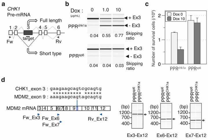
Designer PPR proteins induced exon skipping of CHK1 mRNA and the retardation of cell growth. (a) Exon–intron structure of CHK1 pre-mRNA. The target position was designed in exon 3 of the CHK1 gene. (b) RT-PCR analysis of exon skipping. The amount of the splicing variant with or without exon 3 (+Ex3 or −Ex3) was analyzed using Dox-induced expression of PPRchk1a. PPRsp6 was used as a negative control. The primer position used for RT-PCR is shown in (a). The skipping ratio was estimated by calculating the signal intensity of [−Ex3]/([+Ex3] + [−Ex3]). (c) Analysis of cell number with or without the induction of the designer PPR genes (PPRchk1a or PPRsp6). Cell number was analyzed using trypan blue staining with the cells two days after dox induction (N = 3). (d) Off-target RNA analysis. Two mismatched chk1a sequences located in MDM2 exon 9. The exon was confirmed using RT-PCR with Ex3–Ex12, Ex6–Ex12, and Ex7–Ex12 primer sets. Primer positions are indicated by blue triangles.

