Abstract
Enteropathogenic Escherichia coli (EPEC) was found to exhibit a type III secretion-dependent, contact-mediated, hemolytic activity requiring the EspA, EspB, and EspD secreted proteins. EspB and EspD display homology to pore-forming molecules. Our data suggest a mechanism to explain the requirement for all three Esp proteins in the transfer of EPEC proteins, such as Tir, into target cells.
Many gram-negative pathogens encode a type III secretion apparatus dedicated to the release of a diverse set of virulence-associated proteins (7, 17). Some proteins are secreted into the extracellular environment, while others are translocated directly into target cells to subvert various host processes (7, 17). The injection of proteins into host cells by Shigella and Yersinia spp. has been correlated to their in vitro abilities to lyse erythrocytes (RBC) (5, 6). Enteropathogenic Escherichia coli (EPEC) encodes a similar type III secretion apparatus (8) that is involved in the release of a number of EPEC-secreted proteins (EspA, EspB, EspD, EspF, and Tir) (3, 10–12, 14, 16, 18). EspA has been detected in bacterial-surface appendages that appear to connect the bacteria to host cells and participate in protein delivery (15). A role for EspD, but not EspB, in the correct formation of these EspA-associated appendages has been reported (15), while all three Esp proteins are required for the translocation of Tir into target cells (11). Both EspB and Tir have been detected in the host cytoplasmic and membrane fractions (1a, 9, 11, 12, 23, 25). Whereas the function of EspB in host cells is unknown, Tir is modified in the host cytoplasm and becomes inserted into the membrane, where it acts as a receptor for the bacterial outer membrane protein intimin (9, 11, 22). Tir-intimin interaction triggers host-signalling events and leads to the organization of host cytoskeletal proteins into characteristic pedestal-like structures beneath the adherent bacteria (11, 13, 20). No role for EspF has been found in Tir translocation or pedestal formation (3, 18, 24a).
To determine if the EPEC type III secretion system is associated with a hemolytic activity, EPEC 2348/69 and its type III secretion-defective mutants, the cfm-14 (2) and sepB mutants (8), were analyzed for their abilities to release hemoglobin from RBCs. EPEC strains were grown in Luria broth overnight (without shaking), diluted into Dulbecco’s modified Eagle medium (DMEM) (phenol red minus; GIBCO-BRL), and grown at 37°C without shaking under a 5% CO2 atmosphere to an optical density (A600) of approximately 0.7. One-half milliliter of culture was added to 0.5 ml of a 4% RBC-DMEM solution and centrifuged (at 2,500 × g for 1 min) to initiate EPEC-RBC contact. After a 30-min incubation (at 37°C in 5% CO2), the bacterium-RBC pellet was gently resuspended to facilitate the release of hemoglobin. Cells were repelleted (at 12,000 × g for 1 min), and the supernatant was monitored for the presence of released hemoglobin at the optical wavelength of 543 nm. Similarly treated uninfected RBCs were used as a spectrophotometric zero.
Figure 1A demonstrates that EPEC exhibits a haemolytic activity, lysing approximately 15% of the RBCs. This activity was dependent on the type III secretion apparatus, as no significant hemoglobin release was detected with the cfm-14 or sepB mutant. Such activity was not obtained with bacterium-free (filtered) supernatants or when EPEC organisms were coincubated with the RBCs but separated from them by a 0.4-μm-pore-size filter (12-mm Transwell filter; Costar) (data not shown), implying the absence of a diffusible hemolytic factor. Type III secretion from several pathogens is activated by contact with host cells (7). Lysis of RBCs by EPEC was found to be dependent on de novo protein synthesis, as the addition of chloramphenicol to EPEC prior to incubation with the RBCs prevented hemoglobin release (data not shown). De novo protein synthesis and host cell contact are both required for EPEC to induce host responses leading to Tir-mediated pedestal formation (11, 21, 22), correlating hemolytic activity with the translocation of Tir into host cells.
FIG. 1.
EPEC-induced hemolytic activity. EPEC, various deletion mutants, and complemented mutant strains were assayed for their abilities to lyse sheep RBCs as described in the text. Release of hemoglobin was assessed at an optical wavelength of 543 nm by using RBCs incubated in the absence of bacteria as a spectrophotometeric zero. Results from three independent experiments are shown. Error bars, standard deviations.
To investigate the involvement of type III secreted proteins in hemolysis, EPEC mutants with single deletions in genes encoding secreted proteins were tested for their abilities to lyse RBCs. Whereas the tir and espF mutants efficiently lysed RBCs, no significant lytic activity was detected for the espA, espD, and espB mutants (Fig. 1B). The ability of the latter mutants to lyse RBCs was restored, but not to wild-type levels, by the introduction of plasmids expressing the missing gene product (Fig. 1B). The espF mutant used here was generated by J.W. and has the same phenotype as espF, described in reference 22.
These results can be rationalized in light of recent findings relating to bacterium-host cell interactions. Thus, the absence of hemolytic activity for the espA mutant may be attributable to its role in the formation of the novel appendages that link EPEC to the host cell surface and participate in protein translocation (15). Therefore, EspA may have an indirect effect on hemolytic activity by mediating the delivery of the pore-forming molecule(s) into the host membrane.
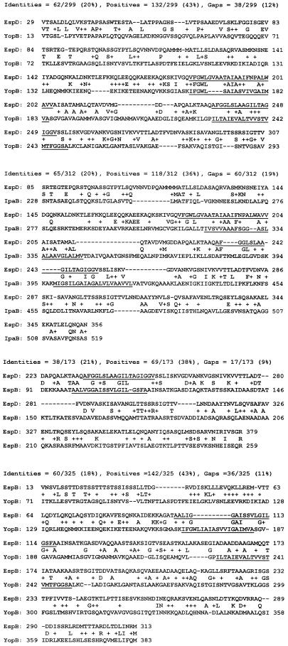
In contrast to EspA, both EspB and EspD display weak homology to the Yersinia YopB and Shigella IpaB proteins, and to each other (Fig. 2). These Shigella and Yersinia type III secreted proteins have pore-forming activity (5, 6). However, only EspB resists digestion by proteases of intact host cells (12, 24), making it the best candidate for a transmembrane pore-forming molecule that allows EPEC proteins to cross the host membrane. This would explain why disruption of espB abolishs hemolytic activity.
FIG. 2.
Homologies of EspB and EspD with each other and with Yersinia YopB and Shigella IpaB. Homologies, cited in reference 4, were obtained by using the Blast Similarity server (1). Protein sequence and residue numbers are given, with gaps introduced during alignment depicted by dashes. Letters in the space between sequences represent identical amino acids, and plus signs indicate similar amino acids. Predicted transmembrane domains (cited in reference 19) are underlined.
EspD could potentially have an indirect, or direct, role in pore formation (hemolytic activity), as on the one hand it is required for the correct formation of the EspA-associated “delivery” appendages (15) while on the other it is found associated with the host membrane (24). EspB, EspD, YopB, and IpaB are all predicted to possess transmembrane domains (4, 19) that are located, and even align, within these homologous regions (Fig. 2). Thus, it is possible that EspB and EspD have similar functions and associate to form a pore structure in the host membrane. The presence of a putative transmembrane domain in EspA (4, 19) could enable it to participate in pore formation also. This raises the possibility that all three Esp proteins could participate, at the tips of the EspA appendages, in the formation of a functional membrane-spanning pore structure to allow the injection of EPEC proteins into the host cell.
Neither the espF nor the tir gene plays a role in hemolytic activity, as no significant reduction in RBC lysis was detected. It is already known that Tir is translocated into the host cell (1a, 9, 11), while the presence of polyproline-rich regions in EspF indicates that this protein may be targeted into host cells (3). This contrasting phenotype supports a role for EspA, EspB, and EspD in forming a structure(s) to mediate the transfer of EPEC molecules into host cells.
In conclusion, we have shown that the EPEC type III secreted proteins EspA, EspB, and EspD are essential for contact-mediated hemolytic activity that is correlated with the ability to translocate EPEC proteins, such as Tir, into host cells.
Acknowledgments
We thank James Kaper and Michael Donnenberg for providing EPEC UMD864 (espB) and UMD870 (espD) and the cfm-14 and sepB mutants.
This work was supported by a Medical Research Council (MRC) of Canada operating grant to B.B.F. and a Wellcome Trust Career Development Fellowship to B.K. B.B.F. is a Howard Hughes Medical Institute Institutional Scholar and an MRC Scientist.
REFERENCES
- 1.BLAST Similarity server. 18 December 1998, revision date. [Online.] National Center for Biotechnology Information. http://www.ncbi.nlm.nih.gov/blast/blast/cgi. [17 August 1999, last date accessed.]
- 1a.Deibel C, Kramer S, Chakraborty T, Ebel F. EspE, a novel secreted protein of attaching and effacing bacteria, is directly translocated into infected host cells, where it appears as a tyrosine-phosphorylated 90 kDa protein. Mol Microbiol. 1998;28:463–474. doi: 10.1046/j.1365-2958.1998.00798.x. [DOI] [PubMed] [Google Scholar]
- 2.Donnenberg M S, Calderwood S B, Donohue-Rolfe A, Keusch G T, Kaper J B. Construction and analysis of TnphoA mutants of enteropathogenic Escherichia coli unable to invade HEp-2 cells. Infect Immun. 1990;58:1565–1571. doi: 10.1128/iai.58.6.1565-1571.1990. [DOI] [PMC free article] [PubMed] [Google Scholar]
- 3.Donnenberg M S, Lai L C, Taylor K A. The locus of enterocyte effacement pathogenicity island of enteropathogenic Escherichia coli encodes secretion functions and remnants of transposons at its extreme right end. Gene. 1997;184:107–114. doi: 10.1016/s0378-1119(96)00581-1. [DOI] [PubMed] [Google Scholar]
- 4.Elliott S J, Wainwright L A, McDaniel T K, Jarvis K G, Deng Y K, Lai L C, McNamara B P, Donnenberg M S, Kaper J B. The complete sequence of the locus of enterocyte effacement (LEE) from enteropathogenic Escherichia coli E2348/69. Mol Microbiol. 1998;28:1–4. doi: 10.1046/j.1365-2958.1998.00783.x. [DOI] [PubMed] [Google Scholar]
- 5.Hakansson S, Schesser K, Persson C, Galyov E E, Rosqvist R, Homble F, Wolf-Watz H. The YopB protein of Yersinia pseudotuberculosis is essential for the translocation of Yop effector proteins across the target cell plasma membrane and displays a contact-dependent membrane disrupting activity. EMBO J. 1996;15:5812–5823. [PMC free article] [PubMed] [Google Scholar]
- 6.High N, Mounier J, Prevost M C, Sansonetti P J. IpaB of Shigella flexneri causes entry into epithelial cells and escape from the phagocytic vacuole. EMBO J. 1992;11:1991–1999. doi: 10.1002/j.1460-2075.1992.tb05253.x. [DOI] [PMC free article] [PubMed] [Google Scholar]
- 7.Hueck C J. Type III protein secretion systems in bacterial pathogens of animals and plants. Microbiol Mol Biol Rev. 1998;62:379–433. doi: 10.1128/mmbr.62.2.379-433.1998. [DOI] [PMC free article] [PubMed] [Google Scholar]
- 8.Jarvis K G, Giron J A, Jerse A E, McDaniel T K, Donnenberg M S, Kaper J B. Enteropathogenic Escherichia coli contains a specialized secretion system necessary for the export of proteins involved in attaching and effacing lesion formation. Proc Natl Acad Sci USA. 1995;92:7996–8000. doi: 10.1073/pnas.92.17.7996. [DOI] [PMC free article] [PubMed] [Google Scholar]
- 9.Kenny B. Phosphorylation of tyrosine 474 of the enteropathogenic E. coli (EPEC) Tir receptor molecule is essential for actin nucleating activity and is preceded by additional host modifications. Mol Microbiol. 1999;31:1229–1241. doi: 10.1046/j.1365-2958.1999.01265.x. [DOI] [PubMed] [Google Scholar]
- 10.Kenny B, Abe A, Stein M, Finlay B B. Enteropathogenic Escherichia coli protein secretion is induced in response to conditions similar to those in the gastrointestinal tract. Infect Immun. 1997;65:2606–2612. doi: 10.1128/iai.65.7.2606-2612.1997. [DOI] [PMC free article] [PubMed] [Google Scholar]
- 11.Kenny B, DeVinney R, Stein M, Reinscheid D J, Frey E A, Finlay B B. Enteropathogenic Escherichia coli (EPEC) transfers its receptor for intimate adherence into mammalian cells. Cell. 1997;91:511–520. doi: 10.1016/s0092-8674(00)80437-7. [DOI] [PubMed] [Google Scholar]
- 12.Kenny B, Finlay B B. Secretion of proteins by enteropathogenic E. coli which mediate signaling in host epithelial cells. Proc Natl Acad Sci USA. 1995;92:7991–7995. doi: 10.1073/pnas.92.17.7991. [DOI] [PMC free article] [PubMed] [Google Scholar]
- 13.Kenny B, Finlay B B. Intimin-dependent binding of enteropathogenic Escherichia coli to host cells triggers novel signaling events, including tyrosine phosphorylation of phospholipase C-γ1. Infect Immun. 1997;65:2528–2536. doi: 10.1128/iai.65.7.2528-2536.1997. [DOI] [PMC free article] [PubMed] [Google Scholar]
- 14.Kenny B, Lai L-C, Finlay B B, Donnenberg M S. EspA, a protein secreted by enteropathogenic Escherichia coli (EPEC), is required to induce signals in epithelial cells. Mol Microbiol. 1996;20:313–323. doi: 10.1111/j.1365-2958.1996.tb02619.x. [DOI] [PubMed] [Google Scholar]
- 15.Knutton S, Rosenshine I, Pallen M J, Nisan I, Neves B C, Bain C, Wolff C, Dougan G, Frankel G. A novel EspA-associated surface organelle of enteropathogenic Escherichia coli involved in protein translocation into epithelial cells. EMBO J. 1998;17:2166–2176. doi: 10.1093/emboj/17.8.2166. [DOI] [PMC free article] [PubMed] [Google Scholar]
- 16.Lai L C, Wainwright L A, Stone K D, Donnenberg M S. A third secreted protein that is encoded by the enteropathogenic Escherichia coli pathogenicity island is required for transduction of signals and for attaching and effacing activities in host cells. Infect Immun. 1997;65:2211–2217. doi: 10.1128/iai.65.6.2211-2217.1997. [DOI] [PMC free article] [PubMed] [Google Scholar]
- 17.Lee C A. Type III secretion systems: machines to deliver bacterial proteins into eukaryotic cells? Trends Microbiol. 1997;5:148–156. doi: 10.1016/S0966-842X(97)01029-9. [DOI] [PubMed] [Google Scholar]
- 18.McNamara B P, Donnenberg M S. A novel proline-rich protein, EspF, is secreted from enteropathogenic Escherichia coli via the type III export pathway. FEMS Microbiol Lett. 1998;166:71–78. doi: 10.1111/j.1574-6968.1998.tb13185.x. [DOI] [PubMed] [Google Scholar]
- 19.Pallen M J, Dougan G, Frankel G. Coiled-coil domains in proteins secreted by type III secretion systems. Mol Microbiol. 1997;25:423–425. doi: 10.1046/j.1365-2958.1997.4901850.x. [DOI] [PubMed] [Google Scholar]
- 20.Rosenshine I, Donnenberg M S, Kaper J B, Finlay B B. Signal transduction between enteropathogenic Escherichia coli (EPEC) and epithelial cells: EPEC induces tyrosine phosphorylation of host cell proteins to initiate cytoskeletal rearrangement and bacterial uptake. EMBO J. 1992;11:3551–3560. doi: 10.1002/j.1460-2075.1992.tb05438.x. [DOI] [PMC free article] [PubMed] [Google Scholar]
- 21.Rosenshine I, Ruschkowski S, Finlay B B. Expression of attaching/effacing activity by enteropathogenic Escherichia coli depends on growth phase, temperature, and protein synthesis upon contact with epithelial cells. Infect Immun. 1996;64:966–973. doi: 10.1128/iai.64.3.966-973.1996. [DOI] [PMC free article] [PubMed] [Google Scholar]
- 22.Rosenshine I, Ruschkowski S, Stein M, Reinscheid D J, Mills S D, Finlay B B. A pathogenic bacterium triggers epithelial signals to form a functional bacterial receptor that mediates actin pseudopod formation. EMBO J. 1996;15:2613–2624. [PMC free article] [PubMed] [Google Scholar]
- 23.Taylor K A, O’Connell C B, Luther P W, Donnenberg M S. The EspB protein of enteropathogenic Escherichia coli is targeted to the cytoplasm of infected HeLa cells. Infect Immun. 1998;66:5501–5507. doi: 10.1128/iai.66.11.5501-5507.1998. [DOI] [PMC free article] [PubMed] [Google Scholar]
- 24.Wachter C, Beinke C, Mattes M, Schmidt M A. Insertion of EspD into epithelial target cell membranes by infecting enteropathogenic Escherichia coli. Mol Microbiol. 1999;31:1695–1707. doi: 10.1046/j.1365-2958.1999.01303.x. [DOI] [PubMed] [Google Scholar]
- 24a.Warawa, J., and B. Kenny. Unpublished data.
- 25.Wolff C, Nisan I, Hanski E, Frankel G, Rosenshine I. Protein translocation into host epithelial cells by infecting enteropathogenic Escherichia coli. Mol Microbiol. 1998;28:143–155. doi: 10.1046/j.1365-2958.1998.00782.x. [DOI] [PubMed] [Google Scholar]


