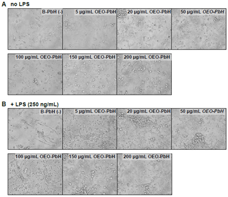
Figure 8.
Brightfield microscopy images of OEO-PbH treated human DCs. DCs were cultured for 7 days with GM-CSF and IL-4. Cells were subsequently stimulated without LPS (A) or with LPS (B) and treated with either OEO-PbH or B-PbH formulations at different concentrations for 24 h. The images were taken under a light microscope using a 20× objective.

