Abstract
Maintaining the structure of protein and peptide drugs has become one of the most important goals of scientists in recent decades. Cold and thermal denaturation conditions, lyophilization and freeze drying, different pH conditions, concentrations, ionic strength, environmental agitation, the interaction between the surface of liquid and air as well as liquid and solid, and even the architectural structure of storage containers are among the factors that affect the stability of these therapeutic biomacromolecules. The use of genetic engineering, side-directed mutagenesis, fusion strategies, solvent engineering, the addition of various preservatives, surfactants, and additives are some of the solutions to overcome these problems. This article will discuss the types of stress that lead to instabilities of different proteins used in pharmaceutics including regulatory proteins, antibodies, and antibody-drug conjugates, and then all the methods for fighting these stresses will be reviewed. New and existing analytical methods that are used to detect the instabilities, mainly changes in their primary and higher order structures, are briefly summarized.
Keywords: protein drugs, antibody drugs, antibody drug conjugates, pharmaceutical proteins and peptides, stabilization, denaturing stresses, protein aggregation, protein folding, protein drug characterizations, LC-MS
1. Introduction
It has been a century since the first peptide drug (insulin) entered the health sector [1]. Currently, there are a variety of proteins and peptides such as growth hormone, green fluorescence protein, insulin analog forms, and many antibodies in the field of therapy, diagnosis, control and management of diseases, some of which are intentionally very different from their native/wild-type counterparts [2]. The protein drug market is divided into several segments: antibody drugs, peptide hormones, blood products, and enzymes [3]. The antibody drugs sector has experienced some of the fastest growth in this marketplace. In addition to being used as therapeutic proteins against various diseases, antibody molecules have also been conjugated with various anticancer drugs, named antibody-drug conjugates (ADCs), to increase drug efficacy by specific targeting. Moreover, the advances made in drug delivery systems using nanotechnology and the rapid increase in chronic diseases are among the growing causes of this market [4]. However, not everything is positive and clear about these protein-based drugs; there are other challenges such as the synthesis or production methods of these drugs [5], purification from expression systems [6], correct folding into native forms in the process of production and consumption [7], and instabilities of these drugs during their manufacturing, storage, and delivery. Among these, structural instability caused by unfolding, misfolding, or covalent/non-covalent modifications, as well as aggregation can degrade drug efficacy and greatly overshadow the bright features of these protein-based drugs.
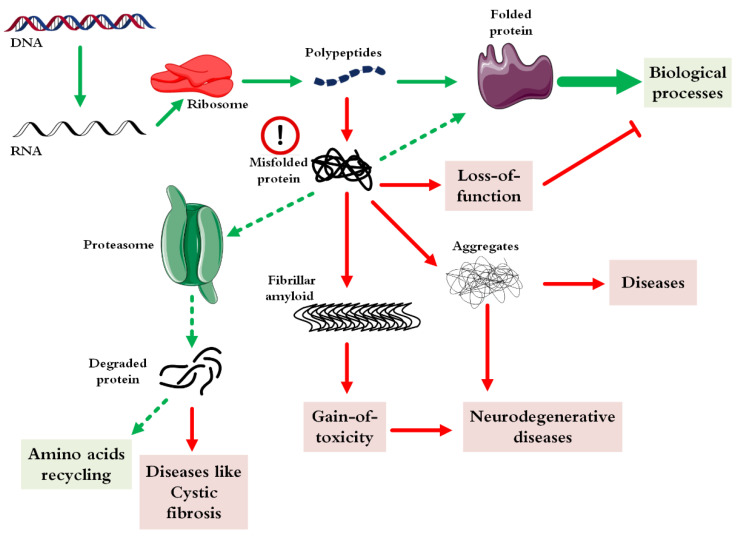
Protein misfolding or incorrect folding is an unwanted instability process in which the protein either gets function (triggering gain-of-function diseases) and/or loses its specific function (initiating loss-of-function diseases) [8]. Likewise, the in vitro misfolding of recombinant proteins/peptides is also another challenge in the field of biotechnology and the production of pharmaceutical proteins. Figure 1 indicates the different fates of a protein after its production by a ribosome [9]. From the causative point of view, as mentioned above, several factors trigger misfolding of a protein [10].
Figure 1.
Possible intracellular pathways of correct and/or incorrect protein folding.
At the beginning of the transcription of a protein, thanks to the intracellular chaperone systems and the biophysical laws governing protein folding, correct folding occurs most of the time. When due to cellular defects and rapid protein expression, protein folding becomes problematic, and several fates may occur for the protein. In cases where misfolding leads to the loss of protein activity (such as enzymes), the corresponding disease will appear directly. As this state continues, the misfolded protein may turn into amorphous clots or aggregates with regular structures, each of which can lead to various neurological diseases or even cancer. In the most optimistic scenario, the misfolded protein enters the proteasome machinery and is initially converted into smaller peptides and finally broken down into building amino acids. In a pessimistic view, in some cases, diseases may occur from the peptides obtained from the proteasome system, which is very rare.
On the other hand, proteins have a great tendency to aggregate under various stresses, especially when they are kept at high concentrations required for effective doses. Therefore, the main problem in the production of pharmaceutical proteins, is the issue of stability with the hope of maintaining their activity [8]. Types of strategies that have been adopted to increase the stability of these biomacromolecules include: direct mutations on the structure of the genome-encoding proteins [9], joining these molecules to inert proteins (such as human serum albumin) [10], their chemical changes after conjugation (like ADCs) or purification from expression systems [11], the use of nanoparticles, polymers, sugars and other additives to maintain the structure of proteins and drug peptides [12], and the engineering of storage containers and packing approaches [13] for these large molecules. Analytical methods to detect and characterize highly heterogeneous structural changes of protein-based drugs are other challenging issues in tackling the instability of these drugs [14].
This review will discuss various stresses causing instabilities and available strategies for improving the stability of pharmaceutical proteins and peptides. Analytical methods used to monitor and characterize pharmaceutical proteins and drug peptides will also be described. Here, pharmaceutical proteins include protein therapeutics and proteins which are not the main therapeutics ingredients but aim to improve the effectiveness of drug targeting (like ADCs) or stabilization in blood circulation (like serum albumin).
2. Stability Issues of Different Protein-Based Drugs
Of the significant aspects in the industrialization of proteins are their stability and solubility [14,15]. The stability of proteins during the process of expression and purification is one of the most important and challenging issues because many recombinant proteins are unstable under the conditions they are expressed and lose their correct folding or undergo proteolytic digestion [16]. Therefore, studying the stability of proteins and identifying the factors that increase their stability is one of the important and interesting topics in scientific societies [17]. Today, numerous plans have been proposed to increase the solubility and stability of proteins, which include guided changes in the protein molecule or optimization in the expression protocols [18], purification and solubility process of proteins [19].
The production of biological medicine is not a direct path, because, on the one hand, this process requires a lot of work, and on the other hand, the issue of product instability during the process and storage is raised. In addition, there are other challenges such as the need for strong laws, scrutinizing Good Manufacturing Practice (GMP) regulations and intense competition in the consumer market (for example due to the emergence of biosimilars) [20]. Proteins and peptides are only stable in a limited range of concentration, temperature, ionic strength and acidity conditions (marginal stability) and are very prone to physical and chemical destabilizations. Many recombinant proteins are also under conditions that become unstable and lose their correct folding. Additionally, medicinal proteins have a great tendency to aggregate, especially when they are in high concentration. When talking about the stability of protein-based drugs, researchers often seek to understand why and when the native structure of proteins tends to become denatured. For more than 50 years, scientists have made great efforts to understand the relationship between protein denaturation and the competition between their reversible and irreversible denaturations [21]. The concern of stability is different for different classes of proteins. Some protein drugs have an enzymatic activity that can replace abnormal and low-expressed counterparts in the body. Since enzymes usually have a high turnover number, a small disorder in their structure will lead to huge changes in the accumulation of their substrate and, on the other hand, the concentration of their products [22]. Consequently, for this category of proteins, not only enzyme aggregation but also the smallest change in the structure can have great effects [23].
Monoclonal antibodies (mAbs) accounted for almost half (48%) of the therapeutics protein sales in recent years [24]. Since all the antibodies have a binding role to the antigen, the most important components in this category are maintaining the structure of complementarity-determining regions (CDRs) which bind their specific antigens. Most of the stability challenges in this field are related to their aggregation and oxidation in different conditions and high on-demanded concentrations [25]. In addition, ADCs [26] are a special class of protein-based drugs made by binding chemical drugs (payloads) to antibodies with an emphasis on payload efficacy. The concept is to achieve high cancer cell specificity and low cytotoxicity to off-target cells through antibody targeting so that a broader therapeutic window of the payload can be gained [27]. This idea was introduced in early 1970. By now, 12 ADCs have been approved by the FDA [28,29] and about 60 other ADCs are also in clinical trials [30]. One of the critical efforts that need to be harmonized in this regard is the issue of stabilization of these important therapies. This is because the binding of a chemical drug may give the antibody a new behavior that overshadows its stability and leads to the changing of native antibodies to unfolded states [29].
3. Causes of Instability
3.1. Physical Instability
3.1.1. Temperature-Induced Instability
Some proteins are very resistant to high temperatures [31], nonetheless, most of them are unable to maintain their natural structure against this physical stress and turn into aggregated states [32]. In this process, it is suggested that the hydrophobic domains of the proteins, which are mainly hidden in the protein structure (by van der Waals and hydrogen forces), are initially removed from the buried state and exposed to the external environment (surface of protein). Due to the hydrophobic nature of these domains, hydrophobic interactions occur and lead to the binding of a large number of protein molecules, which ultimately leads to the formation of aggregated states of the protein. For ADCs, the hydrophobicity of the payload as well as the hydrophobicity of the antibody surface will be one of the most important factors in shortening the shelf life of the final product of ADCs [33]. Nevertheless, unlike antibodies, the nature of payloads tends to be more hydrophobic, which has led to a challenge to connect these two different natures (connecting the hydrophobic payload to the hydrophilic surface of the antibody) [34]. As a result, maintaining hydrophobic structures while inactivated by temperature is very important to suppress or modulate the consequences of the phenomena [35,36].
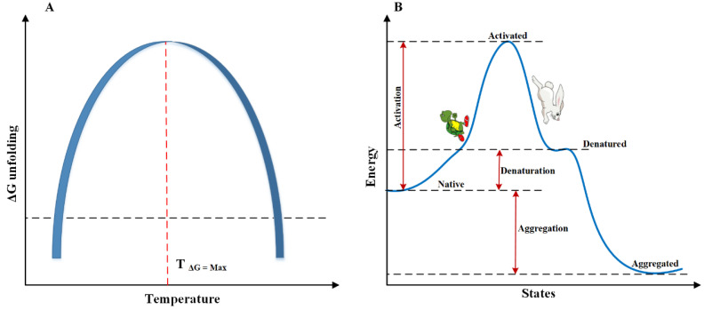
Figure 2 shows the relationship between temperature and protein unfolding. The maximum of the ΔGunfolding is in a small temperature range, after which a protein instability occurs beyond this certain temperature (either below or above this temperature) [37]. The width of the diagram or the sharpness of the peak determines how well the protein can withstand temperature changes. The wider the graph, the higher the resistance of the protein to temperature changes [38]. Not only increasing the temperature of a protein has the potential to unfold it, but lowering it to a critical point can be a lament of protein aggregation [39]. The ribonuclease A enzyme has been shown to precipitate at temperatures as low as −22 °C [40], and temperatures below zero have been reported to be destructive to serum albumin [41]. Nevertheless, thanks to poor hydrophobic interactions, protein unfolding is less likely to occur at low temperatures. This is probably why cold aggregation is usually reversible. It has been observed that the aggregation of serum cryoglobulins protein at a temperature below 37 °C can be reversibly converted to a soluble state and it has also been reported that the human IgM cryoglobulin in the temperature window between 10–12 °C turns into a jelly from which with increasing temperature this state becomes reversible [38]. The discussion of protein aggregation at low temperatures will be interpreted in another section of this article.
Figure 2.
The relationship between protein unfolding and internal energy (the data were extracted from [21,38]). (A) Shows the possible quantities of ΔGunfolding at different temperatures. (B) Indicates the several stages of the protein unfolding pathway. At first, by obtaining energy, the protein is activated to higher levels by increasing the amount of internal energy, and at a critical point, it unfolds by releasing energy to reach a more stable state. In a large number of studies, it is believed that the most stable state of a protein is its aggregated form (fibril form), however, some researchers believe that its natural form is the most stable state.
Whether protein aggregates in the aggregation-driven force (Figure 2B) or can survive as a refolded state is most likely related to the collapsing of hydrophobic domains and the repulsion of electrostatic forces. Thus, at high temperatures, the collapse of hydrophobic forces is overcome in competition with the electrostatic repulsion and finally, the protein will aggregate [32]. Apart from the hydrophobic challenge, it has also been reported that increasing the temperature of the protein solution leads to an increase in the rate of protein oxidation and deamidation, which in itself leads to faster protein aggregation [42].
A quantity index also defines the temperature stability of proteins, which is called the melting temperature (Tm) of proteins. This temperature is equivalent to the temperature at which half of the protein molecules are unfolded. Unsurprisingly, this temperature varies between different proteins, and in nature, it usually fluctuates between 40 and 80 °C [43]. In many stages of production, purification, packaging and transporting of medicinal proteins, it is essential to keep the temperature of the products below the agreed melting point. By reaching the maximum energy state (also the most unstable state of the protein), with a faster rate of activation, the target protein falls into the valley of energy and goes to the point of unfolding. Between these pathways, many intermediate states may happen for the protein, each of which can be individually stable or unstable. Subsequently, as the unfolding process continues, communities of unfolded proteins accumulate regularly or irregularly, eventually turning into morphous and/or amorphous aggregated states [44] (Figure 2B).
3.1.2. Cold Denaturation
The history of studying low temperatures on the structure of proteins probably dates back to the 1930s and Hopkins’s article [45]. In the study, it was found that in the presence of urea, the ovalbumin protein unfolds at 0 °C faster than at 23 °C. Clark later, proved this consequence in his observations, but stated that this result can be seen only in the presence of high concentrations of urea while in low concentrations of urea, the unfolding rate increases with increasing temperature [46].
It has been described that cold-induced unfolding is mainly due to a change in the pattern of interaction between water molecules and non-polar groups in the protein [47]. As the temperature decreases, the free energy of the interaction decreases, which increases the conditions for this unwanted communication and ultimately leads to the hydration of the hydrophobic domains [48]. With the help of in silico studies, it has been proven that with decreasing temperature, the diameter of this aqueous layer increases around non-polar (hydrophobic) groups, and fortunately, with increasing temperature, this harmful aqueous layer disappears, which is a reason for the reversibility of cold denaturation of most proteins [49].
The vast majority of studies have referred to the law of all-or-none during low temperature-induced unfolding. This means that during the process, there are two important states of protein: unfolded and folded state [50]. Nevertheless, low-temperature NMR studies by Wand et al., indicated that this process is step-by-step and produces intermediates of semi-active structures in addition to completely inactive configurations [51]. Aside from all these cold-denaturation issues, fortunately for most proteins, cold-temperatures are dangerous below freezing points [51].
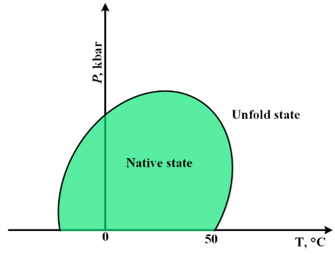
Some early studies in the field of cold unfolding have pointed out the importance of the effect of pressure on this phenomenon [52,53]. As seen at high pressures, the cold unfolding process accelerates. Meanwhile, it has been reported that when the pressure of the bovine pancreatic ribonuclease A solution rises above the pressure where water transforms to the dense ice phase (2 kbars), the local density of water around the polar groups of the protein decreases (by reducing the temperature), leading to the unfolding of the protein [53]. In this section, with the help of Figure 3 diagram, the importance of the relationship between temperature and pressure during protein unfolding is discussed.
Figure 3.
The relationship between pressure and temperature during the folding and unfolding of proteins [54].
Most proteins remain in their natural state (folded) within a certain range of temperature and pressure. For most of them, the critical temperature is 50 °C. In addition, in negative temperatures, the change of the normal state to unfolding has been observed, which, unlike heat-induced unfolding, cold-induced unfolding is reversible. Moving toward this temperature window, protein unfolding will occur with little stress. At room temperature, the highest pressure is required for the protein to transition from the fold to unfold.
3.1.3. Photo-Induced Instability
All aspects of life are constantly and/or temporarily exposed to light, and on the occasion of light-induced problems, living organisms within the cell have a mechanism in place to repel these damages. Although the protective layer of the atmosphere absorbs most of the dangerous UV waves (UVC: less than 295 nm), in the habitable atmosphere around us, UVB and A waves, as well as visible light, is also dangerous for bio-infrastructures. It is generally suggested that light-induced damage in proteins occurs by two general mechanisms; direct and indirect pathways [55,56,57]. The photo-induced damages may also be regarded as chemical instability since chemical conversions are involved in these pathways.
The direct pathway, which occurs mainly with UVB waves (λ 280–320 nm), is restricted to a small number of amino acids in proteins such as tyrosine, tryptophan, cysteine, and histidine. Once the UVB energy is received by these chromophores (the residues), they temporarily change to the first-excited singlet states. This state is transient and quickly turns into a triplet state which is relatively more stable with the release of excess energy. The released energy can be transferred to other functional groups. The resulting intermediates in the triplet state may generate dangerous free electrons that can interact with other functional groups and, as a radical group, disrupt protein function [57,58]. One of the problems is the release of hydrated electrons, which are regarded as one of the reactive oxidizing species (ROS) and are well described in Refs. [59,60]. These hydrated electrons can attack the free carbonyls at the amine end or the side chain of the amino acid lysine, leading to deamination and/or the elimination of H˙. It has also been described that the production of anionic radicals in small peptides originates from these hydrated electrons [61]. On the other hand, the indirect path of photo-induced damage is due to the formation of singlet oxygen, which occurs due to the energy transfer to intra-protein bonds and/or external chromophores. The latter include porphyrins [62], drugs, polyaromatic, and dye-conjugated molecules [57,63]. All of the above can lead to structural changes in the protein and, ultimately, loss of function.
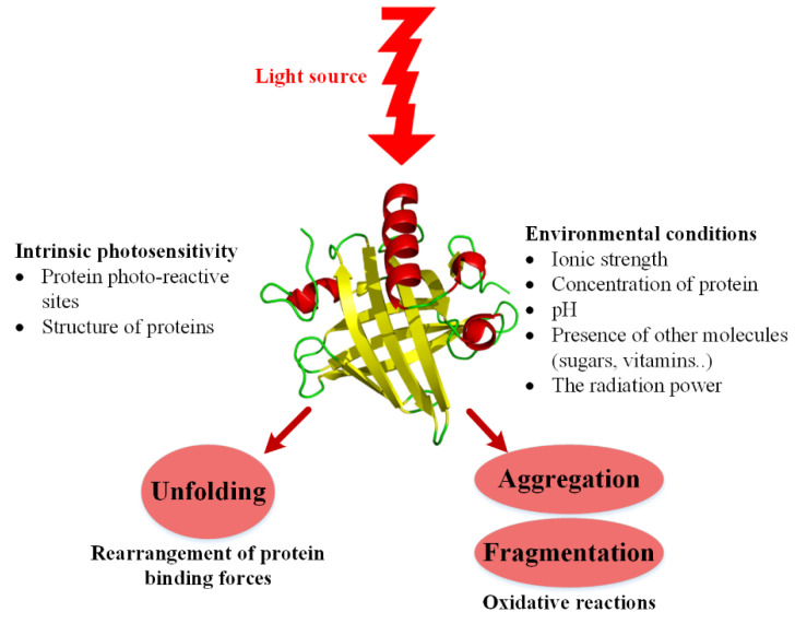
To summarize what has been believed about the effect of photos on proteins/peptides, the following schematic (Figure 4) is presented.
Figure 4.
Determinants of photo-susceptibility of proteins (the data were extracted from [63]).
Some intrinsic factors of proteins such as their amino acid sequence and even their spatial structure can affect vulnerability to photo stress. In addition to the intrinsic properties, environmental factors can also affect the vulnerability of the protein to stress.
In the line with the above information, the effects of photo on ADCs can be much more dangerous, especially through the indirect path of photo-induced damage to the monoclonal structure of antibodies since some conjugated small molecules are themselves chromophores which can interact with UV to make ADCs more susceptible to unfolding compared with non-conjugated antibody counterparts. For example, in the case of trastuzumab, the intact protein itself has long-term physical and chemical stabilities. Conversely, when combined with eosin, as a model for chemical drugs, it becomes more sensitive to photo-induced aggregation (the aggregation was started after 7 min incubation of eosin conjugated trastuzumab under 20 W·h/m2 of UVA light) [56]. Doxorubicin (a topoisomerase II inhibitor), which is used in a wide range of ADCs [64,65], is also very sensitive to light; once exposed to UVA waves, it either breaks down or leads to the production of ROS [66]. The latter byproduct is capable of threatening the protein portion of ADCs for aggregation. There are other drugs similar to Dox used for ADCs, although they have also been shown to be highly sensitive to light. Among these drugs, cyclopropapyrroloind-4-one, CC-1065, and cyclopropabenzindol-4-one are the most important candidates in which light’s destructive role on protein structure should be seriously considered [67].
3.1.4. Agitation-Induced Instability
Agitation during stages of pumping, mixing, shipping, filtering, and filling can affect the structure of the therapeutic proteins and lead to aggregation [68,69].
The role of turbidity/agitation of the aquatic environment around the protein in the kinetics of protein aggregation is very complex and debatable. Many studies mention that different types of turbulence caused in different ways have diverse effects on the amount of aggregation. For example, it has been demonstrated that that the effect of steering on the speed and percentage of aggregation is more pronounced than shaking [70], and even the location of the steering can affect this quantity [71]. In other studies, researchers even went so far as to point out the role of dropping in the process of protein aggregation. A dropping method is sometimes used when filling therapeutic vials and syringes with protein drugs. By dropping the products, bubbles are formed, which are supposed to form oxygen free radicals near these bubbles, which lead to a change in the structure of the protein and eventually causing its aggregation [72,73].
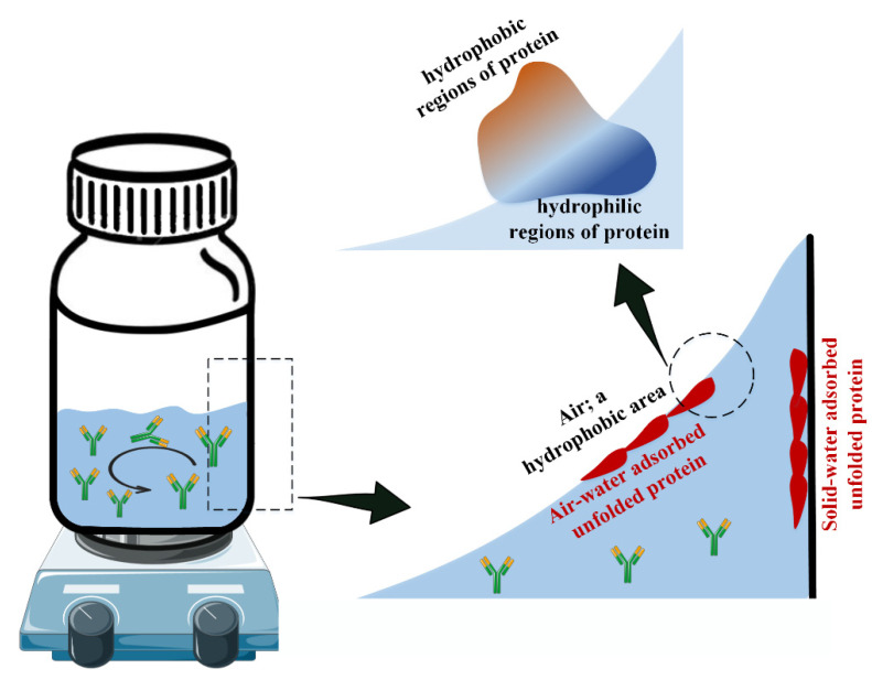
Looking at the mechanism of the effect of agitation on protein aggregation, some researchers believe that due to the increase in the liquid and air interfaces during the enforced agitation, the protein aggregation rate increases [74,75]. This effect has been shown to increase the rate of aggregation and amyloid formation in proteins such as amyloid-β [76,77], insulin [21], and β-lactoglobulin [78] more than three times on average. For a visual look at how environmental agitation affects the unfolding of proteins, Figure 5 is drawn.
Figure 5.
Agitation-induced protein unfolding/aggregation (the concepts were inspired by [79,80]).
With the increase of the environmental agitation, the interface surface between the liquid and the air as well as the liquid with the body’s container (solid) will increase. Air, as a hydrophobic environment, leads to the exposure of the hydrophobic parts of a protein from inside the structure, and the same happens for the interaction between proteins on the solid surface. With the continuation of this process, most of the folded proteins become unfolded, which leads to the reduction of the active protein population, then aggregation due to environmental agitation.
Using hydrophobic excipients, in several independent studies, there are other theories for how proteins are aggregated during agitation stress. For example, in the study of Bam et al., by the use of tween, it was found that hydrophobic interactions can modulate the aggregation rate of human growth hormone, indicating hydrophobic connections between unfolded proteins in agitation stress [81]. Similarly, others have also used additives such as salts [82,83], lipids [84], surfactants [85,86], and various additives [87] to reduce the aggregation of proteins in the hope of reducing the interface of air and solution created during agitation.
3.2. Chemical Instability
3.2.1. Hydrolysis
Since almost all parts of life form and continue to survive in the aquatic environment, the phenomenon of occasional hydrolysis is ubiquitous. In many biological processes, this phenomenon, which is defined as breaking the chemical bond by water molecules, is common and useful, but in some cases, such as the storage of drug proteins in water, it can be dangerous and lead to some unpleasant consequences such as severe allergies [88]. Many proteins, including therapeutic monoclonal antibodies, have been reported to be non-enzymatically hydrolyzed in the hinge region [89,90]. Cordoba et al. found that incubating IgG1 antibody in a neutral, aqueous medium for three months resulted in some non-enzymatic hydrolysis [91]. With the efforts of Dillon et al. using a series of reverse phase chromatography linked with mass spectroscopy (LC-MS), the same result was obtained for the IgG1 antibody [92]. As a result, in both acidic and alkaline conditions, the rate of this reaction increases [93], which is mainly due to the protonation of carbonyl oxygen or the addition of a hydroxyl group to the carbonyl carbon in the peptide bond. However, by studying several dipeptides in neutral conditions, it has been indicated that it is necessary to overcome a large energy barrier equal to 27–30 kcal/mol for hydrolysis to occur [94]. Nutritionally speaking, hydrolysis of a protein can also lead to generating and exposing epitopes in the structure of the protein, which can eventually cause various allergies. It has been revealed that different types of milk proteins with different hydrolysis percentages can bring about percentages of allergenic shocks in consumers [95].
Concerns about the consequences of hydrolysis of protein drugs are not limited to the proteins themselves. Rather, additives are sometimes used during the formulation of these drugs; hydrolysis can lead to the deformation of the protein. As a clear example, when the monoclonal antibody MAB001 was examined in terms of susceptibility to aggregation with different sugars, it was found that this response is higher in the presence of sucrose than sorbitol, which also depends on the temperature and pH of the environment. Further studies have shown that this destructive effect is due to the hydrolysis of sucrose and the effect of the byproducts of hydrolyzed sucrose on the protein [96]. Therefore, in addition to the structural properties of the protein, the formulation of the therapeutic protein should also be considered, avoiding the destructive effects of hydrolysis.
3.2.2. Oxidation
Today, many pharmaceutical proteins are produced by recombinant DNA technology using bacterial or mammalian cells like Chinese hamster ovary (CHO) expression systems [97]. The oxidation and eventual breakdown of proteins may even occur in the expression platform [97]. For example, the G1 antibody in the hamster expression system has been shown to degrade at rapid oxygen delivery rates, producing a pattern similar to the incubation of antibodies to hydrogen peroxide in an in vitro condition [98]. In addition to the challenges of production, the purification process of pharmaceutical proteins sometimes leads to oxidation. For example, during the purification of lactate dehydrogenase using metal affinity chromatography, degradation was observed due to the protein oxidation induced by metal ions [99].
Besides, many other factors can lead to protein oxidation and subsequent decomposition, including exposure to oxygen during shipping and storage, the presence of some oxidants, light oxidation, and the presence of transition metals in formulation steps [100]. Methionine is the most susceptible amino acid residue to oxidation [101,102]. In the case of monoclonal antibodies, there are two methionine residues in their FC (crystallizable fragment) region, which have caused the greatest weakness points of this group of protein drugs. The two residues are located around the neonatal Fc receptor (FcRn) binding region; any change in which could lead to a change in the behavior of the monoclonal receptor or a change in their cytotoxicity [103,104]. In addition to methionine, it has been observed that other amino acids such as tryptophan, histidine, cysteine, tyrosine, and sometimes amino acids arginine, lysine, and proline are also involved in the oxidation of proteins [105]. On top of problems with protein structure, oxidation of these amino acids sometimes results in immunological reactions [106,107]. Some studies have revealed that the oxidation process rapidly takes place between the skin and the blood before it enters the bloodstream, once subcutaneously injected so that dangerous by-products enter the bloodstream [108]. The extent of oxidation could be greatly affected by the conjugation sites of ADCs. The cysteine-conjugated payload showed the highest sensitivity to oxidation damage compared to the lysine-conjugated payload. It was also observed that due to the different structural changes of the cysteine-conjugated payload compared to the lysine-conjugated cargo, the methionine of the FC part in the former sample was oxidized four times more than its latter counterpart [109].
3.2.3. Disulfide Exchanges
It has been observed in many studies that the un-paring and miss-assembly of disulfide bonds in IgG1 can occur in a variety of production processes. One of the earliest stages that initiates this error is the lack of a proper folding system in the endoplasmic reticulum during its production (fermentation) in eukaryotic expression systems [110]. In the case of this antibody, four disulfide bonds between heavy and light chains, as well as two heavy chain disulfide linkages, were found to be more vulnerable than 12 disulfide bonds within the IgG domains [111]. Amano et al., [19] showed that several months need to generate hinge-region cysteine racemization in IgG1, while high pHs and temperature through numerous pathways in a shorter period ultimately can modify the antibody in the same way.
3.2.4. Deamidation
This reaction, which mostly occurs in amino acids Asn and Gln, leads to the transformation of the side chain amide linkage into a carboxyl group [112]. The deamidation of Asn, which mostly occurs in the sequence of Asn-Gly and Asn-Ser, is seen to be more accelerated in neutral and/or alkaline conditions [112]. It has even been understood that the rate of deamidation in these sequences is higher than in free Asn and Gln residues. This shows that the rate of deamidation may be different in the different sites of a protein. Sometimes, if these sequences are in an important position, such as the active site of a pharmaceutical enzyme, it causes the loss of enzyme activity, and if this position is in the structure, it leads to a change in structure, without having a strong effect on the activity. As a result, the consequences of deamidation are different for each protein. By and large, the reaction eventually introduces a negative charge to the protein, which can have different effects on the behavior of the protein. It has been realized that deamidation changes the binding of hemoglobin to oxygen in the sequence of Asn-Gly [113]. It has also been understood that the reaction in the Asn25-Gly26 sequence leads to a change in the activity (decreasing effect) of the porcine adrenocorticotropic hormone (ACTH) [114]. Additionally, incubation of human growth hormone in normal conditions such as a neutral environment (pH 7.4) of natural body temperature (37 °C) for two weeks results in the conversion of 37% of the protein into deamidated form, which will result in a severe decrease in activity [112]. In another study, it was concluded that placing the calmodulin protein at 37 °C (pH 7.4) for one month causes deamidation in such a way that eventually, only 10% of the proteins will remain active [115]. On the other hand, there are controversial results regarding the deamidation of proteins and their immunogenic properties. For example, it has been observed that with the deamidation of mouse epidermal growth factor polypeptide, the altered protein is not significantly different from the control sample in terms of immunogenicity [112]. The same result has been obtained in the mode of deamidation of several antibodies [116]. Nonetheless, in the case of gliadin protein, it has been agreed that the reaction will make the protein more immunogenic [117]. In some studies, even in vivo deamidation has been mentioned in some diseases such as cataracts and Alzheimer’s [118,119]. When a protein is placed in a neutral and/or alkaline environments, the Asn sequence with the succinimide ring intermediate is rapidly converted to products such as isoAsp and n-Asp. It is very important to distinguish these two forms since it is believed that the product isoAsp is responsible for changing the behavior of the protein and, as a result, the occurrence of diseases [120]. In addition to the acidic environment and protein sequence, other factors such as the ionic strength, the tertiary structure of the protein, temperature and buffer content can affect the rate of protein deamidation [121]. Accordingly, investigating this reaction not only can be an answer for some neurodegenerative diseases in the living environment, but it may be a mechanism to prevent this unwanted reaction in the industrial environment and the field of production and storage of medicinal proteins and peptides.
3.2.5. Conjugation-Induced Instability
ADCs and some other protein drugs require conjugation reactions in their manufacture process. In addition to carrying toxic drugs, various linkers such as sugars or polymers [122] are conjugated to improve drug efficacy. The manufacture of conjugation, however, requires several specific steps and sometimes at elevated temperatures (typically 37 °C) which may induce chemical or physical changes. Additional buffer exchange, solvent removal, and concentration steps are also involved in the final manufacture and may further accelerate the changes and instability.
Cysteine and lysine conjugations are so far the most common conjugation residues and both are used an activation step to provide some degree of site-specificity in conjugation. The fabrication of cysteine-linked ADCs uses cysteine conjugation primarily through maleimide-thiol reactive chemistry. Although generally considered to be a chemically stable bioconjugation approach, there is clear evidence that this conjugation is reversible, and this reversibility manifests itself with the concern of free thiols in circulation. For example, the presence of free thiols may induce cysteine racemization in the hinge region of IgG1 19 and CH2/hinge region destabilization is linked to ADC aggregation. The lysine conjugation method involving an activating step with a dual functional linker has also been reported to introduce a risk of cross-linking. It has been stated in the literature that the change may even be in a place far from the active site of a protein, however, eventually the conjugation leads to a decrease in drug activity [123].
It should be noted that there is no general rule to categorize the susceptibility of peptides and proteins to different stresses. For example, the large BSA protein (66.4 kDa), although it is 50% hydrophobic [124] and has 17 disulfide bonds, it is considered a protein resistant to chemical and thermal stresses. Nonetheless, with two disulfide bonds and 22 kDa size, growth hormone has been considered an unstable protein [125]. However, in general, it has been stated that peptides are more sensitive to physicochemical stresses such as temperature instability, microbial digestions, and protease cleavage [1]. Due to fewer intramolecular interactions compared to large proteins, peptides are also vulnerable to those stresses that target non-covalent interactions. Instead, stresses that are directly dependent on the sequence of amino acids, such as light- or chemical-dependent oxidation, glycation, and deamidation may be more dangerous for large proteins due to having more susceptible amino acids. Similar to other characteristics, general principles cannot be stated for agitation-induced aggregation (and hydrophobic susceptibility of proteins and peptides). Depending on the position where the hydrophobic domains of proteins are placed and what percentage of the final structure has an alpha helix or beta sheet structure, the weakness of the target molecule to the damage will be different. Speaking about the secondary structure of proteins, in general, according to the destruction energy of alpha and beta structures, it has been stated that proteins that have a high percentage of beta sheets are more resistant than heta proteins that have a higher percentage of alpha.
As it was implied, the amino acid content of a protein and/or peptide plays an important role in the internal interactions of the protein and finally its resistance level against environmental stresses. For example, most thermostable proteins have both negatively and positively charged amino acids, which give rise to salt bridge and cations π interactions [126,127]. Depending on the distribution of these amino acids in large or small proteins, the resistance of that molecule to environmental stresses (generally caused by temperature) will vary. It should not be overlooked that in larger proteins, the presence of these amino acids may be higher according to statistical principles, but other factors such as hydrophobic domains and amino acids sensitive to deamidation and oxidation are also higher.
4. Strategies in the Stabilization
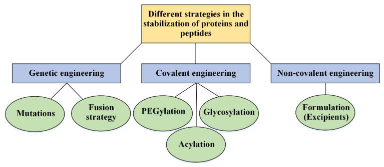
In many cases of the delivery of proteins and peptides for research use only, the powdered/dried form of proteins is preferable to their soluble form. In this delivery route, there is no protein hydrolysis pathways, in-solution decomposition, or air–water interactions caused by the agitation of the protein solution and environmental pH changes. Nevertheless, for the treatment sector and therapeutic usage, the powder form of protein drugs may cause many mistakes and consequences for patients, including mistakes in determining the correct dose of the drug, using solutions that may not have the required standard, and/or the need to send approved solvents from the companies. Thus, it is better to think through the solution form of protein and peptide drugs for therapeutic purposes (that is currently being considered) [10]. Some of these strategies are related to the genetic design to produce more stable protein analogs [128], and some other paths are connected to protein structure and solvent engineering [129]. The changes in genetics and the surrounding environment of medicinal proteins can be done with various goals such as increasing activity, increasing solubility, and protein stability. Figure 6 provides an overview of various approaches of the latter aim to stabilize pharmaceutical proteins.
Figure 6.
A variety of strategies to increase the stability of medicinal proteins and peptides.
Some of these approaches are related to the primary protein sequence (genetic changes) and many of these strategies are linked to protein changes after production and purification; the latter class usually has been divided into two main sub-classes: non-covalent and covalent modifications.
4.1. Genetic Engineering: Protein Analogs
With the discovery of genetic engineering and gaining the knowledge that it is possible to change the DNA sequence in our favor, in 1973, the hope was revived to start producing proteins with properties beyond the natural states. From that year onwards, precisely 1982, and with the creation of the first protein drug with a human genetic source (insulin), it was gradually added to the number of recombinant drugs [1]. Today, with all types of analog insulins, all types of stability and speed of activity are accessible to diabetic patients [14]. The genetic manipulation of medicinal proteins and peptides should be aimed at not harming the protein’s activity as much as possible and only stepping in the valley of its stability must be careful. Meanwhile, two general strategies for the production of analog proteins using direct mutation (site-directed mutagenesis) and random mutation of proteins and the production of fusion proteins will be discussed.
4.1.1. Site-Directed Mutagenesis
There are countless ways to induce a mutation in the genetic structure of the protein to produce a protein analog, which is to create a more resistant or even more active species than the wild form of the protein. For the purposeful creation of protein analog species, one must have sufficient knowledge of the structure of the wild-type protein. Among all changes that lead to the production of analog or mutant proteins, it is worthwhile to refer to substitution, deletion and addition of one or more amino acids in the protein structure. As an example, in our previous study, two amino acids lysine were added at the end of the B chain of insulin, which led to the aggregation of obtained analogs during subcutaneous injection, which increased the in vivo stability of these insulins [130]. Besides, sometimes, some essential amino acids are also added to native proteins to introduce the glycosylation and acylation sites, for increasing the biological and physical stabilities of the target proteins with the help of sugar and fatty acid molecules.
Studying some successful protein drugs such as interferon (IFN) β1b, human fibroblast growth factor (FGF) and granulocyte colony-stimulating factor (G-CSF), has had the result that the substitution of cysteine residue with serine has led to the production of some analogs with a longer half-life [131,132]. Today, with the help of in silico platforms for estimating and predicting domains sensitive to aggregation (such as hydrophobic domains buried in the structure of proteins), researchers can succeed in mutations that make the resulting proteins more stable against temperature and other physical stresses. Human growth hormone (hGH) and G-CSF can be mentioned among these case studies. In the first case, it was reported that the obtained hGH analogs with 10–14 mutations showed a four- to ten-fold increase in shelf life and an increase in temperature stability. Speaking about G-CSF, it was also reported that in addition to an increase in temperature stability up to 16 °C compared to the wild-type protein, the analog has activity in cell proliferation studies, as well [133,134]. As previously reviewed, the digestion/degradation and cleavage of medicinal proteins in many chemical modifications such as oxidation and deamidation can be the coup de grâce to the proteins. Accordingly, efforts to create mutants resistant to protein digestion can be valuable. For example, it is commonly seen that the site of proteolysis of proteins is evolutionarily institutionalized in the flexible loops. Thus, mutations that increase the flexibility of proteins may cause resistance to proteolytic digestion [134]. As an example, it has been found that the mutation of two arginine amino acids in the flexible region of factor VIIIa protein leads to the resistance of this medicinal protein to the proteases of thrombin, activated protein C, and factor VIIIa [135].
4.1.2. Fusion Strategies
Usually, the most important purpose of creating a fusion state in medicinal proteins is to increase their half-life in the blood circulation system and significantly improve pharmacokinetic profiles [136]. For this reason, it is often used to increase the size of protein molecules to escape from the renal filtration system. In this achievement, a protein that can circulate in the blood for a long period is usually used (such as albumin or the FC part of antibodies) [137]. As a famous and appreciated example, the etanercept protein that is used medicinally for rheumatoid arthritis disease, today, is obtained from the addition of the extracellular domain of p75 tumor necrosis factor receptor (TNFR) and the Fc part of IgG antibody. The FC part leads to an increase in the life of the protein in the blood circulation by the mechanisms of increasing the size of the fusion protein and possibly by endosomal recycling. On the other hand, because the Fc part is a dimer, the binding rate of this protein to TNF-alpha is between 50 and 1000 times higher than that of monomeric TNF-alpha samples [134]. Regarding albumin, two major mechanisms have been proposed. One is the direct binding of this protein to the medicinal proteins (e.g., different variants of barbourin [138] and hirudin [139] and the other is the binding of peptides that can act as linkers between the medicinal protein and albumin (peptides that have a high binding tendency to albumin); extending an albumin-binding peptide label to the anti-tissue factor D3H44 Fab that increased the half-life nearly 40-fold [140]. In addition to these two natural protein molecules, other molecules have also been used to produce fusion therapeutic proteins. In a general view, the following table (Table 1) refers to the types of different fusion partners that have been used to produce fusion proteins.
Table 1.
The introduction of different partners in the fusion proteins production [137].
| Partner Type | Marketed Examples | The Effects of the Partner | |
|---|---|---|---|
| Non-peptide partners | Fc | Enbrel®, Eloctate® and Alprolix® (FDA approved). | Enlargement to a size greater than 70 kDa (the critical size to escape renal excretion). |
| Albumin | Tanzeum® (FDA approved). | Increases blood circulation | |
| Transferrin | - | Improve oral administration, no FDA-approved cases, | |
| Carboxyl terminal peptide | Elonva® (produced by Merck and Co., Rahway, NJ, USA, but not FDA approved). LagovaTM (in phase 3 clinical trials). | 31 residues with four O-glycosylation sites | |
| Recombinant polypeptide partners | XTEN | - | Increases blood circulation |
| Elastin-like polypeptides | GlymeraTM, VasomeraTM | Contains V-P-G-x-G, the sequence where x is any residue except proline. | |
| Proline-alanine-serine | XL-Protein GmbH (preclinical state). |
The repetition of Pro, Ala and Ser residues in 100–200 copies. Delaying the renal clearance | |
| Glycine-Serine rich peptides | - | As a linker and a partner for medicinal peptides |
Abbreviations: FDA: food and drug administration, Fc: crystallizable fragment, Tf: transferrin, ELP: Elastin-like polypeptides.
Sometimes, the purpose of creating a fusion is to increase the stability of the medicinal protein in bacterial expression systems. Especially for therapeutic peptides, the expression of such small molecules in prokaryotic systems such as E. coli is always accompanied by the challenge of protease digestion in the bacterial cells, which leads to a low yield of the final product. On the other hand, by designing fusion (carrier protein) to the target peptide, post-expression pathways such as purification will take place more easily. As a previous attempt, in the expression system of E. coli, the genes of insulin A and B chains were separately fused with the chaperone protein carrier alpha-B crystallin of the human eye lens, in addition to increasing the efficiency of the expression in the bacterial platform, the purification pathways were also conducted more simply [141]. Recently, a similar study has been carried out to produce proinsulin in the prokaryotic system [142].
4.2. Covalent Engineering
In this section, the purpose is to explain those changes that are covalently applied to the structure of proteins and peptides to increase their stability. In general, this category of changes includes the connection of synthetic polymer molecules such as polyethylene glycol and some other extracted molecules: sugars and fatty acids.
4.2.1. Protein–Polymer Conjugates
The challenges to the stability of medicinal proteins and peptides are so deep that they cannot be completely solved by the strategies reviewed so far. In many cases, low-stability proteins need to be conjugated with synthetic polymers to brighten the hope of increasing their stability. One of the most prominent examples of protein-polymer conjugation is the PEGylation of medicinal proteins, which leads to an increase in the life of proteins in the circulatory systems [143]. There are currently more than 40 FDA-approved protein drugs that have been conjugated by PEG [144]. In addition to increasing the life expectancy in plasma, other benefits such as protein stability in the digestive system, control of receptor-ligand binding, and improving the storage stability of medicinal proteins using PEG polymer have been reported. It is necessary to take great care about the selection of the type of protein, the type of polymer and even the conjugation method, to follow the paths that the guest protein does not change from its natural state (fold) to an unnatural form (unfold) [143].
In this section, the types of polymers that have been used for the engineering of medicinal proteins will be discussed. In order to correctly and systematically choose a suitable polymer for conjugation to protein, it is necessary to consider several criteria, which are discussed in the table below (Table 2).
Table 2.
Essential considerations in selecting a suitable polymer for protein-polymer conjugation [143].
| Factors Must Be Consider | Example | Description |
|---|---|---|
| Identity | PEG and PEG analogs | Increases the hydrodynamic radius, decreasing immunogenicity |
| Stimuli-responsive polymers | Thermo responsive: p(NIPAAm) pH-responsive: poly(acrylic acid) and p(DMAEMA) |
|
| Biomimetic polymers | Trehalose Heparin-mimicking polymers: poly(styrene sulfonate) and poly(vinyl sulfonate) |
|
| Degradable polymers | Cyclic ketene acetals copolymerized with vinyl monomers: degradable under basic pH Hydroxyethyl starch: a-amylase sensitive |
|
| Toxicity | Hydroxyethyl starch | Plasma volume expander |
| Molecular weight | PEGylated proteins | Decreasing renal filtration |
| Polymer architecture | Branched, brush and linear polymers are accessible to be conjugate with proteins. | Stimulating the immune system |
| Polymer solubility | - | Soluble in the range between 100 and 500 mg/mL |
Abbreviations: PEG: poly ethylene glycol; p(NIPAAm): poly(Nisopropylacrylamide); p(DMAEMA): poly((N,N-dimethylamino)ethyl methacrylate).
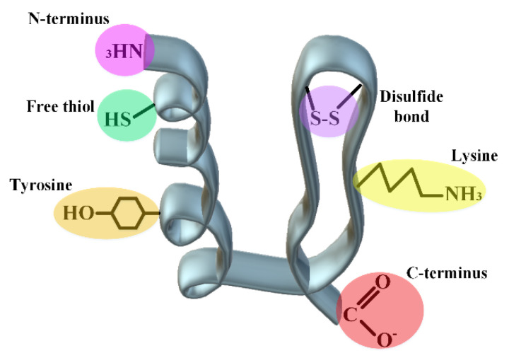
Additionally, in the selection of protein, attention should be paid to the types of functional groups that can react with the selected polymer. These functional groups should not be considered in the sensitive areas or the active site of the protein. The following figure (Figure 7) shows the types of possible active groups in proteins.
Figure 7.
Introducing several active groups on the surface and inside a hypothetical protein structure to introduce desired covalent modifications.
In addition to both C- and N-ends of a protein, disulfide bonds, side chains of lysine and tyrosine as well as free thiol can undergo purposely various chemical changes to increase protein stability.
The suitable environment for the reactivity of lysine (pKa~10.5) is more in the neutral to basic ranges, which is not suitable for some proteins that are not stable in this range [145]. Additionally, the amino acid surface accessibility and the charge around it will be effective in its reactivity. In the case of cysteine, because it is less abundant than lysine in the primary structure of proteins, the residue is more popular for targeted chemical modifications. Nevertheless, the changes in this amino acid are a bit riskier and can lead to changes in the protein structure. So, the structural characterization of proteins should be investigated after cysteine modification. In many medicinal proteins and especially antibodies, there are a large number of disulfide bonds, which with their slight reduction, it has been seen that it does not cause severe damage to the tertiary structure of the proteins. The same method is used for the changes of some proteins in the reduced cysteines. For example, in the brentuximab vedotin antibody model, after partial reduction of the disulfide bonds, the obtained structure is then conjugated to the maleimide functionalized anti-cancer drug [145]. This method cannot be useful for small proteins with a small number of disulfide bonds, since the whole structure may collapse by reducing the bonds. For chemical changes in the disulfide bonds of these proteins, replacing natural disulfide bonds with synthetic ones has obtained hopeful results [146]. Additionally, pka for the N-terminus amine of proteins is usually reported 6–8, which is usually used as a mindset for changes in the amine end (pH-dependent reactions). For example, the pegfilgrastim protein drug with the same method has a PEG at the N-terminus amine. Reductive amination in acidic pH usually leads to N-terminus amine PEGylation of proteins with yields above 90% [145,147]. However, it should be kept in mind that connecting the polymer to the N-terminus amine will be advantageous in proteins that are small and have a few lysine residues. Because the amine group located in the side chain of lysine can win by competing with the amine at the end of the protein and reducing the percentage of the final product. Usually, it is difficult to distinguish between the C-terminus of the protein and the carboxyl present in the side chain of amino acids Asp and Glu, which indicates the necessity of using tags at the C-terminus to confirm the reactivity of this end [148,149]. Tyrosine is also a residue that has a low abundance on the surface of proteins and is usually not followed by high popularity in the field of polymer-protein conjugation due to its cross-reactivity with the amino acid histidine in nucleophilic reactions.
4.2.2. Linker Chemistry
The linker is a very essential part of protein conjugation, especially for ADCs. Linker chemistry can be tuned to impart adequate stability for protein drugs [150]. As mentioned above, PEGylation is an example of using hydrophilic PEG linkers to improve drug efficacy [151]. There are many factors to consider when choosing linkers for ADCs. Nevertheless, one of the most important of them is the stability of the linker throughout its presence in the plasma, while it must break down as soon as it enters the target cell (in the form of a conjugate with ADCs) and leads to the activation of the released anti-cancer drugs [152]. There are cleavable and non-cleavable linkers. The cleavable linkers are cleaved by chemical or biochemical changes in the environment such as acidic changes for hydrozone linkers and glutathione or high pH values for disulfide linkers. Some other cleavable linkers contain enzymatic digestion sites and are sensitive to the enzymes inside the vesicle such as valine-citrulline di-peptides for cathepsin B enzyme. On the other hand, the non-cleavable linkers depend on the complete digestion of the antibody after entering the vesicle [153].
Among the important reasons that led to the removal of Mylotarg from the marketing list in 2010 were items such as the instability of the hydrazine linker and its payload release [154,155]. More recently, Trastuzumab-emtansine (T-DM1) 1 with a non-cleavable linker is the first generation of ADCs approved for the treatment of HER2 (human epidermal growth factor receptor 2)-positive breast cancer. Second-generation ADCs targeting HER2, trastuzumab duocarmazine (SYD985), was characterized to supersede T-DM1 by overcoming all resistance using a cleavable linker and a more potent payload, duocarmycin (DUBA) via different conjugation chemistry. There are, however, no measurable changes observed between the half-life of Adcetris® with a cleavable linker and Kadcyla® with non-cleavable linker at the clinical level [156,157].
The linker property will also determine how the final products act during blood circulation and also touch the sensitivity of the antibody to environmental stresses [158]. Some disulfide-linked ADCs were found to conjugate with blood proteins through cleaved free thiols. Hydrophobic linkers and payloads frequently encourage the aggregation of ADC molecules, e.g., King et al. showed that multiple loading of the BR96 antibody that is simultaneously attached to doxorubicin drug leads to non-covalent dimerization of the antibody, which will eventually lead to hepatotoxicity [159,160]. It has also been seen that the linkers that are more hydrophobic are not able to significantly affect the MDR1+ cells. Instead, linkers with a more polar and amphiphilic nature were able to appear with a greater effect such as mal-PEG4-N-hydroxysuccinimide and N-Hydroxysuccinimidyl-4-(2-pyridyldithio)-2-sulfobutanoate (sulfo-SPDB) [161]. In addition to the MDR1 challenge, it has also been found that reducing the percentage of hydrophobicity by employing hydrophilic linkers containing negatively charged sulfonate groups, polyethylene glycol (PEG) groups, or pyrophosphate diester groups can also help in improving therapeutic index level and pharmacokinetics [162].
4.2.3. Acylation
This strategy has mostly been used in the case of medicinal peptides; liraglutide and insulin detemir are among the most successful cases of lipidated peptides in the world market. It has been reported that lipidation in the backbone of peptides can give positive characteristics to the obtained product, such as impressive stability against enzymatic digestion, and specificity to the receptor and also to the bioavailability of peptides. In addition to the issues related to the stabilization of medicinal peptides, this process has received much attention in the field of drug delivery of medicinal peptides [163]. This process occurs unsurprisingly in nature. In the living system, various enzymes help a protein or peptide to react with a chain of saturated or unsaturated fatty acids in the form of N- and/or O-links [164]. The binding site of lipids can be the terminal amine and carboxyl, and even in the middle part of the primary protein/peptide structure, which occurs through residues such as serine, threonine, glycine, lysine, and cysteine [165]. It is interesting to note that this addition of lipids can be irreversible (when it is through the connection of the N-terminal of glycine/cysteine) or reversible (when a thioester bond occurs between the fatty acid and thiol group of cysteine) [166,167]. Regarding the medicinal aspects of lipidopeptides, some of these peptides are naturally extracted from microorganism sources such as fungi and bacteria. The use cases of this type of peptides have been proven in anti-cancer, anti-fungal and anti-bacterial fields [2]. However, from another line of view, in silico addition of lipids to peptides is also of special interest [168]. In addition to the fact that the presence of peptides can lead to the appearance of some new features in the peptide, it is reported that the type of spacer between the lipid and the peptide, the type of lipid and its chemical nature (saturated and unsaturated) are also effective in the activity of the lipidopeptide [169,170]. The table below (Table 3) mentions some of the peptides that have been joined with lipids as encouraging options for treatment.
Table 3.
The list of some peptide-lipid conjugates.
| Lipidopeptide/Protein | Lipid | Attachment Strategy | Effects | Ref. |
|---|---|---|---|---|
| Liraglutide | Palmitic acid | γ-glutamic acid | Extension of the half-life, reducing renal clearance | [171] |
| Salmon calcitonin | N-palmitoylated | Cys-1 and Cys-7 of the peptide | Extension of the half-life, reducing renal clearance | [172] |
| Opioid peptide leu-enkephalin | 3,4 bis(decylthiomethyl)- 2,5-furandione 16 |
N-terminus binding | Reducing receptor binding, increasing protease stability | [173] |
| Thyrotropin-releasing hormone | lauric acid |
N-terminus binding | Peptide penetration across the small intestine |
[174,175] |
| H-Ras | 1,2-dioleoyl-sn-glycero-3-phosphoethanolamine | maleimide-functionalized phospholipids/ S-palmitoylation at Cys181 and Cys184 | Dimerization | [176] |
| Caveolin-3 | octanethiol | cysteine S-fatty acylation | As a small protein model in the study of lipidation of proteins | [177] |
| Interferon-induced transmembrane protein 3 (IFITM3) |
maleimide palmitate | maleimide-functionalized phospholipids/ S-palmitoylation at Cys72 and Cys105 | Enhancing the conserved amphipathic domain | [178] |
| Cathepsin-B inhibitor | Palmitic acid | Palmitoylation of the amino groups (ε-lysyl amino groups) |
Increasing the inhibitory effect on cathepsin-B | [179] |
4.2.4. Cyclization
Cyclization is another solution that has been proposed to increase the stability of medicinal peptides, especially against enzymatic digestions. The idea of this strategy is taken from nature by discovering peptides such as mammalian theta-defensins [180] and plant cyclotides [181]. The best example of an engineered cyclic peptide is a 29 amino acids peptide, kalata-B1, which is resistant to a wide range of environmental stresses such as 8 M urea, acidic environments (0.5 M HCl), 6 M GnHCl, and the presence of various proteases [182]. Taken as a whole, the high stability of these cyclic peptides has caused special attention in recent years to search for faster synthesis methods and the purification of cyclized peptides.
At first, in the following table (Table 4), several circular medicinal peptides will be introduced, and then, due to the importance of this section, various methods of peptide ringing will be discussed. As can be deduced from the information in the table, most cyclic peptides belong to non-synthetic peptides, extracted from microorganisms. This is due to the complex routes of cyclization of peptides and finally their purification to an acceptable level [183].
Table 4.
Some examples of naturally extracted and chemically synthesized cyclic peptides with their applications.
| Name | Source | Application | Ref. |
|---|---|---|---|
| Sunflower trypsin inhibitor 1 (SFTI-1) |
Helianthus annuus | Trypsin inhibitor, angiogenic activity | [184] |
| Gramicidin S | Bacillus brevis | Antibiotic activity towards Gram-negative, Gram-positive and several pathogenic fungi | [185,186] |
| Plitidepsin | Aplidium albicans | In vitro anticancer activity | [187,188] |
| Tyrocidine | Bacillus brevis | Antibiotic action | [189] |
| Rakicidin F | Streptomyces | Antibacterial effect | [190] |
| Plitidepsin | Aplidium albicans | Antitumor, antiviral and immunosuppressive activities | [191] |
| Asperpeptide A cyclo(-Pro-Ala-Ala-Tyr-5-OHAA) | Aspergillus sp. XS-20090B15 | Antibacterial activity | [192] |
| Cyclosporin A | Tolypocladium inflatum | Calcineurin inhibitor, decreasing the function of lymphocytes | [193] |
| Depsilipopeptide colisporifungin | Colispora cavincola | Antifungal activity | [194] |
| Cryptophycin | Nostoc | Fungicide and anticancer | [195] |
| Romidepsin | Chromobacterium violaceum | Apoptotic activity, anticancer activity | [196,197] |
| Theonellamide G | Theonella swinhoei | Cytotoxic activity | [198] |
| Ziconotide | Conus magus | Analgesic agent; a strong pain killer. | [199] |
| Kahalalide F | Elysia rufescens | Antitumor | [200] |
| Vancomycin | Amycolatopsis orientalis | Antibacterial | [201] |
| Alisporivir | Chemically synthesized from ciclosporin. | Inhibits cyclophilin A, the potential effect on Alzheimer’s disease and hepatitis C | [202,203] |
| Istodax (Romidepsin) | Head–tail lactone cyclization which is stabilized by a pair of disulfide bonds. | Anticancer | [204] |
| Lupkynis (Voclosporin) | Head–tail cyclization | Calcineurin like activity | [205] |
| Vasostrict (Vasopressin) | Disulfide mediated cyclization | Used in Anti-diuretic hormone deficiency | |
| Signifor (Pasireotide) | Head–tail cyclization | Activating a broad spectrum of somatostatin receptors, reducing cortisol secretion | [206] |
| Prialt (Ziconotide) | Disulfide mediated cyclization | Pain killer | [207] |
Based on the nature of the linkage between the two residues in the peptide structure that ultimately ensures the cyclicity, there are two major classes of cyclic peptides: homodetic (involving only a conventional peptide bond) and heterodetic (using a variety of functional groups that required for looping the peptides) [208].
4.2.5. Nanoparticles; Double-Blade Sword
In most past studies, nanoparticles have been used as a platform for the delivery of protein and peptide drugs. The oral route of using biological agents is the easiest way to use such drugs, but due to the destructive conditions of the digestive system (acidic environment, the presence of many protease enzymes, and a small percentage of surface absorption in the small intestine), this route has always been associated with many challenges. For this reason, the use of nanoparticles has been focused on facilitating the delivery of protein drugs in the alimentary tract. However, in some studies, nanoparticle systems have also been used for the intravenous routes of drugs. In any case, for the use of nanoplatforms for the delivery of protein or peptide drugs, the surface and nanoparticle size are factors that should be taken into consideration [209]. From the surface chemistry point of view, firstly, nanoparticles should ideally have hydrophilic surfaces that can be easily dissolved in living systems and secondly escape from the macrophage system [210].
Given particle size, in living systems of vertebrates, macrophages are responsible for ingesting large foreign particles (size around 0.5 µm). The selected nanoparticles should be small enough to escape from this system on the one hand, and on the other hand, they should be large enough to not quickly enter the outside environment of the vessels through the vascular leakage system. It has been agreed that this size is between 150–200 nanometers in the spleen and liver on the one hand, and on the other hand, the gap junctions between endothelial cells are between 100 and 600 nanometers [211,212]. According to this evidence, choosing the right size is a challenging factor. However, looking at the successful studies in this field, in general, it is understood that the useful nanoparticles that have been used so far have been between 3 and 200 nanometers in size. A wide range of nanoparticles have been used to deliver all kinds of drugs, nevertheless, in the case of protein and peptide drugs, most of the attention has been on polymer nanoparticles and solid lipid nanoparticles. Of these two groups, the latter has also been pursued more seriously due to the controlled release of the cargo and its ability to be engineered for a specific organ [213]. To summarize the contents of this section, it is necessary to mention the following table (Table 5), which presents the types of nanoparticles, the cargo, and the delivery method used in previous years.
Table 5.
Examples of those nanoparticles have been used to stabilize proteins and peptides.
| Nanoparticle | Protein Cargo | Delivery Rout | Ref. |
|---|---|---|---|
| Chitosan functionalized lipid nanoparticle (LN) | Insulin | Oral | [214] |
| Chitosan-modified mesoporous silica | Insulin | Suggested for oral delivery | [215] |
| Chitosan-modified carboxymethyl-β-cyclodextrin | BSA | Oral | [216] |
| Poly(lactide)-tocopheryl polyethyde glycol succinate |
BSA | Oral | [217] |
| Alginate | Osteogenic protein-1 | Intranasal | [218] |
| DOPE-liposome | E75 peptide (HER-2/neu-369–377) | Intravenous | [219] |
| DOTAP-liposome | HSV-derivated E7 oncoprotein | Intravenous | [220] |
| Chitosan-coated liposomal system | Ghrelin (a peptide hormone that can regulate appetite and body weight changing) | Nose-to-brain delivery | [221] |
| Chitosan-covered liposomes | Rainbow trout skin-derived peptides |
Suggested for oral delivery | [222] |
| PEGylated liposomes | Opiorphin | Intravenous | [223] |
| Polyacrylate-coated superparamagnetic Fe3O4 | Elastin-like VPGVG pentapeptides | Subcutaneous injections | [223] |
| G4 OH-terminated PAMAM |
AFPQFRSATLLL | Subcutaneous injections | [224] |
| Thiolated chitosan nanoparticles | Insulin | Subcutaneous injections | [225] |
| Chitosan-modified PLGA nanospheres | Elcatonin | Pulmonary | [226] |
| Chitosan nanoparticles | RGD peptide (Arg-Gly-Asp) | Intravenous | [227] |
Abbreviations: DOPE: 1,2-Dioleoyl-sn-glycero-3-phosphoethanolamine, HER: Herceptin, GLP-1: glucacon-like peptide-1, DOTAP: N-(2,3-Dioleoyloxy-1-propyl)trimethylammonium methyl sulfate, HSV: herpes simplex virus, PAMAM: Poly(amidoamine), PLGA: poly(lactic-co-glycolic acid).
4.3. Non-Covalent Engineering of Therapeutic Proteins and Peptides; Solvent Engineering Pathways
4.3.1. Formulation: Solvent Engineering Pathways
Another solution that has been used to preserve the integrity of medicinal proteins and peptides is the use of excipients during storage and even through the use of such drugs. According to the types of these agents, their mechanisms of action are also diverse [228]. In the following table (Table 6), the types of these excipients are presented along with their mechanism to protect proteins. Some of these excipients should be used for a certain amount of window with specific considerations such as salts that can lead to the aggregation of proteins after a critical concentration [229]. Or as another important example, in the category of sugars, usually non-reducing sugars are used because reducing sugars via Millard’s reactions may cause destructive glycosylation of proteins [230].
Table 6.
| Excipient | Mechanism | General Comments |
|---|---|---|
| Buffering agents | Keeping the pH of protein solutions | Usually, the buffers work in the range of 3–10 for proteins. In certain conditions, some buffers may be decomposed, and their by-products destroy the protein structure. |
| Chelators and antioxidants | The roles of antioxidants and chelators are to prevent and/or remove oxidazing factors. | Some reducing agents such as glutathione and ascorbic acid in the presence of metals and enhancing oxidation stresses can have a negative/destructive role on protein structure, although these agents are used in the pharmaceutical sector of proteins. |
| Proteins | By interacting with therapeutic peptides, excipient proteins can increase the blood circulation time. | Nowadays, chaperones have been given a special look as a preservative for medicinal proteins. |
| Polymers | Maintaining the structure of proteins | The main examples of this group include polyvinyl alcohol, dextran and hydroxyethyl starch. |
| Amino acids | Buffering properties, preferential interactions, favored hydration, antioxidant effect and strong binding to protein regions | Glycine (buffering agent and bulking agent during lyophilization), arginine (solubilizing agent and works as chaperone) and histidine (antioxidant and buffering agent) |
| Sugars and carbohydrates | Forming a crystal network with preferential interactions | Sorbitol in lyophilization and liquid formulation conditions, has a stabilizing role, however, in freezing conditions, due to the formation of sorbitol crystals, it assumes a stabilizing role. |
| Salts | Tonicifying agent, reacting with the charged surfaces of proteins and take two stabilizing or destabilizing paths. | Among the cations and anions of the Hofmeister series, the latter has dual protective/destructive effects on proteins. |
| Antimicrobial preservatives | Preventing the growth of bacteria in solution-rich medicinal proteins | m-cresol, phenol and benzyl alcohol are among the popular. |
| Surfactants | Reducing the interface area of solution and air during purification (inner wall of purification column and dialysis bags, etc.). |
In this group, polysorbate 20 (PS20) and polysorbate 80 (PS80) are the most used in protein drugs and especially antibodies. |
| Osmolytes | Generating favored hydration, preferential interactions, and polar interactions | Sorbitol, sucrose, glycine, and trehalose have been able to reach the pharmaceutical sector |
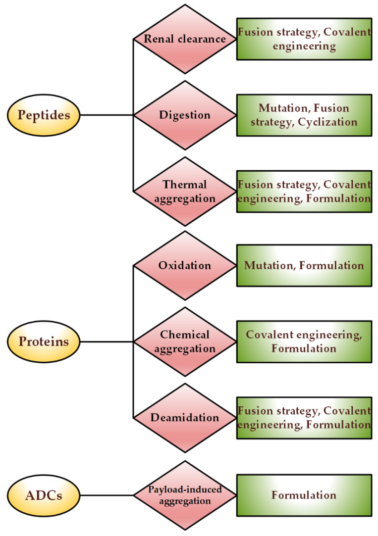
According to all the information that was reviewed about the stabilization of proteins and peptides, at the end of this section, it is necessary to briefly suggest which types of proteins usually suffer from which stress and what are the proposed solutions to overcome the challenges (Figure 8).
Figure 8.
Proposed solutions to the challenges faced by medicinal proteins and peptides.
4.3.2. Choice of Container
Depending on the type and even the architecture of the protein containers, proteins may react with the containers’ surfaces, be adsorbed without or with a change in the tertiary structure, and lead to protein aggregation that is either reversible or irreversible. For example, it has been detected that rhFVIII proteins undergo tertiary structure changes in contact with hydrophobic silica surfaces, and changing the nature of the surface to a negative charge has led to a slight change in the protein structure [233]. It has also resulted in some cases where no significant structural change occurs in the protein, but a severe aggregation behavior occurs during protein binding to the surfaces. For instance, rhPAF-AH protein does not show a measurable change in contact with the hydrophilic surface of silica, though, at a pH 6.5, it showed a very high aggregation rate [234].
Based on past efforts in this field, scientists have generally agreed that glass containers are not ideal, but suitable, options for transporting and storing these therapeutic macromolecules [235,236]. Some studies have also pointed out that even these glass containers can interact with proteins and cause structural changes in them, which in itself can reduce the stability of the protein and other consequences such as ectopic responses in the patient’s body [236]. It is worth mentioning a few studies on the changes that have been made to the surfaces of glass containers to help stabilize drug proteins. From about 1850, therapeutic glass containers were introduced to world markets. Then, in the 1950s, due to problems such as the reactivity of pharmaceutical proteins and some other chemicals with glassware, efforts began to engineer glassware using beneficial polymers [237]. Although glass vials are mainly used for the storage of medications, in some of them, the presence of glass particles, separated from the bottom of the container, has been reported [117]. Container delamination is an undesirable process in which a thin layer of glass is separated from the main body glass and the separated particles have the potential to destroy the drug [238]. For such cases, the US Pharmaceutical Association (USP) has guided assessment glass delamination called USP <1660> [237]. USP <1660> lists most of the possible factors: the composition of the glass, the conditions under which the container is made, the subsequent displacement of the container and the medicinal product in the container. Thus, assessing the inner surface of glass vials, including examining the surface of the glass using an electron microscope (to analyze the degree of the surface cavity or depth), measuring the total composition that may have been separated from the glass using inductively coupled plasma-mass spectrometry, or examination of visible particles using dynamic light diffraction or scanning electron microscopy-energy scattering (SEM-EDX) is essential [237]. Combining all mentioned studies, for industrial technology, glass vials are preferred over plastic vials due to the standard depyrogenation, operations of washing and possibly terminal autoclaving and in another view, glass containers have less immunogenicity in comparison with plastic counterparts [239]. Although in recent years, different particles with an outer layer of silica have also been invented, which can be an alternative option for current protein drugs containers. However, it is still necessary to go through several confirmation stages to industrialize these candidates, [240,241].
5. Different Approaches in Characterizing Protein Drugs and Induced Structural Changes
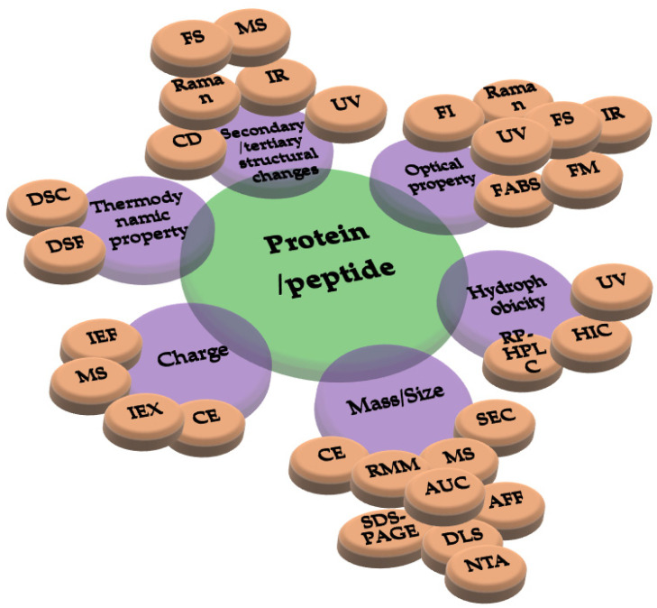
The characterization of the chemical and physical stabilities of medicinal proteins is vital in drug development and must directly link to a number of critical quality attributes (CQAs) that must be checked to guarantee final-product quality [242]. Advances in analytical techniques have given researchers and biopharmaceutical companies new and increasingly sensitive methods for characterizing their products. Due to the complexity of pharmaceutical proteins, it is regularly essential to study the stability of them using several techniques [243] based on their changes in mass/size, folding, charge, and hydrophobilicity, as well as optical and thermodynamic properties as drawn in Figure 9.
Figure 9.
Different analytical techniques that are currently applied for characterization of protein and peptide drugs. Abbreviations: AUC: analytical ultracentrifugation, DSC: differential calorimetry, CE: Capillary electrophoresis, CD: circular dichroism, DSF: differential scanning fluorimetry, UV: ultraviolet, DLS: dynamic light scattering, FAPS: fluorescence-activated particle sorter, AFF: asymmetrical flow-field flow fractionation, NTA: nanoparticle tracking analysis, RMM: resonant mass measurement, SEC: size-exclusion chromatography, SDS-PAGE: sodium dodecyl sulfate-polyacrylamide gel electrophoresis, MS: mass spectrometry, RP-HPLC: reversed-phase high-performance liquid chromatography, IEF: isoelectric focusing, IEX: ion exchange chromatography, FM: Fluorescence microspcopy, FI: Fluorescence imaging, and FS: Fluorescence signal.
Although the first sign of the validity of the protein may be its transparent state and its solubility, which can be easily seen even with the naked eye [244], most CQAs require more sensitive monitoring at the molecular level which needs to be examined by analytical instruments. Using LC-MS, subunit and intact protein masses can be directly measured to confirm product identity and quantify different sub-units, as well as to monitor conjugated species and the drug-to-antibody ratio of ADCs at the same time [245]. Moreover, a bottom-up approach relying on the enzymatic digestion of the protein into peptides before LC-MS is used to identify chemical modifications such as oxidation and deamidation on a specific residue site as well as to estimate their modification levels [246]. Due to the varied range of post-translational modifications and different steps during industrialization, pharmaceutical proteins are severely heterogeneous and need high-resolution mass spectrometry (HRMS) for identifying, characterizing, and monitoring these attributes [247]. Disulfide linkages can also be solved by bottom-up LC-MS approach with non-reducing digestion using gas phase [248] and/or solution phase [249] fragmentations. However, bottom-up processing can result in considerable information loss or errors due to mis-cleavages or data mismatching. Combining intact, middle-up, and bottom-up approaches of LCMS or capillary electrophoresis-MS (CE-MS) appears to be one of the main platforms in characterizing and monitoring the chemical stability of pharmaceutical proteins [250].
The challenge of protein aggregation has always been discussed to a high degree in industrialization. Regarding this, the size exclusion chromatography (SEC) technique has been known for years, however nowadays, due to the innovation in the field of monitoring protein aggregates, many institutions request orthogonal analytical methods for the accuracy and reliability of the SEC data [251,252]. In addition to SEC, protein aggregation can be detected in various ways such as small-angle X-ray scattering and dynamic light scattering. Fortunately, during the structural changes of proteins and their folding, many signals can be monitored to ensure whether the protein in question is folded or not. Sometimes the size of the protein is considered, in some cases the tertiary structure or the chemical composition of the protein will determine the amount of damage done to the protein. The size, changes in the three-dimensional shape of a protein, and changes in its chemical nature are among the general criteria that can be considered [253]. Native MS, a new technique to simultaneously determine molecular weight and detect the exposed charge profile of a native protein, has been applied to study protein folding and stabilities by varying temperature [254] with unparalleled sensitivity, dynamic range, and selectivity for studies of both cold- and heat-induced chemical processes.
There are many challenges in determining the properties of protein drugs and especially ADCs whose synthesis normally leads to heterogeneous drug loads depending on the kinetics of conjugation chemistry and linkers [255]. Consequently, it is necessary to make a very good purification of ADCs with a specific drug-to-antibody ratio [256]. Hydrophobic interaction chromatography (HIC) has emerged as a central technique for separating protein conjugates with different drug loads for cysteine or related conjugates where the linker-payload molecules add appreciable hydrophobicity to the ADC. Several orthogonal separations techniques have been developed to purify ADCs with different drug loads. For example, two-dimensional liquid chromatography (2D-LC) via two different separation techniques such as ion exchange followed by a reversed-phase was used to intensify the separation influence [257]. The ultra-high voltage capillary electrophoresis (UHV-CE) technique, which usually uses at least 120 kV and an electric field over 2000 V/cm, was initially used to separate peptides and sugars, nevertheless, recently it has also been used to separate ADCs based on their charges [258].
Likewise, sometimes the dynamics of proteins become important. In the dynamic environment of the cell, proteins do not exist in a static physical form and are constantly undergoing structural changes to fit and bind to their receptors. Instead, some proteins with oligomers with larger structures can be present in the environment, and techniques such as X-ray crystallography and NMR are not able to monitor such changes. Hydrogen deuterium exchange MS (HDX–MS) [259,260] or other MS-compatible chemical labeling methods such as hydroxyl radicals [261] or reductive amination [262] have been used before MS detection to reveal solvent exposure domain in a protein since these regions are more amiable to chemical instability. Although these techniques require a lot of time to analyze the data, they give a good experimental view of molecular dynamics. To summarize all the contents in this section, the following table (Table 7) tries to categorize the various techniques and methods of analyzing the structure of pharmaceutical proteins coherently. Many of these applied techniques have international standards from authorities such as the World Health Organization (WHO), the Food and Drug Administration (FDA) and the National Institutes of Health (NIH) to determine the accuracy of size [10]. Additionally, these techniques must be evaluated and validated from the aspects of linearity, specificity, limit of detection, precision sensitivity recovery rate, and accuracy [263].
Table 7.
| Technique | Output | Destructive | QC Method | General Comments | |
|---|---|---|---|---|---|
| Conformational assessments | DSC | Thermal parameter (Tm, ΔG unfolding) | Yes | No | The best option for checking temperature-dependent parameters; not high throughput. |
| CD | Secondary and tertiary statures | No | No | Sensitive to the polarity of the solution. | |
| Fluorescence spectroscopy | Tertiary statures/ general view on the protein structure | Yes | No | Sensitive to small structural changes of proteins and peptides; the need for relatively small amounts of material; relatively high speed; takes a general view of structural changes and cannot go into details. | |
| DSF | Tm, aggregation onset | Yes | No | During the thermal unfolding of a protein, a dye (commonly Sypro Orange) is added to the sample, which, by connecting to the unfolded parts, leads to an increase in its fluorescence emission. | |
| UV | Tertiary structure | No | No | Makes a general view of structural changes. | |
| Raman | Secondary structure/ chemical characterization | Yes | No | It has a high overlap with FTIR, but on the contrary, a wide range of solvents can be used in Raman analysis to examine samples. | |
| Infrared | Secondary structure/chemical integrity | Yes | No | By examining the amine-I and II regions of proteins and their deconvolution, it is possible to reach the percentage of the secondary structure in the protein (in solution and solid states) either in the form of fold and/or aggregation/fibrils. ATR facility requires very small amounts of substance without the need to prepare KBR tablets. | |
| Oligomerization studies | AUC | Molecular weight/shape | No | Yes | Determining the size of particle aggregation. |
| DLS | Hydrodynamic size | No | High range of particle detection (between 1nm to 5 µM), reliable within a certain range of polydispersity | ||
| FAPS | Particles/Serum interactions | No | Extracting fluorescence data from the aggregated samples. One of its limitations is the use of dyes to identify aggregates, which may affect the protein structure. | ||
| AF4 | Hydrodynamic size | Yes | Yes | AF4 technique separates particles based on their diffusion coefficients. | |
| NTA | Hydrodynamic size | No | It can measure the size of particles, imaging and quantifying them. | ||
| RMM | Concentration/size/mass | Yes | No | In a microfluidic way, it can calculate particle size. | |
| SEC | Hydrodynamic size | Yes | Yes | Determine the molecular weight, aggregation rate, and interactions between proteins. | |
| SDS-PAGE (all types) | Molecular weight/ interactions between proteins | Yes | Yes | Covalent interactions between proteins and also protein digests; in reducing and non-reducing types, it can observe disulfide bonds. | |
| Optical microscopy | Size/morphology | No | No | Detecting large particles (larger than 1 µm). | |
| Native MS | Fragments/aggregates | Yes | No | With principles similar to MS, it investigates non-covalent interactions and post-translational changes in proteins. | |
| Light obscuration | Concentration/size | No | Yes | This technique, which is also known as Single Particle Optical Sensing (SPOS), is not very sensitive to small sizes (detecting sizes more than 1 µm). | |
| Fluorescence microscopy | Particles/amorphous and morphous aggregates | Yes | No | A fluorophore molecule is needed that can provide the output signal. Some fluorophore dyes change fluorescence intensity by interacting with proteins and being buried in their structure, which can be a pattern of protein folding and even aggregation. | |
| Flow imaging | Concentration/size/morphology | No | Yes | The basis is similar to optical microscopy, except that it can provide data qualitatively. | |
| CE-SDS | Molecular weight | Yes | Yes | Advantages such as quicker analysis, the facility for quantification, full automation, the need for low sample, and better resolution | |
| Electrical zone sensing | Concentration/size | Yes | No | By applying electric force and migration of the two-pole magnifier, it can achieve the size of the particles. | |
| Turbidity | Optical density > 360 nm | No | Yes | It is a low-cost, rough method that can be used to detect large particles. Its high speed and simplicity are its positive points. | |
| Chemical changes | RP-HPLC | Hydrophobicity | Yes | Yes | Sensitive to slight changes in surface hydrophobicity of proteins, requiring small amounts of samples. |
| cIEF | Charge | Yes | Yes | Similar to IEF, it can separate proteins based on their PI. Compared to its traditional sample, i.e., IEF, it requires much less material, and due to its capillary nature, a higher voltage can be applied to the sample, which leads to a reduction in the test time. This test is successful in the case of samples higher than 150,000 Daltons that dissolve well in aqueous solutions. | |
| IEX chromatography | Charge | Yes | Yes | Separating proteins by considering their charge. High speed and acceptable accuracy. | |
| MS | By the difference in weight molecular changes | Yes | No | Differentiation of diverse components with considering mass-to-charge ratio (m/z). | |
| LC-MS | By the difference in weight molecular changes | Yes | No | In cases where a protein complex is present, initial separation by HPLC enables MS to obtain more details of the sample by removing noise. | |
| Zeta potential | Charge | No | Yes | Measuring the particle charges; the types of solvents and even the percentage of ions in water strongly affect the data. |
Abbreviations: can: acetonitrile, AUC: analytical ultracentrifugation, DSC: differential calorimetry, Tm: melting temperature, CD: circular dichroism, DSF: differential scanning fluorimetry, UV: ultraviolet, DLS: dynamic light scattering, FAPS: fluorescence-activated particle sorter, AF4: asymmetrical flow-field flow fractionation, NTA: nanoparticle tracking analysis, RMM: resonant mass measurement, SEC: size-exclusion chromatography, SDS-PAGE: sodium dodecyl sulfate-polyacrylamide gel electrophoresis, MS: mass spectrometry, CE-SDS: capillary electrophoresis sodium dodecyl sulfate, RP-HPLC: reversed-phase high-performance liquid chromatography, cIEF: capillary isoelectric focusing, IEX: ion exchange chromatography, and LC: liquid chromatography.
6. Conclusions and the Future of Protein and Peptide Drugs
Looking at the sales share of protein and peptide drugs in recent years and their fast-growing trend, it is not far from expected that these macromolecules will cover most of the future therapeutic markets. Considering the positive features of this class of drugs compared to conventional chemical-synthetic drugs, such as high specificity, reasonable biological lifetime and low toxicity in the body, as well as the need for very low doses to create therapeutic responses, large pharmaceutical companies are willing to invest in this field. However, the problem with these drugs, since the beginning of their arrival, is their instability in different environmental conditions. As mentioned, many efforts have been made to solve this problem, but so far no promising strategy has been achieved in this field. For some therapeutic proteins (such as insulin hormone analogs), the binding of chemical molecules such as fatty acids has been a good option to increase their stability. Similar to the new approach used to produce antibody drug-conjugated, it is expected that a similar strategy will be adopted for some other therapeutic macromolecules such as DNA or RNA molecules in the future. Besides, some efforts have been focused not on the structure of the proteins, but on their storage containers. Nowadays, most of the containers for storing therapeutic proteins and peptides are made of borosilicate glasses, which bring challenges such as the crushing of microscopic pieces of them in the solution and diverse interactions of proteins with their surfaces. In the future, it is expected that this part of science will also reach its evolution, meeting the need for stable medicinal proteins. Until now, a huge part of the application of nanoparticles and polymers has been used in the direction of drug (protein/peptides) delivery, however, in the field of engineering containers for medicinal proteins, no coherent effort has been made.
As reviewed, many pathways threaten the folding and shape of proteins/peptides. Likewise, fortunately, many solutions have been implemented to stabilize these important medicinal macromolecules. Consequently, it should be noted that the choice of which stabilization path to use is strongly related to the type of protein/peptide and even the route of administration of the protein in the body. Furthermore, sometimes it is necessary to apply two or even three strategies in combination to a protein to achieve the desired stability. Nevertheless, whatever the plan of stabilization is, it must ensure that the resulting protein remains non-immunogenic and active. Thanks to bio-analytical chemistry, various methods have been obtained to confirm the final structure of the engineered protein/peptide.
Acknowledgments
For MA: that was an honor to be chosen for the NCKU90 & beyond scholarship at National Cheng Kung University. He wishes to eternally thank and appreciates all the support and attention of Professor Shu-Hui Chen and the NCKU90 & beyond scholarship for his first-year research at the university.
Author Contributions
Conceptualization, S.-H.C.; methodology, M.A. and S.-H.C.; software, M.A. and S.-H.C.; validation, S.-H.C.; formal analysis, M.A. and S.-H.C.; investigation, M.A. and S.-H.C.; resources, S.-H.C.; data curation, M.A. and S.-H.C.; writing—original draft preparation, M.A. and S.-H.C.; writing—review and editing, M.A. and S.-H.C.; visualization, M.A. and S.-H.C.; supervision, S.-H.C.; project administration, S.-H.C.; funding acquisition, S.-H.C. All authors have read and agreed to the published version of the manuscript.
Institutional Review Board Statement
Not applicable.
Informed Consent Statement
Not applicable.
Data Availability Statement
Not applicable.
Conflicts of Interest
The authors declare no conflict of interest.
Funding Statement
This research was funded by National Science and Technology Council, Taiwan, Republic of China, grant number 111-2113-M-00600130MY3.
Footnotes
Publisher’s Note: MDPI stays neutral with regard to jurisdictional claims in published maps and institutional affiliations.
References
- 1.Akbarian M. Insulin therapy: A valuable legacy and its future perspective. Int. J. Biol. Macromol. 2021;181:1224–1230. doi: 10.1016/j.ijbiomac.2021.05.052. [DOI] [PubMed] [Google Scholar]
- 2.Akbarian M., Khani A., Eghbalpour S., Uversky V.N. Bioactive Peptides: Synthesis, Sources, Applications, and Proposed Mechanisms of Action. Int. J. Mol. Sci. 2022;23:1445. doi: 10.3390/ijms23031445. [DOI] [PMC free article] [PubMed] [Google Scholar]
- 3.Karst D.J., Steinebach F., Morbidelli M. Continuous integrated manufacturing of therapeutic proteins. Curr. Opin. Biotechnol. 2018;53:76–84. doi: 10.1016/j.copbio.2017.12.015. [DOI] [PubMed] [Google Scholar]
- 4.Radhakrishnan D., Wells E.A., Robinson A.S. Strategies to enhance productivity and modify product quality in therapeutic proteins. Curr. Opin. Chem. Eng. 2018;22:81–88. doi: 10.1016/j.coche.2018.09.005. [DOI] [Google Scholar]
- 5.Hong M.S., Severson K.A., Jiang M., Lu A.E., Love J.C., Braatz R.D. Challenges and opportunities in biopharmaceutical manufacturing control. Comput. Chem. Eng. 2018;110:106–114. doi: 10.1016/j.compchemeng.2017.12.007. [DOI] [Google Scholar]
- 6.Burnett M.J., Burnett A.C. Therapeutic recombinant protein production in plants: Challenges and opportunities. Plants People Planet. 2020;2:121–132. doi: 10.1002/ppp3.10073. [DOI] [Google Scholar]
- 7.Zaman R., Islam R.A., Ibnat N., Othman I., Zaini A., Lee C.Y., Chowdhury E.H. Current strategies in extending half-lives of therapeutic proteins. J. Control. Release. 2019;301:176–189. doi: 10.1016/j.jconrel.2019.02.016. [DOI] [PubMed] [Google Scholar]
- 8.Hartl F.U. Protein misfolding diseases. Annu. Rev. Biochem. 2017;86:21–26. doi: 10.1146/annurev-biochem-061516-044518. [DOI] [PubMed] [Google Scholar]
- 9.Jackson M.P., Hewitt E.W. Cellular proteostasis: Degradation of misfolded proteins by lysosomes. Essays Biochem. 2016;60:173–180. doi: 10.1042/EBC20160005. [DOI] [PMC free article] [PubMed] [Google Scholar]
- 10.Butreddy A., Janga K.Y., Ajjarapu S., Sarabu S., Dudhipala N. Instability of therapeutic proteins—An overview of stresses, stabilization mechanisms and analytical techniques involved in Lyophilized proteins. Int. J. Biol. Macromol. 2021;167:309–325. doi: 10.1016/j.ijbiomac.2020.11.188. [DOI] [PubMed] [Google Scholar]
- 11.Emami F., Vatanara A., Park E.J., Na D.H. Drying technologies for the stability and bioavailability of biopharmaceuticals. Pharmaceutics. 2018;10:131. doi: 10.3390/pharmaceutics10030131. [DOI] [PMC free article] [PubMed] [Google Scholar]
- 12.Braun A.C., Gutmann M., Lühmann T., Meinel L. Bioorthogonal strategies for site-directed decoration of biomaterials with therapeutic proteins. J. Control. Release. 2018;273:68–85. doi: 10.1016/j.jconrel.2018.01.018. [DOI] [PubMed] [Google Scholar]
- 13.Duivelshof B.L., Murisier A., Camperi J., Fekete S., Beck A., Guillarme D., D’Atri V. Therapeutic Fc-fusion proteins: Current analytical strategies. J. Sep. Sci. 2021;44:35–62. doi: 10.1002/jssc.202000765. [DOI] [PubMed] [Google Scholar]
- 14.Akbarian M., Ghasemi Y., Uversky V.N., Yousefi R. Chemical modifications of insulin: Finding a compromise between stability and pharmaceutical performance. Int. J. Pharm. 2018;547:450–468. doi: 10.1016/j.ijpharm.2018.06.023. [DOI] [PubMed] [Google Scholar]
- 15.Falconer R.J. Advances in liquid formulations of parenteral therapeutic proteins. Biotechnol. Adv. 2019;37:107412. doi: 10.1016/j.biotechadv.2019.06.011. [DOI] [PubMed] [Google Scholar]
- 16.Whitfield C. Packing in the proteins. Nat. Rev. Chem. 2021;5:670. doi: 10.1038/s41570-021-00330-0. [DOI] [PubMed] [Google Scholar]
- 17.Lundahl M.L., Fogli S., Colavita P.E., Scanlan E.M. Aggregation of protein therapeutics enhances their immunogenicity: Causes and mitigation strategies. RSC Chem. Biol. 2021;2:1004–1020. doi: 10.1039/D1CB00067E. [DOI] [PMC free article] [PubMed] [Google Scholar]
- 18.Paraskevopoulou V., Falcone F.H. Polyionic tags as enhancers of protein solubility in recombinant protein expression. Microorganisms. 2018;6:47. doi: 10.3390/microorganisms6020047. [DOI] [PMC free article] [PubMed] [Google Scholar]
- 19.Ekladious I., Colson Y.L., Grinstaff M.W. Polymer–drug conjugate therapeutics: Advances, insights and prospects. Nat. Rev. Drug Discov. 2019;18:273–294. doi: 10.1038/s41573-018-0005-0. [DOI] [PMC free article] [PubMed] [Google Scholar]
- 20.Burcham C.L., Florence A.J., Johnson M.D. Continuous manufacturing in pharmaceutical process development and manufacturing. Annu. Rev. Chem. Biomol. Eng. 2018;9:253–281. doi: 10.1146/annurev-chembioeng-060817-084355. [DOI] [PubMed] [Google Scholar]
- 21.Akbarian M., Yousefi R., Farjadian F., Uversky V.N. Insulin fibrillation: Toward strategies for attenuating the process. Chem. Commun. 2020;56:11354–11373. doi: 10.1039/D0CC05171C. [DOI] [PubMed] [Google Scholar]
- 22.Younesi F.S., Pazhang M., Najavand S., Rahimizadeh P., Akbarian M., Mohammadian M., Khajeh K. Deleting the Ig-like domain of Alicyclobacillus acidocaldarius endoglucanase Cel9A causes a simultaneous increase in the activity and stability. Mol. Biotechnol. 2016;58:12–21. doi: 10.1007/s12033-015-9900-3. [DOI] [PubMed] [Google Scholar]
- 23.Chapman R., Stenzel M.H. All wrapped up: Stabilization of enzymes within single enzyme nanoparticles. J. Am. Chem. Soc. 2019;141:2754–2769. doi: 10.1021/jacs.8b10338. [DOI] [PubMed] [Google Scholar]
- 24.Dimitrov D.S. Therapeutic proteins. Methods Mol. Biol. 2012;899:1–26. doi: 10.1007/978-1-61779-921-1_1. [DOI] [PMC free article] [PubMed] [Google Scholar]
- 25.Guo J., Carta G. Unfolding and aggregation of monoclonal antibodies on cation exchange columns: Effects of resin type, load buffer, and protein stability. J. Chromatogr. A. 2015;1388:184–194. doi: 10.1016/j.chroma.2015.02.047. [DOI] [PubMed] [Google Scholar]
- 26.Chau C.H., Steeg P.S., Figg W.D. Antibody–drug conjugates for cancer. The Lancet. 2019;394:793–804. doi: 10.1016/S0140-6736(19)31774-X. [DOI] [PubMed] [Google Scholar]
- 27.Joubert N., Beck A., Dumontet C., Denevault-Sabourin C. Antibody–drug conjugates: The last decade. Pharmaceuticals. 2020;13:245. doi: 10.3390/ph13090245. [DOI] [PMC free article] [PubMed] [Google Scholar]
- 28.Sau S., Alsaab H.O., Kashaw S.K., Tatiparti K., Iyer A.K. Advances in antibody–drug conjugates: A new era of targeted cancer therapy. Drug Discov. Today. 2017;22:1547–1556. doi: 10.1016/j.drudis.2017.05.011. [DOI] [PMC free article] [PubMed] [Google Scholar]
- 29.Duerr C., Friess W. Antibody-drug conjugates-stability and formulation. Eur. J. Pharm. Biopharm. 2019;139:168–176. doi: 10.1016/j.ejpb.2019.03.021. [DOI] [PubMed] [Google Scholar]
- 30.Eustáquio A.S., Janso J.E., Ratnayake A.S., O’Donnell C.J., Koehn F.E. Spliceostatin hemiketal biosynthesis in Burkholderia spp. is catalyzed by an iron/α-ketoglutarate–dependent dioxygenase. Proc. Natl. Acad. Sci. USA. 2014;111:E3376–E3385. doi: 10.1073/pnas.1408300111. [DOI] [PMC free article] [PubMed] [Google Scholar]
- 31.Coleman R.G., Sharp K.A. Shape and evolution of thermostable protein structure. Proteins Struct. Funct. Bioinform. 2010;78:420–433. doi: 10.1002/prot.22558. [DOI] [PMC free article] [PubMed] [Google Scholar]
- 32.Delahaije R.J., Wierenga P.A., Giuseppin M.L., Gruppen H. Comparison of heat-induced aggregation of globular proteins. J. Agric. Food Chem. 2015;63:5257–5265. doi: 10.1021/acs.jafc.5b00927. [DOI] [PubMed] [Google Scholar]
- 33.Wakankar A., Chen Y., Gokarn Y., Jacobson F.S. Analytical methods for physicochemical characterization of antibody drug conjugates. MAbs. 2011;3:161–172. doi: 10.4161/mabs.3.2.14960. [DOI] [PMC free article] [PubMed] [Google Scholar]
- 34.Tumey L.N., Li F., Rago B., Han X., Loganzo F., Musto S., Graziani E.I., Puthenveetil S., Casavant J., Marquette K., et al. Site selection: A case study in the identification of optimal cysteine engineered antibody drug conjugates. AAPS J. 2017;19:1123–1135. doi: 10.1208/s12248-017-0083-7. [DOI] [PubMed] [Google Scholar]
- 35.Pauling L. Protein interactions. Aggregation of globular proteins. Discuss. Faraday Soc. 1953;13:170–176. doi: 10.1039/df9531300170. [DOI] [Google Scholar]
- 36.March D., Bianco V., Franzese G. Protein unfolding and aggregation near a hydrophobic interface. Polymers. 2021;13:156. doi: 10.3390/polym13010156. [DOI] [PMC free article] [PubMed] [Google Scholar]
- 37.Jha S.K., Udgaonkar J.B. Free energy barriers in protein folding and unfolding reactions. Curr. Sci. 2010:457–475. [Google Scholar]
- 38.Wang W., Nema S., Teagarden D. Protein aggregation—Pathways and influencing factors. Int. J. Pharm. 2010;390:89–99. doi: 10.1016/j.ijpharm.2010.02.025. [DOI] [PubMed] [Google Scholar]
- 39.Mahler H.-C., Friess W., Grauschopf U., Kiese S. Protein aggregation: Pathways, induction factors and analysis. J. Pharm. Sci. 2009;98:2909–2934. doi: 10.1002/jps.21566. [DOI] [PubMed] [Google Scholar]
- 40.Zhang J., Peng X., Jonas A., Jonas J. NMR study of the cold, heat, and pressure unfolding of ribonuclease A. Biochemistry. 1995;34:8631–8641. doi: 10.1021/bi00027a012. [DOI] [PubMed] [Google Scholar]
- 41.Kosa T., Maruyama T., Otagiri M. Species differences of serum albumins: II. Chemical and thermal stability. Pharm. Res. 1998;15:449–454. doi: 10.1023/A:1011932516717. [DOI] [PubMed] [Google Scholar]
- 42.Makin O.S., Serpell L.C. Structures for amyloid fibrils. FEBS J. 2005;272:5950–5961. doi: 10.1111/j.1742-4658.2005.05025.x. [DOI] [PubMed] [Google Scholar]
- 43.Wang W. Instability, stabilization, and formulation of liquid protein pharmaceuticals. Int. J. Pharm. 1999;185:129–188. doi: 10.1016/S0378-5173(99)00152-0. [DOI] [PubMed] [Google Scholar]
- 44.Bischof J.C., He X. Thermal stability of proteins. Ann. N. Y. Acad. Sci. 2006;1066:12–33. doi: 10.1196/annals.1363.003. [DOI] [PubMed] [Google Scholar]
- 45.Hopkins F.G. Denaturation of Proteins by Urea and Related Substances. Nature. 1930;126:328–330. doi: 10.1038/126328a0. [DOI] [Google Scholar]
- 46.Clark J.H. The temperature coefficient of the urea denaturation of egg albumin. J. Gen. Physiol. 1945;28:539–545. doi: 10.1085/jgp.28.6.539. [DOI] [PMC free article] [PubMed] [Google Scholar]
- 47.Ervin J., Larios E., Osváth S., Schulten K., Gruebele M. What causes hyperfluorescence: Folding intermediates or conformationally flexible native states? Biophys. J. 2002;83:473–483. doi: 10.1016/S0006-3495(02)75183-7. [DOI] [PMC free article] [PubMed] [Google Scholar]
- 48.Privalov P.L. Cold denaturation of protein. Crit. Rev. Biochem. Mol. Biol. 1990;25:281–306. doi: 10.3109/10409239009090612. [DOI] [PubMed] [Google Scholar]
- 49.Menon S., Sengupta N. The cold thermal response of an amyloid oligomer differs from typical globular protein cold denaturation. J. Phys. Chem. Lett. 2019;10:2453–2457. doi: 10.1021/acs.jpclett.9b00709. [DOI] [PubMed] [Google Scholar]
- 50.Sabelko J., Ervin J., Gruebele M. Cold-denatured ensemble of apomyoglobin: Implications for the early steps of folding. J. Phys. Chem. B. 1998;102:1806–1819. doi: 10.1021/jp973178p. [DOI] [Google Scholar]
- 51.Babu C.R., Hilser V.J., Wand A.J. Direct access to the cooperative substructure of proteins and the protein ensemble via cold denaturation. Nat. Struct. Mol. Biol. 2004;11:352–357. doi: 10.1038/nsmb739. [DOI] [PubMed] [Google Scholar]
- 52.Hawley S. Methods in Enzymology. Vol. 49. Elsevier; Amsterdam, The Netherlands: 1978. High-pressure techniques; pp. 14–24. [DOI] [PubMed] [Google Scholar]
- 53.Marqués M.I., Borreguero J.M., Stanley H.E., Dokholyan N.V. Possible mechanism for cold denaturation of proteins at high pressure. Phys. Rev. Lett. 2003;91:138103. doi: 10.1103/PhysRevLett.91.138103. [DOI] [PubMed] [Google Scholar]
- 54.Mishra R., Winter R. Cold-and Pressure-Induced Dissociation of Protein Aggregates and Amyloid Fibrils. Angew. Chem. Int. Ed. 2008;47:6518–6521. doi: 10.1002/anie.200802027. [DOI] [PubMed] [Google Scholar]
- 55.Bensasson R., Land E., Truscott T. Pulse Radiolysis and Flash Photolysis: Contributions to the Chemistry of Biology and Medicine. Pergamon Press; Oxford, UK: 1983. [Google Scholar]
- 56.Cockrell G.M., Wolfe M.S., Wolfe J.L., Schöneich C. Photoinduced aggregation of a model antibody–drug conjugate. Mol. Pharm. 2015;12:1784–1797. doi: 10.1021/mp5006799. [DOI] [PubMed] [Google Scholar]
- 57.Pattison D.I., Rahmanto A.S., Davies M.J. Photo-oxidation of proteins. Photochem. Photobiol. Sci. 2012;11:38–53. doi: 10.1039/c1pp05164d. [DOI] [PubMed] [Google Scholar]
- 58.Davies M.J., Truscott R.J. Photo-oxidation of proteins and its role in cataractogenesis. J. Photochem. Photobiol. B Biol. 2001;63:114–125. doi: 10.1016/S1011-1344(01)00208-1. [DOI] [PubMed] [Google Scholar]
- 59.Herbert J.M., Coons M.P. The hydrated electron. Annu. Rev. Phys. Chem. 2017;68:447–472. doi: 10.1146/annurev-physchem-052516-050816. [DOI] [PubMed] [Google Scholar]
- 60.O’Neill P. The Chemical Basis of Radiation Biology. Taylor & Francis; Oxfordshire, UK: 1987. [Google Scholar]
- 61.Davies M., Dean R., Davies D. Radical-Mediated Protein Oxidation: From Chemistry to Medicine. Oxford University Press; New York, NY, USA: 1997. [Google Scholar]
- 62.Afonso S., Enriquez de Salamanca R., Batlle A.d.C. The photodynamic and non-photodynamic actions of porphyrins. Braz. J. Med. Biol. Res. 1999;32:255–266. doi: 10.1590/S0100-879X1999000300002. [DOI] [PubMed] [Google Scholar]
- 63.Manzocco L. Photo-induced modification of food protein structure and functionality. Food Eng. Rev. 2015;7:346–356. doi: 10.1007/s12393-015-9110-6. [DOI] [Google Scholar]
- 64.Chen X., Ding G., Gao Q., Sun J., Zhang Q., Du L., Qiu Z., Wang C., Zheng F., Sun B., et al. A human anti-c-Met Fab fragment conjugated with doxorubicin as targeted chemotherapy for hepatocellular carcinoma. PLoS ONE. 2013;8:e63093. doi: 10.1371/journal.pone.0063093. [DOI] [PMC free article] [PubMed] [Google Scholar]
- 65.Trail P.A., Willner D., Lasch S.J., Henderson A.J., Hofstead S., Casazza A.M., Firestone R.A., Hellström I., Hellström K.E. Cure of xenografted human carcinomas by BR96-doxorubicin immunoconjugates. Science. 1993;261:212–215. doi: 10.1126/science.8327892. [DOI] [PubMed] [Google Scholar]
- 66.Nawara K., Krysinski P., Blanchard G. Photoinduced reactivity of doxorubicin: Catalysis and degradation. J. Phys. Chem. A. 2012;116:4330–4337. doi: 10.1021/jp303218r. [DOI] [PubMed] [Google Scholar]
- 67.Ducry L., Stump B. Antibody− drug conjugates: Linking cytotoxic payloads to monoclonal antibodies. Bioconjugate Chem. 2010;21:5–13. doi: 10.1021/bc9002019. [DOI] [PubMed] [Google Scholar]
- 68.Carpenter J.F., Kendrick B.S., Chang B.S., Manning M.C., Randolph T.W. Methods Enzymol. Volume 309. Elsevier; Amsterdam, The Netherlands: 1999. Inhibition of stress-induced aggregation of protein therapeutics; pp. 236–255. [DOI] [PubMed] [Google Scholar]
- 69.Mahler H.-C., Müller R., Frieβ W., Delille A., Matheus S. Induction and analysis of aggregates in a liquid IgG1-antibody formulation. Eur. J. Pharm. Biopharm. 2005;59:407–417. doi: 10.1016/j.ejpb.2004.12.004. [DOI] [PubMed] [Google Scholar]
- 70.Kiese S., Papppenberger A., Friess W., Mahler H.-C. Shaken, not stirred: Mechanical stress testing of an IgG1 antibody. J. Pharm. Sci. 2008;97:4347–4366. doi: 10.1002/jps.21328. [DOI] [PubMed] [Google Scholar]
- 71.Ishikawa T., Kobayashi N., Osawa C., Sawa E., Wakamatsu K. Prevention of stirring-induced microparticle formation in monoclonal antibody solutions. Biol. Pharm. Bull. 2010;33:1043–1046. doi: 10.1248/bpb.33.1043. [DOI] [PubMed] [Google Scholar]
- 72.Randolph T.W., Schiltz E., Sederstrom D., Steinmann D., Mozziconacci O., Schöneich C., Freund E., Ricci M.S., Carpenter J.F., Lengsfeld C.S. Do not drop: Mechanical shock in vials causes cavitation, protein aggregation, and particle formation. J. Pharm. Sci. 2015;104:602–611. doi: 10.1002/jps.24259. [DOI] [PMC free article] [PubMed] [Google Scholar]
- 73.Lentz Y., Anchordoquy T., Lengsfeld C. DNA acts as a nucleation site for transient cavitation in the ultrasonic nebulizer. J. Pharm. Sci. 2006;95:607–619. doi: 10.1002/jps.20511. [DOI] [PubMed] [Google Scholar]
- 74.Wiesbauer J., Cardinale M., Nidetzky B. Shaking and stirring: Comparison of controlled laboratory stress conditions applied to the human growth hormone. Process Biochem. 2013;48:33–40. doi: 10.1016/j.procbio.2012.11.007. [DOI] [Google Scholar]
- 75.Biddlecombe J.G., Smith G., Uddin S., Mulot S., Spencer D., Gee C., Fish B.C., Bracewell D.G. Factors influencing antibody stability at solid–liquid interfaces in a high shear environment. Biotechnol. Prog. 2009;25:1499–1507. doi: 10.1002/btpr.211. [DOI] [PubMed] [Google Scholar]
- 76.Dunstan D.E., Hamilton-Brown P., Asimakis P., Ducker W., Bertolini J. Shear flow promotes amyloid-β fibrilization. Protein Eng. Des. Sel. 2009;22:741–746. doi: 10.1093/protein/gzp059. [DOI] [PubMed] [Google Scholar]
- 77.Hamilton-Brown P., Bekard I., Ducker W.A., Dunstan D.E. How does shear affect Aβ fibrillogenesis? J. Phys. Chem. B. 2008;112:16249–16252. doi: 10.1021/jp805257n. [DOI] [PubMed] [Google Scholar]
- 78.Hill E.K., Krebs B., Goodall D.G., Howlett G.J., Dunstan D.E. Shear flow induces amyloid fibril formation. Biomacromolecules. 2006;7:10–13. doi: 10.1021/bm0505078. [DOI] [PubMed] [Google Scholar]
- 79.Kiyoshi M., Tada M., Shibata H., Aoyama M., Ishii-Watabe A. Characterization of aggregated antibody-silicone oil complexes: From perspectives of morphology, 3D image, and Fcγ receptor activation. J. Pharm. Sci. 2021;110:1189–1196. doi: 10.1016/j.xphs.2020.10.022. [DOI] [PubMed] [Google Scholar]
- 80.Torisu T., Maruno T., Hamaji Y., Ohkubo T., Uchiyama S. Synergistic effect of cavitation and agitation on protein aggregation. J. Pharm. Sci. 2017;106:521–529. doi: 10.1016/j.xphs.2016.10.015. [DOI] [PubMed] [Google Scholar]
- 81.Bam N.B., Cleland J.L., Yang J., Manning M.C., Carpenter J.F., Kelley R.F., Randolph T.W. Tween protects recombinant human growth hormone against agitation-induced damage via hydrophobic interactions. J. Pharm. Sci. 1998;87:1554–1559. doi: 10.1021/js980175v. [DOI] [PubMed] [Google Scholar]
- 82.Yano Y.F., Uruga T., Tanida H., Toyokawa H., Terada Y., Takagaki M., Yamada H. Driving force behind adsorption-induced protein unfolding: A time-resolved X-ray reflectivity study on lysozyme adsorbed at an air/water interface. Langmuir. 2009;25:32–35. doi: 10.1021/la803235x. [DOI] [PubMed] [Google Scholar]
- 83.Yano Y.F., Kobayashi Y., Ina T., Nitta K., Uruga T. Hofmeister anion effects on protein adsorption at an air–water interface. Langmuir. 2016;32:9892–9898. doi: 10.1021/acs.langmuir.6b02352. [DOI] [PubMed] [Google Scholar]
- 84.Miller R., Fainerman V., Makievski A., Krägel J., Grigoriev D.O., Kazakov V., Sinyachenko O. Dynamics of protein and mixed protein/surfactant adsorption layers at the water/fluid interface. Adv. Colloid Interface Sci. 2000;86:39–82. doi: 10.1016/S0001-8686(00)00032-4. [DOI] [PubMed] [Google Scholar]
- 85.Bos M.A., Van Vliet T. Interfacial rheological properties of adsorbed protein layers and surfactants: A review. Adv. Colloid Interface Sci. 2001;91:437–471. doi: 10.1016/S0001-8686(00)00077-4. [DOI] [PubMed] [Google Scholar]
- 86.Dickinson E. Adsorbed protein layers at fluid interfaces: Interactions, structure and surface rheology. Colloids Surf. B Biointerfaces. 1999;15:161–176. doi: 10.1016/S0927-7765(99)00042-9. [DOI] [Google Scholar]
- 87.Hong T., Iwashita K., Han J., Nishinami S., Handa A., Shiraki K. Aggregation of hen egg white proteins with additives during agitation. LWT. 2021;146:111378. doi: 10.1016/j.lwt.2021.111378. [DOI] [Google Scholar]
- 88.Salvatore S., Vandenplas Y. Protein in Neonatal and Infant Nutrition: Recent Updates. Volume 86. Karger Publishers; Basel, Switzerland: 2016. Hydrolyzed proteins in allergy; pp. 11–27. [DOI] [PubMed] [Google Scholar]
- 89.Kahne D., Still W.C. Hydrolysis of a peptide bond in neutral water. J. Am. Chem. Soc. 1988;110:7529–7534. doi: 10.1021/ja00230a041. [DOI] [Google Scholar]
- 90.Almela L., Sánchez-Muñoz B., Fernández-López J.A., Roca M.J., Rabe V. Liquid chromatograpic–mass spectrometric analysis of phenolics and free radical scavenging activity of rosemary extract from different raw material. J. Chromatogr. A. 2006;1120:322–326. doi: 10.1016/j.chroma.2006.02.056. [DOI] [PubMed] [Google Scholar]
- 91.Cordoba A.J., Shyong B.-J., Breen D., Harris R.J. Non-enzymatic hinge region fragmentation of antibodies in solution. J. Chromatogr. B. 2005;818:115–121. doi: 10.1016/j.jchromb.2004.12.033. [DOI] [PubMed] [Google Scholar]
- 92.Dillon T.M., Bondarenko P.V., Rehder D.S., Pipes G.D., Kleemann G.R., Ricci M.S. Optimization of a reversed-phase high-performance liquid chromatography/mass spectrometry method for characterizing recombinant antibody heterogeneity and stability. J. Chromatogr. A. 2006;1120:112–120. doi: 10.1016/j.chroma.2006.01.016. [DOI] [PubMed] [Google Scholar]
- 93.Smith R.M., Hansen D.E. The pH-rate profile for the hydrolysis of a peptide bond. J. Am. Chem. Soc. 1998;120:8910–8913. doi: 10.1021/ja9804565. [DOI] [Google Scholar]
- 94.Pan B., Ricci M.S., Trout B.L. A molecular mechanism of hydrolysis of peptide bonds at neutral pH using a model compound. J. Phys. Chem. B. 2011;115:5958–5970. doi: 10.1021/jp1076802. [DOI] [PubMed] [Google Scholar]
- 95.Tsabouri S., Douros K., Priftis K.N. Cow’s milk allergenicity. Endocr. Metab. Immune Disord.-Drug Targets. 2014;14:16–26. doi: 10.2174/1871530314666140121144224. [DOI] [PubMed] [Google Scholar]
- 96.Banks D.D., Hambly D.M., Scavezze J.L., Siska C.C., Stackhouse N.L., Gadgil H.S. The effect of sucrose hydrolysis on the stability of protein therapeutics during accelerated formulation studies. J. Pharm. Sci. 2009;98:4501–4510. doi: 10.1002/jps.21749. [DOI] [PubMed] [Google Scholar]
- 97.Torosantucci R., Schöneich C., Jiskoot W. Oxidation of therapeutic proteins and peptides: Structural and biological consequences. Pharm. Res. 2014;31:541–553. doi: 10.1007/s11095-013-1199-9. [DOI] [PubMed] [Google Scholar]
- 98.Yan B., Yates Z., Balland A., Kleemann G.R. Human IgG1 hinge fragmentation as the result of H2O2-mediated radical cleavage. J. Biol. Chem. 2009;284:35390–35402. doi: 10.1074/jbc.M109.064147. [DOI] [PMC free article] [PubMed] [Google Scholar]
- 99.Krishnamurthy R., Madurawe R.D., Bush K.D., Lumpkin J.A. Conditions Promoting Metal-Catalyzed Oxidations during Immobilized Cu—Iminodiacetic Acid Metal Affinity Chromatography. Biotechnol. Prog. 1995;11:643–650. doi: 10.1021/bp00036a007. [DOI] [Google Scholar]
- 100.Men L., Wang Y. The oxidation of yeast alcohol dehydrogenase-1 by hydrogen peroxide in vitro. J. Proteome Res. 2007;6:216–225. doi: 10.1021/pr0603809. [DOI] [PubMed] [Google Scholar]
- 101.Vogt W. Oxidation of methionyl residues in proteins: Tools, targets, and reversal. Free. Radic. Biol. Med. 1995;18:93–105. doi: 10.1016/0891-5849(94)00158-G. [DOI] [PubMed] [Google Scholar]
- 102.Li S., Schöneich C., Borchardt R.T. Chemical instability of protein pharmaceuticals: Mechanisms of oxidation and strategies for stabilization. Biotechnol. Bioeng. 1995;48:490–500. doi: 10.1002/bit.260480511. [DOI] [PubMed] [Google Scholar]
- 103.Tada M., Ishii-Watabe A., Suzuki T., Kawasaki N. Development of a cell-based assay measuring the activation of FcγRIIa for the characterization of therapeutic monoclonal antibodies. PLoS ONE. 2014;9:e95787. doi: 10.1371/journal.pone.0095787. [DOI] [PMC free article] [PubMed] [Google Scholar]
- 104.Bertolotti-Ciarlet A., Wang W., Lownes R., Pristatsky P., Fang Y., McKelvey T., Li Y., Li Y., Drummond J., Prueksaritanont T., et al. Impact of methionine oxidation on the binding of human IgG1 to FcRn and Fcγ receptors. Mol. Immunol. 2009;46:1878–1882. doi: 10.1016/j.molimm.2009.02.002. [DOI] [PubMed] [Google Scholar]
- 105.Amici A., Levine R., Tsai L., Stadtman E. Conversion of amino acid residues in proteins and amino acid homopolymers to carbonyl derivatives by metal-catalyzed oxidation reactions. J. Biol. Chem. 1989;264:3341–3346. doi: 10.1016/S0021-9258(18)94071-8. [DOI] [PubMed] [Google Scholar]
- 106.Turyan I., Khatwani N., Sosic Z., Jayawickreme S., Mandler D. A novel approach for oxidation analysis of therapeutic proteins. Anal. Biochem. 2016;494:108–113. doi: 10.1016/j.ab.2015.10.015. [DOI] [PubMed] [Google Scholar]
- 107.Moussa E.M., Panchal J.P., Moorthy B.S., Blum J.S., Joubert M.K., Narhi L.O., Topp E.M. Immunogenicity of therapeutic protein aggregates. J. Pharm. Sci. 2016;105:417–430. doi: 10.1016/j.xphs.2015.11.002. [DOI] [PubMed] [Google Scholar]
- 108.Varkhede N., Bommana R., Schöneich C., Forrest M.L. Proteolysis and oxidation of therapeutic proteins after intradermal or subcutaneous administration. J. Pharm. Sci. 2020;109:191–205. doi: 10.1016/j.xphs.2019.08.005. [DOI] [PMC free article] [PubMed] [Google Scholar]
- 109.Buecheler J.W., Winzer M., Weber C., Gieseler H. Oxidation-induced destabilization of model antibody-drug conjugates. J. Pharm. Sci. 2019;108:1236–1245. doi: 10.1016/j.xphs.2018.10.039. [DOI] [PubMed] [Google Scholar]
- 110.Liu H., May K. Disulfide bond structures of IgG molecules: Structural variations, chemical modifications and possible impacts to stability and biological function. MAbs. 2012;4:17–23. doi: 10.4161/mabs.4.1.18347. [DOI] [PMC free article] [PubMed] [Google Scholar]
- 111.Liu H., Chumsae C., Gaza-Bulseco G., Hurkmans K., Radziejewski C.H. Ranking the susceptibility of disulfide bonds in human IgG1 antibodies by reduction, differential alkylation, and LC−MS analysis. Anal. Chem. 2010;82:5219–5226. doi: 10.1021/ac100575n. [DOI] [PubMed] [Google Scholar]
- 112.Liu D.T.-Y. Deamidation: A source of microheterogeneity in pharmaceutical proteins. Trends Biotechnol. 1992;10:364–369. doi: 10.1016/0167-7799(92)90269-2. [DOI] [PubMed] [Google Scholar]
- 113.Charache SFox J., McCurdy P., Kazazian H., Jr., Winslow R., Hathaway P., van Beneden R., Jessop M. Postsynthetic deamidation of hemoglobin Providence (Beta 82 Lys replaced by Asn, Asp) and its effect on oxygen transport. J. Clin. Invest. 1977;59:652–658. doi: 10.1172/JCI108683. [DOI] [PMC free article] [PubMed] [Google Scholar]
- 114.Gráf L., Hajós G., Patthy A., Cseh G. The influence of deamidation on the biological activity of porcine adrenocorticotropic hormone (ACTH) Horm. Metab. Res. 1973;5:142–143. doi: 10.1055/s-0028-1096726. [DOI] [PubMed] [Google Scholar]
- 115.Johnson B.A., Shirokawa J.M., Aswad D.W. Deamidation of calmodulin at neutral and alkaline pH: Quantitative relationships between ammonia loss and the susceptibility of calmodulin to modification by protein carboxyl methyltransferase. Arch. Biochem. Biophys. 1989;268:276–286. doi: 10.1016/0003-9861(89)90589-4. [DOI] [PubMed] [Google Scholar]
- 116.Zeunik R., Ryuzoji A.F., Peariso A., Wang X., Lannan M., Spindler L.J., Knierman M., Copeland V., Patel C., Wen Y. Investigation of immune responses to oxidation, deamidation, and isomerization in therapeutic antibodies using preclinical immunogenicity risk assessment assays. J. Pharm. Sci. 2022 doi: 10.1016/j.xphs.2022.05.005. [DOI] [PubMed] [Google Scholar]
- 117.Srinivasan C., Ma Y., Liu Y., Wang Y., Hengst L., Liu X., Toth R., Rodriguez J., Mohammad A., Bandaranayake B.M., et al. Quality attributes and evaluation of pharmaceutical glass containers for parenterals. Int. J. Pharm. 2019;568:118510. doi: 10.1016/j.ijpharm.2019.118510. [DOI] [PubMed] [Google Scholar]
- 118.Roher A., Lowenson J., Clarke S., Wolkow C., Wang R., Cotter R., Reardon I., Zürcher-Neely H., Heinrikson R., Ball M. Structural alterations in the peptide backbone of beta-amyloid core protein may account for its deposition and stability in Alzheimer’s disease. J. Biol. Chem. 1993;268:3072–3083. doi: 10.1016/S0021-9258(18)53661-9. [DOI] [PubMed] [Google Scholar]
- 119.Takata T., Oxford J.T., Demeler B., Lampi K.J. Deamidation destabilizes and triggers aggregation of a lens protein, βA3-crystallin. Protein Sci. 2008;17:1565–1575. doi: 10.1110/ps.035410.108. [DOI] [PMC free article] [PubMed] [Google Scholar]
- 120.Shimizu T., Fukuda H., Murayama S., Izumiyama N., Shirasawa T. Isoaspartate formation at position 23 of amyloid beta peptide enhanced fibril formation and deposited onto senile plaques and vascular amyloids in Alzheimer’s disease. J. Neurosci. Res. 2002;70:451–461. doi: 10.1002/jnr.10350. [DOI] [PubMed] [Google Scholar]
- 121.Hao P., Adav S.S., Gallart-Palau X., Sze S.K. Recent advances in mass spectrometric analysis of protein deamidation. Mass Spectrom. Rev. 2017;36:677–692. doi: 10.1002/mas.21491. [DOI] [PubMed] [Google Scholar]
- 122.Milczek E.M. Commercial applications for enzyme-mediated protein conjugation: New developments in enzymatic processes to deliver functionalized proteins on the commercial scale. Chem. Rev. 2018;118:119–141. doi: 10.1021/acs.chemrev.6b00832. [DOI] [PubMed] [Google Scholar]
- 123.Ciepluch K., Radulescu A., Hoffmann I., Raba A., Allgaier J., Richter D., Biehl R. Influence of PEGylation on domain dynamics of phosphoglycerate kinase: PEG acts like entropic spring for the protein. Bioconjugate Chem. 2018;29:1950–1960. doi: 10.1021/acs.bioconjchem.8b00203. [DOI] [PubMed] [Google Scholar]
- 124.Jeyachandran Y.L., Mielczarski E., Rai B., Mielczarski J.A. Quantitative and qualitative evaluation of adsorption/desorption of bovine serum albumin on hydrophilic and hydrophobic surfaces. Langmuir. 2009;25:11614–11620. doi: 10.1021/la901453a. [DOI] [PubMed] [Google Scholar]
- 125.Mohammadpanah H., Rastegar H., Ramazani M.R., Jaafari M.R. Effects of different buffers and pH on the stability of recombinant human growth hormone. Biosci. Biotechnol. Res. Asia. 2013;10:193–203. doi: 10.13005/bbra/1110. [DOI] [Google Scholar]
- 126.Kumar S., Nussinov R. Salt bridge stability in monomeric proteins. J. Mol. Biol. 1999;293:1241–1255. doi: 10.1006/jmbi.1999.3218. [DOI] [PubMed] [Google Scholar]
- 127.Chakravarty S., Varadarajan R. Elucidation of factors responsible for enhanced thermal stability of proteins: A structural genomics based study. Biochemistry. 2002;41:8152–8161. doi: 10.1021/bi025523t. [DOI] [PubMed] [Google Scholar]
- 128.Kimchi-Sarfaty C., Schiller T., Hamasaki-Katagiri N., Khan M.A., Yanover C., Sauna Z.E. Building better drugs: Developing and regulating engineered therapeutic proteins. Trends Pharmacol. Sci. 2013;34:534–548. doi: 10.1016/j.tips.2013.08.005. [DOI] [PubMed] [Google Scholar]
- 129.Stolnik S., Shakesheff K. Formulations for delivery of therapeutic proteins. Biotechnol. Lett. 2009;31:1–11. doi: 10.1007/s10529-008-9834-y. [DOI] [PubMed] [Google Scholar]
- 130.Akbarian M., Yousefi R., Moosavi-Movahedi A.A., Ahmad A., Uversky V.N. Modulating insulin fibrillation using engineered B-chains with mutated C-termini. Biophys. J. 2019;117:1626–1641. doi: 10.1016/j.bpj.2019.09.022. [DOI] [PMC free article] [PubMed] [Google Scholar]
- 131.Arakawa T., Prestrelski S.J., Narhi L.O., Boone T.C., Kenney W.C. Cysteine 17 of recombinant human granulocyte-colony stimulating factor is partially solvent-exposed. J. Protein Chem. 1993;12:525–531. doi: 10.1007/BF01025117. [DOI] [PubMed] [Google Scholar]
- 132.Culajay J.F., Blaber S.I., Khurana A., Blaber M. Thermodynamic characterization of mutants of human fibroblast growth factor 1 with an increased physiological half-life. Biochemistry. 2000;39:7153–7158. doi: 10.1021/bi9927742. [DOI] [PubMed] [Google Scholar]
- 133.Luo P., Hayes R.J., Chan C., Stark D.M., Hwang M.Y., Jacinto J.M., Juvvadi P., Chung H.S., Kundu A., Ary M.L., et al. Development of a cytokine analog with enhanced stability using computational ultrahigh throughput screening. Protein Sci. 2002;11:1218–1226. doi: 10.1110/ps.4580102. [DOI] [PMC free article] [PubMed] [Google Scholar]
- 134.Marshall S.A., Lazar G.A., Chirino A.J., Desjarlais J.R. Rational design and engineering of therapeutic proteins. Drug Discov. Today. 2003;8:212–221. doi: 10.1016/S1359-6446(03)02610-2. [DOI] [PubMed] [Google Scholar]
- 135.Pipe S.W., Kaufman R.J. Characterization of a genetically engineered inactivation-resistant coagulation factor VIIIa. Proc. Natl. Acad. Sci. USA. 1997;94:11851–11856. doi: 10.1073/pnas.94.22.11851. [DOI] [PMC free article] [PubMed] [Google Scholar]
- 136.Sytkowski A.J., Lunn E.D., Risinger M.A., Davis K.L. An erythropoietin fusion protein comprised of identical repeating domains exhibits enhanced biological properties. J. Biol. Chem. 1999;274:24773–24778. doi: 10.1074/jbc.274.35.24773. [DOI] [PubMed] [Google Scholar]
- 137.Strohl W.R. Fusion proteins for half-life extension of biologics as a strategy to make biobetters. Biodrugs. 2015;29:215–239. doi: 10.1007/s40259-015-0133-6. [DOI] [PMC free article] [PubMed] [Google Scholar]
- 138.Marques J.A., George J.K., Smith I.J., Bhakta V., Sheffield W.P. A barbourin-albumin fusion protein that is slowly cleared in vivo retains the ability to inhibit platelet aggregation in vitro. Thromb. Haemost. 2001;86:902–908. doi: 10.1055/s-0037-1616148. [DOI] [PubMed] [Google Scholar]
- 139.Syed S., Schuyler P.D., Kulczycky M., Sheffield W.P. Potent antithrombin activity and delayed clearance from the circulation characterize recombinant hirudin genetically fused to albumin. Blood J. Am. Soc. Hematol. 1997;89:3243–3252. doi: 10.1182/blood.V89.9.3243. [DOI] [PubMed] [Google Scholar]
- 140.Dennis M.S., Zhang M., Meng Y.G., Kadkhodayan M., Kirchhofer D., Combs D., Damico L.A. Albumin binding as a general strategy for improving the pharmacokinetics of proteins. J. Biol. Chem. 2002;277:35035–35043. doi: 10.1074/jbc.M205854200. [DOI] [PubMed] [Google Scholar]
- 141.Akbarian M., Yousefi R. Human αB-crystallin as fusion protein and molecular chaperone increases the expression and folding efficiency of recombinant insulin. PLoS ONE. 2018;13:e0206169. doi: 10.1371/journal.pone.0206169. [DOI] [PMC free article] [PubMed] [Google Scholar]
- 142.Khosravi F., Upadhyay M., Kumar A., Shahsavani M.B., Akbarian M., Najafi H., Tamaddon A.M., Yousefi R. A novel method for the chaperone aided and efficient production of human proinsulin in the prokaryotic system. J. Biotechnol. 2022;346:35–46. doi: 10.1016/j.jbiotec.2022.01.002. [DOI] [PubMed] [Google Scholar]
- 143.Ko J.H., Maynard H.D. A guide to maximizing the therapeutic potential of protein–polymer conjugates by rational design. Chem. Soc. Rev. 2018;47:8998–9014. doi: 10.1039/C8CS00606G. [DOI] [PMC free article] [PubMed] [Google Scholar]
- 144.Pelegri-O’Day E.M., Lin E.-W., Maynard H.D. Therapeutic protein–polymer conjugates: Advancing beyond PEGylation. J. Am. Chem. Soc. 2014;136:14323–14332. doi: 10.1021/ja504390x. [DOI] [PubMed] [Google Scholar]
- 145.Rosen C.B., Francis M.B. Targeting the N terminus for site-selective protein modification. Nat. Chem. Biol. 2017;13:697–705. doi: 10.1038/nchembio.2416. [DOI] [PubMed] [Google Scholar]
- 146.Kuan S.L., Wang T., Weil T. Site-Selective Disulfide Modification of Proteins: Expanding Diversity beyond the Proteome. Chem.–A Eur. J. 2016;22:17112–17129. doi: 10.1002/chem.201602298. [DOI] [PMC free article] [PubMed] [Google Scholar]
- 147.Kinstler O., Molineux G., Treuheit M., Ladd D., Gegg C. Mono-N-terminal poly (ethylene glycol)–protein conjugates. Adv. Drug Deliv. Rev. 2002;54:477–485. doi: 10.1016/S0169-409X(02)00023-6. [DOI] [PubMed] [Google Scholar]
- 148.Qi Y., Amiram M., Gao W., McCafferty D.G., Chilkoti A. Sortase-catalyzed initiator attachment enables high yield growth of a stealth polymer from the C terminus of a protein. Macromol. Rapid Commun. 2013;34:1256–1260. doi: 10.1002/marc.201300460. [DOI] [PMC free article] [PubMed] [Google Scholar]
- 149.Stephanopoulos N., Francis M.B. Choosing an effective protein bioconjugation strategy. Nat. Chem. Biol. 2011;7:876–884. doi: 10.1038/nchembio.720. [DOI] [PubMed] [Google Scholar]
- 150.Su Z., Xiao D., Xie F., Liu L., Wang Y., Fan S., Zhou X., Li S. Antibody–drug conjugates: Recent advances in linker chemistry. Acta Pharm. Sin. B. 2021;11:3889–3907. doi: 10.1016/j.apsb.2021.03.042. [DOI] [PMC free article] [PubMed] [Google Scholar]
- 151.Sano K., Nakajima T., Miyazaki K., Ohuchi Y., Ikegami T., Choyke P.L., Kobayashi H. Short PEG-linkers improve the performance of targeted, activatable monoclonal antibody-indocyanine green optical imaging probes. Bioconjugate Chem. 2013;24:811–816. doi: 10.1021/bc400050k. [DOI] [PMC free article] [PubMed] [Google Scholar]
- 152.Kostova V., Désos P., Starck J.-B., Kotschy A. The chemistry behind ADCs. Pharmaceuticals. 2021;14:442. doi: 10.3390/ph14050442. [DOI] [PMC free article] [PubMed] [Google Scholar]
- 153.Perez H.L., Cardarelli P.M., Deshpande S., Gangwar S., Schroeder G.M., Vite G.D., Borzilleri R.M. Antibody–drug conjugates: Current status and future directions. Drug Discov. Today. 2014;19:869–881. doi: 10.1016/j.drudis.2013.11.004. [DOI] [PubMed] [Google Scholar]
- 154.Petersdorf S.H., Kopecky K.J., Slovak M., Willman C., Nevill T., Brandwein J., Larson R.A., Erba H.P., Stiff P.J., Stuart R.K., et al. A phase 3 study of gemtuzumab ozogamicin during induction and postconsolidation therapy in younger patients with acute myeloid leukemia. Blood. 2013;121:4854–4860. doi: 10.1182/blood-2013-01-466706. [DOI] [PMC free article] [PubMed] [Google Scholar]
- 155.Abaza Y., Fathi A. Monoclonal Antibodies in Acute Myeloid Leukemia—Are We There Yet? Cancer J. 2022;28:37–42. doi: 10.1097/PPO.0000000000000577. [DOI] [PubMed] [Google Scholar]
- 156.Younes A., Bartlett N.L., Leonard J.P., Kennedy D.A., Lynch C.M., Sievers E.L., Forero-Torres A. Brentuximab vedotin (SGN-35) for relapsed CD30-positive lymphomas. N. Engl. J. Med. 2010;363:1812–1821. doi: 10.1056/NEJMoa1002965. [DOI] [PubMed] [Google Scholar]
- 157.LoRusso P.M., Weiss D., Guardino E., Girish S., Sliwkowski M.X. Trastuzumab emtansine: A unique antibody-drug conjugate in development for human epidermal growth factor receptor 2–positive cancer. Clin. Cancer Res. 2011;17:6437–6447. doi: 10.1158/1078-0432.CCR-11-0762. [DOI] [PubMed] [Google Scholar]
- 158.Tsuchikama K., An Z. Antibody-drug conjugates: Recent advances in conjugation and linker chemistries. Protein Cell. 2018;9:33–46. doi: 10.1007/s13238-016-0323-0. [DOI] [PMC free article] [PubMed] [Google Scholar]
- 159.King H.D., Dubowchik G.M., Mastalerz H., Willner D., Hofstead S.J., Firestone R.A., Lasch S.J., Trail P.A. Monoclonal antibody conjugates of doxorubicin prepared with branched peptide linkers: Inhibition of aggregation by methoxytriethyleneglycol chains. J. Med. Chem. 2002;45:4336–4343. doi: 10.1021/jm020149g. [DOI] [PubMed] [Google Scholar]
- 160.Finbloom D.S., Abeles D., Rifai A., Plotz P.H. The specificity of uptake of model immune complexes and other protein aggregates by the murine reticuloendothelial system. J. Immunol. 1980;125:1060–1065. [PubMed] [Google Scholar]
- 161.Chari R.V., Miller M.L., Widdison W.C. Antibody–drug conjugates: An emerging concept in cancer therapy. Angew. Chem. Int. Ed. 2014;53:3796–3827. doi: 10.1002/anie.201307628. [DOI] [PubMed] [Google Scholar]
- 162.Lyon R.P., Bovee T.D., Doronina S.O., Burke P.J., Hunter J.H., Neff-LaFord H.D., Jonas M., Anderson M.E., Setter J.R., Senter P.D. Reducing hydrophobicity of homogeneous antibody-drug conjugates improves pharmacokinetics and therapeutic index. Nat. Biotechnol. 2015;33:733–735. doi: 10.1038/nbt.3212. [DOI] [PubMed] [Google Scholar]
- 163.Sunna A., Care A., Bergquist P.L. Peptides and Peptide-Based Biomaterials and Their Biomedical Applications. Springer; Berlin/Heidelberg, Germany: 2017. [Google Scholar]
- 164.Resh M. Fatty acylation of proteins: The long and the short of it. Prog. Lipid Res. 2016;63:120–131. doi: 10.1016/j.plipres.2016.05.002. [DOI] [PMC free article] [PubMed] [Google Scholar]
- 165.Diallo I., Seve M., Cunin V., Minassian F., Poisson J.-F., Michelland S., Bourgoin-Voillard S. Current trends in protein acetylation analysis. Expert Rev. Proteomic. 2019;16:139–159. doi: 10.1080/14789450.2019.1559061. [DOI] [PubMed] [Google Scholar]
- 166.Driessen H., De Jong W., Tesser G., Bloemendal H. The mechanism of N-terminal acetylation of protein. Crit. Rev. Biochem. 1985;18:281–325. doi: 10.3109/10409238509086784. [DOI] [PubMed] [Google Scholar]
- 167.Lee K.K., Workman J. Histone acetyltransferase complexes: One size doesn’t fit all. Nat. Rev. Mol. Cell Biol. 2007;8:284–295. doi: 10.1038/nrm2145. [DOI] [PubMed] [Google Scholar]
- 168.Silva O., Alves E.S.F., De La Fuente-Núñez C., Ribeiro S., Mandal S.M., Gaspar D., Veiga A.S., Castanho M., de Andrade C.A.S., Nascimento J.M., et al. Structural studies of a lipid-binding peptide from tunicate hemocytes with anti-biofilm activity. Sci. Rep. 2016;6:27128. doi: 10.1038/srep27128. [DOI] [PMC free article] [PubMed] [Google Scholar]
- 169.Zhang L., Bulaj G. Converting peptides into drug leads by lipidation. Curr. Med. Chem. 2012;19:1602–1618. doi: 10.2174/092986712799945003. [DOI] [PubMed] [Google Scholar]
- 170.Grimsey E., Collis D.W., Mikut R., Hilpert K. The effect of lipidation and glycosylation on short cationic antimicrobial peptides. Biochim. Biophys. Acta-Biomembr. 2020;1862:183195. doi: 10.1016/j.bbamem.2020.183195. [DOI] [PubMed] [Google Scholar]
- 171.Drucker D.J., Dritselis A., Kirkpatrick P. Liraglutide. Nat. Rev. Drug Discov. 2010;9:267–268. doi: 10.1038/nrd3148. [DOI] [PubMed] [Google Scholar]
- 172.Wang J., Chow D., Heiati H., Shen W.-C. Reversible lipidization for the oral delivery of salmon calcitonin. J. Control. Release. 2003;88:369–380. doi: 10.1016/S0168-3659(03)00008-7. [DOI] [PubMed] [Google Scholar]
- 173.Wang J., Hogenkamp D.J., Tran M., Li W.-Y., Yoshimura R.F., Johnstone T.B., Shen W.-C., Gee K.W. Reversible lipidization for the oral delivery of leu-enkephalin. J. Drug Target. 2006;14:127–136. doi: 10.1080/10611860600648221. [DOI] [PubMed] [Google Scholar]
- 174.Tanaka K., Fujita T., Yamamoto Y., Murakami M., Yamamoto A., Muranishi S. Enhancement of intestinal transport of thyrotropin-releasing hormone via a carrier-mediated transport system by chemical modification with lauric acid. Biochim. Biophys. Acta-Biomembr. 1996;1283:119–126. doi: 10.1016/0005-2736(96)00087-9. [DOI] [PubMed] [Google Scholar]
- 175.Muranishi S., Sakai A., Yamada K., Murakami M., Takada K., Kiso Y. Lipophilic peptides: Synthesis of lauroyl thyrotropin-releasing hormone and its biological activity. Pharm. Res. 1991;8:649–652. doi: 10.1023/A:1015869126771. [DOI] [PubMed] [Google Scholar]
- 176.Lin W.-C., Iversen L., Tu H.-L., Rhodes C., Christensen S.M., Iwig J.S., Hansen S.D., Huang W.Y., Groves J.T. H-Ras forms dimers on membrane surfaces via a protein–protein interface. Proc. Natl. Acad. Sci. 2014;111:2996–3001. doi: 10.1073/pnas.1321155111. [DOI] [PMC free article] [PubMed] [Google Scholar]
- 177.Kim J.-H., Peng D., Schlebach J.P., Hadziselimovic A., Sanders C.R. Modest effects of lipid modifications on the structure of caveolin-3. Biochemistry. 2014;53:4320–4322. doi: 10.1021/bi5005238. [DOI] [PMC free article] [PubMed] [Google Scholar]
- 178.Garst E.H., Lee H., Das T., Bhattacharya S., Percher A., Wiewiora R., Witte I.P., Li Y., Peng T., Im W., et al. Site-specific lipidation enhances IFITM3 membrane interactions and antiviral activity. ACS Chem. Biol. 2021;16:844–856. doi: 10.1021/acschembio.1c00013. [DOI] [PMC free article] [PubMed] [Google Scholar]
- 179.Kočevar N., Obermajer N., Štrukelj B., Kos J., Kreft S. Improved acylation method enables efficient delivery of functional palmitoylated cystatin into epithelial cells. Chem. Biol. Drug Des. 2007;69:124–131. doi: 10.1111/j.1747-0285.2007.00477.x. [DOI] [PubMed] [Google Scholar]
- 180.Tang Y.-Q., Yuan J., Osapay G., Osapay K., Tran D., Miller C.J., Ouellette A.J., Selsted M.E. A cyclic antimicrobial peptide produced in primate leukocytes by the ligation of two truncated α-defensins. Science. 1999;286:498–502. doi: 10.1126/science.286.5439.498. [DOI] [PubMed] [Google Scholar]
- 181.Craik D.J., Daly N.L., Bond T., Waine C. Plant cyclotides: A unique family of cyclic and knotted proteins that defines the cyclic cystine knot structural motif. J. Mol. Biol. 1999;294:1327–1336. doi: 10.1006/jmbi.1999.3383. [DOI] [PubMed] [Google Scholar]
- 182.Colgrave M.L., Craik D.J. Thermal, chemical, and enzymatic stability of the cyclotide kalata B1: The importance of the cyclic cystine knot. Biochemistry. 2004;43:5965–5975. doi: 10.1021/bi049711q. [DOI] [PubMed] [Google Scholar]
- 183.Abdalla M.A., McGaw L.J. Natural cyclic peptides as an attractive modality for therapeutics: A mini review. Molecules. 2018;23:2080. doi: 10.3390/molecules23082080. [DOI] [PMC free article] [PubMed] [Google Scholar]
- 184.Luckett S., Garcia-Fernandez R., Barker J., Konarev A., Shewry P., Clarke A., Brady R. High-resolution structure of a potent, cyclic proteinase inhibitor from sunflower seeds. J. Mol. Biol. 1999;290:525–533. doi: 10.1006/jmbi.1999.2891. [DOI] [PubMed] [Google Scholar]
- 185.Kondejewski L.H., Farmer S.W., Wishart D.S., Kay C.M., Hancock R.E.W., Hodges R.S. Modulation of structure and antibacterial and hemolytic activity by ring size in cyclic gramicidin S analogs. J. Biol. Chem. 1996;271:25261–25268. doi: 10.1074/jbc.271.41.25261. [DOI] [PubMed] [Google Scholar]
- 186.Gause G.F., Brazhnikova M.G. Gramicidin S and its use in the treatment of infected wounds. Nature. 1944;154:703. doi: 10.1038/154703a0. [DOI] [Google Scholar]
- 187.Adrio J., Cuevas C., Manzanares I., Joullié M.M. Total synthesis and biological evaluation of tamandarin B analogues. J. Org. Chem. 2007;72:5129–5138. doi: 10.1021/jo070412r. [DOI] [PubMed] [Google Scholar]
- 188.Nuijen B., Bouma M., Manada C., Jimeno J., Schellens J.H., Bult A., Beijnen J. Pharmaceutical development of anticancer agents derived from marine sources. Anti-Cancer Drugs. 2000;11:793–811. doi: 10.1097/00001813-200011000-00003. [DOI] [PubMed] [Google Scholar]
- 189.Qin C., Bu X., Wu X., Guo Z. A chemical approach to generate molecular diversity based on the scaffold of cyclic decapeptide antibiotic tyrocidine A. J. Comb. Chem. 2003;5:353–355. doi: 10.1021/cc0300255. [DOI] [PubMed] [Google Scholar]
- 190.Kitani S., Ueguchi T., Igarashi Y., Leetanasaksakul K., Thamchaipenet A., Nihira T. Rakicidin F, a new antibacterial cyclic depsipeptide from a marine sponge-derived Streptomyces sp. J. Antibiot. 2018;71:139–141. doi: 10.1038/ja.2017.92. [DOI] [PubMed] [Google Scholar]
- 191.Alonso-Álvarez S., Pardal E., Sánchez-Nieto D., Navarro M., Caballero M.D., Mateos M.V., Martin A. Plitidepsin: Design, development, and potential place in therapy. Drug Des. Dev. Ther. 2017;11:253. doi: 10.2147/DDDT.S94165. [DOI] [PMC free article] [PubMed] [Google Scholar]
- 192.Chen M., Shao C.-L., Fu X.-M., Kong C.-J., She Z.-G., Wang C.-Y. Lumazine peptides penilumamides B–D and the cyclic pentapeptide asperpeptide A from a gorgonian-derived Aspergillus sp. fungus. J. Nat. Prod. 2014;77:1601–1606. doi: 10.1021/np5001686. [DOI] [PubMed] [Google Scholar]
- 193.Schreiber S.L., Crabtree G. The mechanism of action of cyclosporin A and FK506. Immunol. Today. 1992;13:136–142. doi: 10.1016/0167-5699(92)90111-J. [DOI] [PubMed] [Google Scholar]
- 194.Ortiz-Lopez F.J., Monteiro M.C.n., Gonzalez-Menendez V., Tormo J.R., Genilloud O., Bills G.F., Vicente F., Zhang C., Roemer T., Singh S.B. Cyclic colisporifungin and linear cavinafungins, antifungal lipopeptides isolated from Colispora cavincola. J. Nat. Prod. 2015;78:468–475. doi: 10.1021/np500854j. [DOI] [PubMed] [Google Scholar]
- 195.Smith C.D., Zhang X., Mooberry S.L., Patterson G.M., Moore R.E. Cryptophycin: A new antimicrotubule agent active against drug-resistant cells. Cancer Res. 1994;54:3779–3784. [PubMed] [Google Scholar]
- 196.Campas-Moya C. Romidepsin for the treatment of cutaneous T-cell lymphoma. Drugs Today. 2009;45:787–795. doi: 10.1358/dot.2009.45.11.1437052. [DOI] [PubMed] [Google Scholar]
- 197.Bertino E.M., Otterson G. Romidepsin: A novel histone deacetylase inhibitor for cancer. Expert Opin. Investig. Drugs. 2011;20:1151–1158. doi: 10.1517/13543784.2011.594437. [DOI] [PubMed] [Google Scholar]
- 198.Youssef D.T., Shaala L.A., Mohamed G.A., Badr J.M., Bamanie F.H., Ibrahim S.R. Theonellamide G, a potent antifungal and cytotoxic bicyclic glycopeptide from the Red Sea marine sponge Theonella swinhoei. Mar. Drugs. 2014;12:1911–1923. doi: 10.3390/md12041911. [DOI] [PMC free article] [PubMed] [Google Scholar]
- 199.Schmidtko A., Lötsch J., Freynhagen R., Geisslinger G. Ziconotide for treatment of severe chronic pain. Lancet. 2010;375:1569–1577. doi: 10.1016/S0140-6736(10)60354-6. [DOI] [PubMed] [Google Scholar]
- 200.Suárez Y., González L., Cuadrado A., Berciano M., Lafarga M., Muñoz A. Kahalalide F, a new marine-derived compound, induces oncosis in human prostate and breast cancer cells. Mol. Cancer Ther. 2003;2:863–872. [PubMed] [Google Scholar]
- 201.De Hoog M., Mouton J.W., van den Anker J. Vancomycin: Pharmacokinetics and administration regimens in neonates. Clin Pharm. 2004;43:417–440. doi: 10.2165/00003088-200443070-00001. [DOI] [PubMed] [Google Scholar]
- 202.Gallay P.A., Lin K. Profile of alisporivir and its potential in the treatment of hepatitis C. Drug Des. Dev. Ther. 2013;7:105. doi: 10.2147/DDDT.S30946. [DOI] [PMC free article] [PubMed] [Google Scholar]
- 203.Dubinin M.V., Starinets V.S., Talanov E.Y., Mikheeva I.B., Belosludtseva N.V., Belosludtsev K. Alisporivir improves mitochondrial function in skeletal muscle of mdx mice but suppresses mitochondrial dynamics and biogenesis. Int. J. Mol. Sci. 2021;22:9780. doi: 10.3390/ijms22189780. [DOI] [PMC free article] [PubMed] [Google Scholar]
- 204.VanderMolen K.M., McCulloch W., Pearce C.J., Oberlies N.H. Romidepsin (Istodax, NSC 630176, FR901228, FK228, depsipeptide): A natural product recently approved for cutaneous T-cell lymphoma. J. Antibiot. 2011;64:525–531. doi: 10.1038/ja.2011.35. [DOI] [PMC free article] [PubMed] [Google Scholar]
- 205.Heo Y.-A. Voclosporin: First approval. Drugs. 2021;81:605–610. doi: 10.1007/s40265-021-01488-z. [DOI] [PMC free article] [PubMed] [Google Scholar]
- 206.Feelders R.A., Yasothan U., Kirkpatrick P. Pasireotide. Nat. Rev. Drug Discov. 2012;11:597–599. doi: 10.1038/nrd3788. [DOI] [PubMed] [Google Scholar]
- 207.McGivern J.G. Ziconotide: A review of its pharmacology and use in the treatment of pain. Neuropsychiatr. Dis. Treat. 2007;3:69–85. doi: 10.2147/nedt.2007.3.1.69. [DOI] [PMC free article] [PubMed] [Google Scholar]
- 208.Claro B., Bastos M., Garcia-Fandino R. Peptide Applications in Biomedicine, Biotechnology and Bioengineering. Elsevier; Amsterdam, The Netherlands: 2018. Design and applications of cyclic peptides; pp. 87–129. [Google Scholar]
- 209.Kianpour M., Akbarian M., Uversky V.N. Nanoparticles for Coronavirus Control. Nanomaterials. 2022;12:1602. doi: 10.3390/nano12091602. [DOI] [PMC free article] [PubMed] [Google Scholar]
- 210.Albanese A., Tang P.S., Chan W.C. The effect of nanoparticle size, shape, and surface chemistry on biological systems. Annu. Rev. Biomed. Eng. 2012;14:1–16. doi: 10.1146/annurev-bioeng-071811-150124. [DOI] [PubMed] [Google Scholar]
- 211.Wisse E., Braet F., Luo D., De Zanger R., Jans D., Crabbe E., Vermoesen A. Structure and function of sinusoidal lining cells in the liver. Toxicol. Pathol. 1996;24:100–111. doi: 10.1177/019262339602400114. [DOI] [PubMed] [Google Scholar]
- 212.Yuan F., Dellian M., Fukumura D., Leunig M., Berk D.A., Torchilin V.P., Jain R.K. Vascular permeability in a human tumor xenograft: Molecular size dependence and cutoff size. Cancer Res. 1995;55:3752–3756. [PubMed] [Google Scholar]
- 213.Batista P., Castro P.M., Madureira A.R., Sarmento B., Pintado M. Recent insights in the use of nanocarriers for the oral delivery of bioactive proteins and peptides. Peptides. 2018;101:112–123. doi: 10.1016/j.peptides.2018.01.002. [DOI] [PubMed] [Google Scholar]
- 214.Fonte P., Nogueira T., Gehm C., Ferreira D., Sarmento B. Chitosan-coated solid lipid nanoparticles enhance the oral absorption of insulin. Drug Deliv. Transl. Res. 2011;1:299–308. doi: 10.1007/s13346-011-0023-5. [DOI] [PubMed] [Google Scholar]
- 215.Shrestha N., Shahbazi M.-A., Araújo F., Zhang H., Mäkilä E.M., Kauppila J., Sarmento B., Salonen J.J., Hirvonen J.T., Santos H.A. Chitosan-modified porous silicon microparticles for enhanced permeability of insulin across intestinal cell monolayers. Biomaterials. 2014;35:7172–7179. doi: 10.1016/j.biomaterials.2014.04.104. [DOI] [PubMed] [Google Scholar]
- 216.Song M., Li L., Zhang Y., Chen K., Wang H., Gong R. Carboxymethyl-β-cyclodextrin grafted chitosan nanoparticles as oral delivery carrier of protein drugs. React. Funct. Polym. 2017;117:10–15. doi: 10.1016/j.reactfunctpolym.2017.05.008. [DOI] [Google Scholar]
- 217.Lee S.H., Zhang Z., Feng S.-S. Nanoparticles of poly(lactide)—Tocopheryl polyethylene glycol succinate (PLA-TPGS) copolymers for protein drug delivery. Biomaterials. 2007;28:2041–2050. doi: 10.1016/j.biomaterials.2007.01.003. [DOI] [PubMed] [Google Scholar]
- 218.Masuda K., Takegami K., An H., Kumano F., Chiba K., Andersson G., Schmid T., Thonar E. Recombinant osteogenic protein-1 upregulates extracellular matrix metabolism by rabbit annulus fibrosus and nucleus pulposus cells cultured in alginate beads. J. Orthop. Res. 2003;21:922–930. doi: 10.1016/S0736-0266(03)00037-8. [DOI] [PubMed] [Google Scholar]
- 219.Arab A., Behravan J., Razazan A., Gholizadeh Z., Nikpoor A.R., Barati N., Mosaffa F., Badiee A., Jaafari M.R. A nano-liposome vaccine carrying E75, a HER-2/neu-derived peptide, exhibits significant antitumour activity in mice. J. Drug Target. 2018;26:365–372. doi: 10.1080/1061186X.2017.1387788. [DOI] [PubMed] [Google Scholar]
- 220.Varypataki E.M., Benne N., Bouwstra J., Jiskoot W., Ossendorp F. Efficient Eradication of Established Tumors in Mice with Cationic Liposome-Based Synthetic Long-Peptide VaccinesLiposomal Peptide Vaccines for Cancer Immunotherapy. Cancer Immunol. Res. 2017;5:222–233. doi: 10.1158/2326-6066.CIR-16-0283. [DOI] [PubMed] [Google Scholar]
- 221.Salade L., Wauthoz N., Deleu M., Vermeersch M., De Vriese C., Amighi K., Goole J. Development of coated liposomes loaded with ghrelin for nose-to-brain delivery for the treatment of cachexia. Int. J. Nanomed. 2017;12:8531. doi: 10.2147/IJN.S147650. [DOI] [PMC free article] [PubMed] [Google Scholar]
- 222.Ramezanzade L., Hosseini S.F., Nikkhah M. Biopolymer-coated nanoliposomes as carriers of rainbow trout skin-derived antioxidant peptides. Food Chem. 2017;234:220–229. doi: 10.1016/j.foodchem.2017.04.177. [DOI] [PubMed] [Google Scholar]
- 223.Mura P., Mennini N., Nativi C., Richichi B. In situ mucoadhesive-thermosensitive liposomal gel as a novel vehicle for nasal extended delivery of opiorphin. Eur. J. Pharm. Biopharm. 2018;122:54–61. doi: 10.1016/j.ejpb.2017.10.008. [DOI] [PubMed] [Google Scholar]
- 224.Ganda I.S., Zhong Q., Hali M., Albuquerque R.L., Padilha F.F., da Rocha S.R., Whittum-Hudson J.A. Dendrimer-conjugated peptide vaccine enhances clearance of Chlamydia trachomatis genital infection. Int. J. Pharm. 2017;527:79–91. doi: 10.1016/j.ijpharm.2017.05.045. [DOI] [PMC free article] [PubMed] [Google Scholar]
- 225.Sudhakar S., Chandran S.V., Selvamurugan N., Nazeer R.A. Biodistribution and pharmacokinetics of thiolated chitosan nanoparticles for oral delivery of insulin in vivo. Int. J. Biol. Macromol. 2020;150:281–288. doi: 10.1016/j.ijbiomac.2020.02.079. [DOI] [PubMed] [Google Scholar]
- 226.Yamamoto H., Kuno Y., Sugimoto S., Takeuchi H., Kawashima Y. Surface-modified PLGA nanosphere with chitosan improved pulmonary delivery of calcitonin by mucoadhesion and opening of the intercellular tight junctions. J. Control. Release. 2005;102:373–381. doi: 10.1016/j.jconrel.2004.10.010. [DOI] [PubMed] [Google Scholar]
- 227.Kim J.-H., Kim Y.-S., Park K., Kang E., Lee S., Nam H.Y., Kim K., Park J.H., Chi D.Y., Park R.-W., et al. Self-assembled glycol chitosan nanoparticles for the sustained and prolonged delivery of antiangiogenic small peptide drugs in cancer therapy. Biomaterials. 2008;29:1920–1930. doi: 10.1016/j.biomaterials.2007.12.038. [DOI] [PubMed] [Google Scholar]
- 228.Rayaprolu B.M., Strawser J.J., Anyarambhatla G. Excipients in parenteral formulations: Selection considerations and effective utilization with small molecules and biologics. Drug Dev. Ind. Pharm. 2018;44:1565–1571. doi: 10.1080/03639045.2018.1483392. [DOI] [PubMed] [Google Scholar]
- 229.Rubin J., Linden L., Coco W.M., Bommarius A.S., Behrens S. Salt-induced aggregation of a monoclonal human immunoglobulin G1. J. Pharm. Sci. 2013;102:377–386. doi: 10.1002/jps.23363. [DOI] [PubMed] [Google Scholar]
- 230.Zhou P., Guo M., Liu D., Liu X., Labuza T. Maillard-reaction-induced modification and aggregation of proteins and hardening of texture in protein bar model systems. J. Food Sci. 2013;78:C437–C444. doi: 10.1111/1750-3841.12061. [DOI] [PubMed] [Google Scholar]
- 231.Izutsu K.-i. Therapeutic Proteins. Springer; Berlin/Heidelberg, Germany: 2005. Stabilization of therapeutic proteins by chemical and physical methods; pp. 287–292. [DOI] [PubMed] [Google Scholar]
- 232.Manning M.C., Chou D.K., Murphy B.M., Payne R.W., Katayama D.S. Stability of protein pharmaceuticals: An update. Pharm. Res. 2010;27:544–575. doi: 10.1007/s11095-009-0045-6. [DOI] [PubMed] [Google Scholar]
- 233.Joshi O., McGuire J., Wang D. Adsorption and function of recombinant factor VIII at solid–water interfaces in the presence of Tween-80. J. Pharm. Sci. 2008;97:4741–4755. doi: 10.1002/jps.21333. [DOI] [PubMed] [Google Scholar]
- 234.Chi E.Y., Weickmann J., Carpenter J.F., Manning M.C., Randolph T.W. Heterogeneous nucleation-controlled particulate formation of recombinant human platelet-activating factor acetylhydrolase in pharmaceutical formulation. J. Pharm. Sci. 2005;94:256–274. doi: 10.1002/jps.20237. [DOI] [PubMed] [Google Scholar]
- 235.Allmendinger A., Lebouc V., Bonati L., Woehr A., Kishore R.S., Abstiens K. Glass leachables as a nucleation factor for free fatty acid particle formation in biopharmaceutical formulations. J. Pharm. Sci. 2021;110:785–795. doi: 10.1016/j.xphs.2020.09.050. [DOI] [PubMed] [Google Scholar]
- 236.Sharma B. Immunogenicity of therapeutic proteins. Part 2: Impact of container closures. Biotechnol. Adv. 2007;25:318–324. doi: 10.1016/j.biotechadv.2007.01.006. [DOI] [PubMed] [Google Scholar]
- 237.Yoneda S., Torisu T., Uchiyama S. Development of syringes and vials for delivery of biologics: Current challenges and innovative solutions. Expert Opin. Drug Deliv. 2021;18:459–470. doi: 10.1080/17425247.2021.1853699. [DOI] [PubMed] [Google Scholar]
- 238.Ditter D., Nieto A., Mahler H.-C., Roehl H., Wahl M., Huwyler J., Allmendinger A. Evaluation of glass delamination risk in pharmaceutical 10 mL/10R vials. J. Pharm. Sci. 2018;107:624–637. doi: 10.1016/j.xphs.2017.09.016. [DOI] [PubMed] [Google Scholar]
- 239.Jenke D.R., Jene J.M., Poss M., Story J., Tsilipetros T., Odufu A., Terbush W. Accumulation of extractables in buffer solutions from a polyolefin plastic container. Int. J. Pharm. 2005;297:120–133. doi: 10.1016/j.ijpharm.2005.03.010. [DOI] [PubMed] [Google Scholar]
- 240.Stan M.S., Cinteză L.O., Petrescu L., Mernea M.A., Călborean O., Mihailescu D.F., Sima C., Dinischiotu A. Dynamic analysis of the interactions between Si/SiO2 quantum dots and biomolecules for improving applications based on nano-bio interfaces. Sci. Rep. 2018;8:5289. doi: 10.1038/s41598-018-23621-x. [DOI] [PMC free article] [PubMed] [Google Scholar]
- 241.Buchman J.T., Elmer W.H., Ma C., Landy K.M., White J.C., Haynes C.L. Chitosan-Coated mesoporous silica nanoparticle treatment of Citrullus lanatus (watermelon): Enhanced fungal disease suppression and modulated expression of stress-related genes. ACS Sustain. Chem. Eng. 2019;7:19649–19659. doi: 10.1021/acssuschemeng.9b04800. [DOI] [Google Scholar]
- 242.Patten P., Schellekens H. The immunogenicity of biopharmaceuticals. Lessons learned and consequences for protein drug development. Dev. Biol. 2003;112:81–97. [PubMed] [Google Scholar]
- 243.Ross P.L., Wolfe J. Physical and chemical stability of antibody drug conjugates: Current status. J. Pharm. Sci. 2016;105:391–397. doi: 10.1016/j.xphs.2015.11.037. [DOI] [PubMed] [Google Scholar]
- 244.Knowles T.P., Vendruscolo M., Dobson C. The amyloid state and its association with protein misfolding diseases. Nat. Rev. Mol. Cell Biol. 2014;15:384–396. doi: 10.1038/nrm3810. [DOI] [PubMed] [Google Scholar]
- 245.Zhang Y., Guo J.J.B. Characterization and QC of biopharmaceuticals by MS-based ‘multi-attribute method’: Advantages and challenges. Bioanalysis. 2017;9:499–502. doi: 10.4155/bio-2017-0004. [DOI] [PubMed] [Google Scholar]
- 246.Parr M.K., Montacir O., Montacir H. Physicochemical characterization of biopharmaceuticals. J. Pharm. Biomed. Anal. 2016;130:366–389. doi: 10.1016/j.jpba.2016.05.028. [DOI] [PubMed] [Google Scholar]
- 247.Rogstad S., Yan H., Wang X., Powers D., Brorson K., Damdinsuren B., Lee S. Multi-attribute method for quality control of therapeutic proteins. Anal. Chem. 2019;91:14170–14177. doi: 10.1021/acs.analchem.9b03808. [DOI] [PubMed] [Google Scholar]
- 248.Huang L.-J., Chiang C.-W., Chen S.-L., Wei S.-Y., Chen S.-H. Complete mapping of disulfide linkages for etanercept products by multi-enzyme digestion coupled with LC-MS/MS using multi-fragmentations including CID and ETD. J. Food Drug Anal. 2019;27:531–541. doi: 10.1016/j.jfda.2018.11.007. [DOI] [PMC free article] [PubMed] [Google Scholar]
- 249.Kuo C.-M., Wei S.-Y., Du S.-H., Lin J.-L., Chu C.-H., Chen C.-H., Tai J.-H., Chen S.-H. Comprehensive workflow for mapping disulfide linkages including free thiols and error checking by on-line UV-induced precolumn reduction and spiked control. Anal. Chem. 2020;93:1544–1552. doi: 10.1021/acs.analchem.0c03866. [DOI] [PubMed] [Google Scholar]
- 250.Said N., Gahoual R., Kuhn L., Beck A., François Y.-N., Leize-Wagner E. Structural characterization of antibody drug conjugate by a combination of intact, middle-up and bottom-up techniques using sheathless capillary electrophoresis–Tandem mass spectrometry as nanoESI infusion platform and separation method. Anal. Chim. Acta. 2016;918:50–59. doi: 10.1016/j.aca.2016.03.006. [DOI] [PubMed] [Google Scholar]
- 251.Mohamed H.E., Mohamed A.A., Al-Ghobashy M.A., Fathalla F.A., Abbas S.S. Stability assessment of antibody-drug conjugate Trastuzumab emtansine in comparison to parent monoclonal antibody using orthogonal testing protocol. J. Pharm. Biomed. Anal. 2018;150:268–277. doi: 10.1016/j.jpba.2017.12.022. [DOI] [PubMed] [Google Scholar]
- 252.Wagh A., Song H., Zeng M., Tao L., Das T.K. Challenges and new frontiers in analytical characterization of antibody-drug conjugates. MAbs. 2018;10:222–243. doi: 10.1080/19420862.2017.1412025. [DOI] [PMC free article] [PubMed] [Google Scholar]
- 253.Filipe V., Hawe A., Carpenter J.F., Jiskoot W. Analytical approaches to assess the degradation of therapeutic proteins. TrAC Trends Anal. Chem. 2013;49:118–125. doi: 10.1016/j.trac.2013.05.005. [DOI] [Google Scholar]
- 254.Laganowsky A., Clemmer D.E., Russell D.H. Variable-temperature native mass spectrometry for studies of protein folding, stabilities, assembly, and molecular interactions. Annu. Rev. Biophys. 2022;51:63–77. doi: 10.1146/annurev-biophys-102221-101121. [DOI] [PMC free article] [PubMed] [Google Scholar]
- 255.Huang W.-C., Huang L.-J., Hsu L.-S., Huang S.-T., Lo W.-T., Wang T.-F., Sun W.-T., Wei W.-Y., Lee Y.-S., Chuang S.-H., et al. Selective and predicable amine conjugation sites by kinetic characterization under excess reagents. Sci. Rep. 2021;11:21222. doi: 10.1038/s41598-021-00743-3. [DOI] [PMC free article] [PubMed] [Google Scholar]
- 256.Matsuda Y. Current approaches for the purification of antibody–drug conjugates. J. Sep. Sci. 2022;45:27–37. doi: 10.1002/jssc.202100575. [DOI] [PubMed] [Google Scholar]
- 257.Stoll D.R., Carr P.W. Two-dimensional liquid chromatography: A state of the art tutorial. Anal. Chem. 2017;89:519–531. doi: 10.1021/acs.analchem.6b03506. [DOI] [PubMed] [Google Scholar]
- 258.Kumar R., Guttman A., Rathore A.S. Applications of capillary electrophoresis for biopharmaceutical product characterization. Electrophoresis. 2022;43:143–166. doi: 10.1002/elps.202100182. [DOI] [PubMed] [Google Scholar]
- 259.Cho E., Mayhugh B.M., Srinivasan J.M., Sacha G.A., Nail S.L., Topp E.M. Stability of antibody drug conjugate formulations evaluated using solid-state hydrogen-deuterium exchange mass spectrometry. J. Pharm. Sci. 2021;110:2379–2385. doi: 10.1016/j.xphs.2021.03.006. [DOI] [PubMed] [Google Scholar]
- 260.Xu G., Chance M.R. Hydroxyl radical-mediated modification of proteins as probes for structural proteomics. Chem. Rev. 2007;107:3514–3543. doi: 10.1021/cr0682047. [DOI] [PubMed] [Google Scholar]
- 261.Maleknia S.D., Downard K.M. Advances in radical probe mass spectrometry for protein footprinting in chemical biology applications. Chem. Soc. Rev. 2014;43:3244–3258. doi: 10.1039/c3cs60432b. [DOI] [PubMed] [Google Scholar]
- 262.Jhan S.-Y., Huang L.-J., Wang T.-F., Chou H.-H., Chen S.-H. Dimethyl labeling coupled with mass spectrometry for topographical characterization of primary amines on monoclonal antibodies. Anal. Chem. 2017;89:4255–4263. doi: 10.1021/acs.analchem.7b00320. [DOI] [PubMed] [Google Scholar]
- 263.Ravisankar P., Gowthami S., Rao G. A review on analytical method development. Indian J. Res. Pharm. Biotechnol. 2014;2:1183. [Google Scholar]
- 264.Zölls S., Tantipolphan R., Wiggenhorn M., Winter G., Jiskoot W., Friess W., Hawe A. Particles in therapeutic protein formulations, Part 1: Overview of analytical methods. J. Pharm. Sci. 2012;101:914–935. doi: 10.1002/jps.23001. [DOI] [PubMed] [Google Scholar]
- 265.Kaur H. Characterization of glycosylation in monoclonal antibodies and its importance in therapeutic antibody development. Crit. Rev. Biotechnol. 2021;41:300–315. doi: 10.1080/07388551.2020.1869684. [DOI] [PubMed] [Google Scholar]
Associated Data
This section collects any data citations, data availability statements, or supplementary materials included in this article.
Data Availability Statement
Not applicable.


