Abstract
The inelastic interaction between flying particles and optical nanocavities gives rise to entangled states in which some excitations of the latter are paired with momentum changes in the former. Specifically, free-electron entanglement with nanocavity modes opens appealing opportunities associated with the strong interaction capabilities of the electrons. However, the achievable degree of entanglement is currently limited by the lack of control over the resulting state mixtures. Here, we propose a scheme to generate pure entanglement between designated optical-cavity excitations and separable free-electron states. We shape the electron wave function profile to select the accessible cavity modes and simultaneously associate them with targeted electron scattering directions. This concept is exemplified through theoretical calculations of free-electron entanglement with degenerate and nondegenerate plasmon modes in silver nanoparticles and atomic vibrations in an inorganic molecule. The generated entanglement can be further propagated through its electron component to extend quantum interactions beyond existing protocols.
Quantum entanglement between free electrons and nanoscale optical excitations is achieved by shaping the electron wave function.
INTRODUCTION
Although entangled states in the context of quantum optics are generally relying on photons (1, 2), the exploration of entanglement with other types of information carriers could open a wealth of possibilities to find previously unexplored phenomena and materialize disruptive protocols for quantum metrology and microscopy (3–5). In particular, free electrons are advantageous candidates because they can undergo substantial inelastic scattering by nanostructures (6), which is an attribute enabling electron energy-loss spectroscopy (EELS) performed in electron microscopes to reveal the presence, strength, and spatial distribution of optical excitations down to the atomic scale (7–13). Actually, low-loss EELS has been extensively used to study atomic vibrations in low-dimensional materials (14, 15) and molecules (16–19), collective excitations such as plasmons (20–24) and phonon polaritons (11, 25–27), and photon confinement in optical cavities (28–30).
In momentum-resolved EELS, each excitation event produced by a traversing electron is individually identified through an electron measurement as a function of the deflection angle and energy loss (31–33), and therefore, this configuration already generates entanglement between electron states with different energy/momentum and excitations in the sampled structure. Consequently, the post-interaction electron-sample state has the form
| (1) |
where n and Qf run over final sample and electron-wave-vector states, respectively, and are complex scattering amplitudes (13). In some simple scenarios, such as the interaction with translationally invariant structures supporting surface polaritons, the excitation of these modes is associated with the transfer of definite amounts of energy and momentum, thus producing electron entanglement with a continuum of optical modes, which reveals their dispersion relation when measuring the electrons as a function of energy loss and deflection angle (31, 33–36). Entangled states produced by interaction with a polaritonic band in a translationally invariant specimen can even include the creation of multiple surfaces modes (32, 37, 38), as reported in the first experimental evidence of surface plasmons (37). However, for localized modes and, generally, in the interaction with photonic cavities, the resulting electron-specimen mixture of states is too complex to be of practical interest for quantum technologies.
Free-electron waves can be manipulated with great precision thanks to an impressive series of advances that occurred in electron microscopy over the past decades. Now, electron beams (e-beams) can be collimated and focused with sub-ångstrom spatial precision (39), as well as monochromatized and energy-filtered within a few millielectronvolt energy resolution (11, 25). In addition to traditional electron-optics elements such as lenses (40) and beam splitters (41–43), electron waves can be laterally shaped into on-demand profiles through static (44, 45) and programmable (46) plates, as well as through interaction with spatially modulated optical fields (47–51). The manipulation of the longitudinal electron wave function component is also possible in ultrafast electron microscopes (52), which enable temporal electron-pulse compression down to attosecond (53–55) time scales, and further endows free electrons with the ability to transfer quantum coherence among different systems (56, 57). The field is thus ripe for the exploitation of free electrons as additional elements in the quantum technology Lego, but as impressive as these advances may seem, they have not yet been leveraged to generate pure entanglement between light and free electrons in which only a few quantum states are involved in Eq. 1.
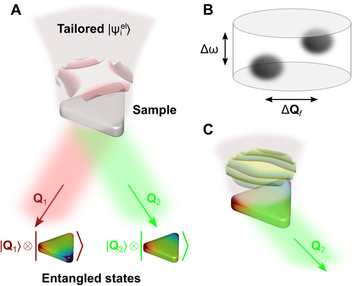
Here, we demonstrate through rigorous quantum theory that pure entanglement between electrons and confined optical modes can be generated by suitably patterning the transverse incident electron wave function. As schematically illustrated in Fig. 1A, the electron undergoes a change in the direction of propagation after being inelastically scattered by a specimen, and we prepare the incident electron wave profile (amplitude and phase) in such a way that only a few sample excitations are accessible (two in the figure), leading to separable transmission directions (transverse wave vectors Q1 and Q2). The two possible excitations created by the electron together with their different associated scattering directions form an entangled state. In essence, we specify a finite volume in the configuration space of transmitted electrons defined by an energy-loss window Δħω and a transverse momentum area ΔħQf in which the final state only populates two well-defined spots (Fig. 1B). As we demonstrate below, this approach can also be used to create heralded single sample excitations (Fig. 1C). In addition, manipulation of the electron component in electron–sample entangled states through, for example, electron interference could be used to process quantum information and imprint it on other (eventually macroscopic) objects via subsequent interactions.
Fig. 1. Proposed scheme for the generation of entangled electron–cavity states.
(A) A preshaped electron interacts with a nanostructure (a triangular plasmonic cavity) supporting well-defined optical or vibrational modes. The incident electron wave function is tailored such that we obtain entangled states after interaction, correlating different specimen excitations (colored triangles) with separated electron scattering directions (final electron state having components of transverse wave vectors Q1 and Q2). A maximally entangled electron-specimen state is thus produced as the sample is in a superposition of excited states correlated with different electron scattering directions. (B) Electrons are emerging along separate spots within a finite region of size Δħω × ΔħQf in the configuration space of energy-loss and transverse-momentum transfers. (C) Momentum filtering at the electron detector allows us to project on the desired sample mode and eventually explore its dynamics through subsequent interrogation, for example, by exposure to a synchronized light pulse.
RESULTS
Free-electron interaction with confined optical modes
We intend to synthesize an electron–sample state, as described by Eq. 1, with the free-electron component piled up at separate regions in momentum–energy space (Fig. 1B) and a different sample excitation associated with each of those regions. The starting point is the initial combined state
where the specimen is in its ground state ∣0〉 and the incident electron wave function, whose spatial dependence is given by
| (2) |
is prepared as a combination of momentum states with coefficients determined through the use of customized transmission masks (44, 45, 58) or phase imprinting based on electrostatic (46) and optical (47, 49) fields. We consider incident monochromatic electrons, such that the dependence of the electron wave function on two-dimensional (2D) transverse coordinates R and its decomposition in 2D wave vectors Qi is everything we need to describe the electron in the interaction region without loss of generality.
The electron–specimen interaction operates a linear transformation relating the final coefficients in Eq. 1 to in Eq. 2. More precisely,
| (3) |
where MQf −Qi,n only depends on the momentum transfer ħ(Qi − Qf) for each excited state n (see Methods).
A connection can be readily established to EELS experiments, in which electron counts are recorded as a function of the energy loss ħω, thus yielding a frequency- and momentum-resolved loss probability , where ħωn is the excitation energy of the sample mode n. Within first-order perturbation theory and further adopting the electrostatic and nonrecoil approximations, the angle-resolved EELS probability can be expressed in terms of mode-dependent dimensionless spectral functions gn(ω) as
| (4) |
where v is the electron velocity and
| (5) |
describes the spatial profile of mode n [see details in Methods, including expressions for the quantities gn(ω) and wn(R, ω) associated with plasmons and atomic vibrations].
Here, we are interested in determining the incident electron wave function profile (i.e., the momentum-dependent coefficients ) such that different sample modes n are associated with final wave function coefficients within well-separated regions in momentum space (see Fig. 1B). To demonstrate the feasibility of this concept in the synthesis of electron–sample entanglement, we invert Eq. 3 with a predetermined choice of , which we set to designated values for each sample excitation n within a targeted finite-size region in Qf space (see details in Methods). This simple procedure is sufficient for the proof-of-principle demonstration that we pursue in this work. However, in one of the examples, we present further improvement of the results when using an iterative method. Other schemes for incident electron wave function optimization could rely on neural-network training (59).
Selected excitation of individual plamons
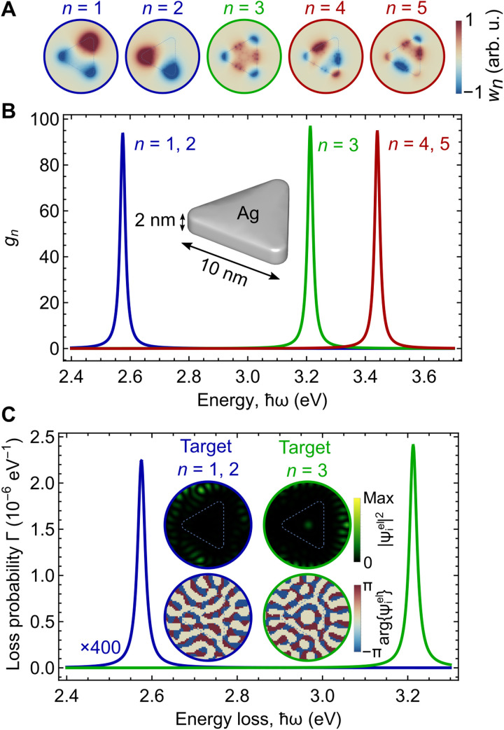
As a preliminary step before addressing electron–sample entanglement, we tackle the problem of selectively exciting a single plasmon in a metallic nanoparticle. Although this can be achieved through post-selection of a small range of scattered electron wave vectors (42), we formulate a solution in which the plasmon-exciting electrons emerge within a relatively large region in momentum space, and this solution is generalized below to create entanglement. We consider a silver triangle that sustains five plasmon modes in the spectral region between 2.4 and 3.7-eV spectral region (60): two sets of doubly degenerate dipolar (blue curve and circles, n = 1,2) and quadrupolar (red, n = 4,5) plasmons and one nondegenerate hexapolar mode (green, n = 3), as revealed by the spatial and spectral functions plotted in Fig. 2 (A and B) (see details of the calculation in Methods). We then optimize the incident electron wave function over a finite Qi region discretized with 1257 pixels and defined by a convergence half-angle φi = 1.5 mrad, such that either n = 1,2 or n = 3 is the only mode excited when the scattered electrons are collected over a Qf region spanning a half-angle φf = 0.75 mrad (discretized with 49 pixels) and energy-filtered between 2.4 and 3.3 eV. Incidentally, modes n = 1 and 2 are dipolar, so they can be effectively excited in the aloof configuration, while mode n = 3 is hexapolar and requires the electron to pass closer to the particle to be excited.
Fig. 2. Selective excitation of plasmon modes in a silver nanotriangle.
(A and B) Spatial profiles (A) and spectral functions (B) associated with plasmons in a silver nanotriangle with a thickness of 2 nm and a side length of 10 nm. We find two sets of degenerate modes (blue and red peaks) and one nondegenerate mode (green; see color labels matched with the index n). (C) Electron energy-loss spectra for two optimized incident electron wave function profiles . The insets show maps of the probability density and phase of the incident wave functions in the space of transverse coordinates R, framed in color-matched circumferences. The optimization is carried out for 100 keV electrons, an electron detector consisting of 49 pixels, an incident convergence half-angle φi = 1.5 mrad, and a collection half-angle φf = 0.75 mrad. The nanotriangle contour is indicated by thin dashed lines in (A) and (C).
The resulting real-space profiles of are shown in the insets of Fig. 2C (circular color plots; see also fig. S6A for the incident electron wave functions in Qi space), along with the color-matched EELS probability curves obtained from Eq. 4 by collecting only electrons that emerge within the indicated Qf and energy region. In a typical experimental scenario with an unshaped electron beam, multiple modes are excited by the incident electron because they have overlapping spatial distributions (Fig. 2A), and the EELS probability integrated over all possible Qf′s is rigorously given by the incoherent average over incident electron positions R, weighted by the electron probability (see Methods) (6, 61). However, our simple optimization procedure is capable of placing the weight of the excitation of either n = 1,2 or n = 3 mode preferably inside the Qf region defined by a collection half-angle φf = 0.75 mrad. Such an optimization can be performed for smaller or larger convergence and collection angles as shown in fig. S1.
Generation of electron–plasmon entangled states
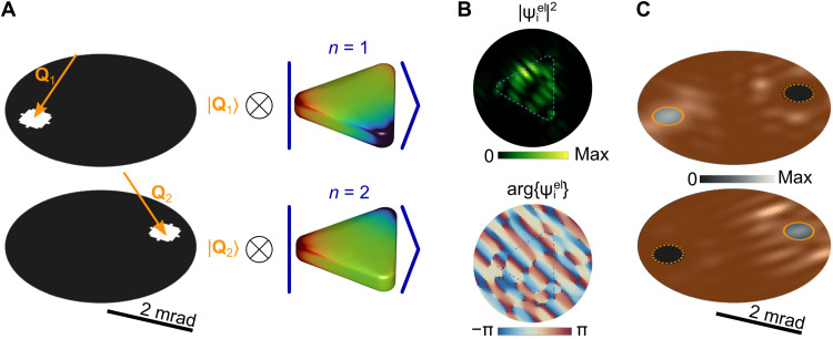
We now apply the principle of shaping to demonstrate the generation of electron–sample entanglement for the same triangular particle as considered above. Specifically, we focus on the lowest-energy degenerate plasmons n = 1,2 (i.e., we consider post-selection of these final states by an energy filter) and aim at correlating these excitations with final electron momentum states along separate Qf directions (Fig. 3A). By maximizing the fraction of the signal associated with the targeted excitation in each respective Qf direction through the steepest-descent method, we find the optimized electron wave function shown in real space in Fig. 3B (and in momentum space in fig. S6B), from which we obtain the actual scattered electron distribution plotted in Fig. 3C in Qf space for components corresponding to the excitation of n = 1 (top) and n = 2 (bottom) modes.
Fig. 3. Creation of electron–sample states with a high degree of entanglement.
(A) Pursued electron–sample entangled state, consisting of the superposition of selected (by means of two apertures) electron momentum states within the white pixels in Qf space (left) and correlated degenerate dipolar plasmons in the silver nanotriangle sample considered in Fig. 2 (right). (B) Spatial profile of the optimized incident wave function required to produce the final state in (A). The contour of the triangular nanoparticle is indicated with dashed lines. (C) Resulting probability distributions ∣〈Qf, n∣Ψf〉∣2 with n = 1 (top) and n = 2 (bottom) in Qf space, where each aperture (small circles) transmits nearly 100% of the targeted excitation n. The area outside the apertures (intended to be masked) is colored to emphasize the selected momentum regions of interest. The optimization is carried out for 100 keV electrons, 81 pixels in each of the detector apertures, φi = 4 mrad, and φf = 2 mrad.
When examining the resulting degree of entanglement, we express the final electron–sample state after energy filtering and momentum post-selection by two apertures defined by the orange circles in Fig. 3C as
| (6) |
where we assume that the apertures are small enough so that each of them captures coherently scattered electrons characterized by a well-defined state ∣Qf〉 with f = 1,2 (in practice, each of them can be a coherent superposition of plane waves transmitted through the finite solid angle region spanned by each aperture). To achieve pure entanglement, we require pnm → 0 for n ≠ m terms in Eq. 6, which happens after the noted numerical optimization: We obtain values and , confirming a high degree of entanglement (62). Incidentally, a more direct inversion procedure without optimization still yields a level of entanglement exceeding 90% (fig. S2).
We note that the symmetry of the selected degenerate plasmons plays a similar role as photon polarization in light-based entanglement schemes (1). In the present instance, the electron wave function profiles are strongly affected by the threefold symmetry of the plasmonic nanoparticles and the choice of correlated electron output angles. Optimized profiles for more symmetric nanoparticles also become more symmetric, as shown for silver disks in fig. S3, where a high degree of entanglement (>99% mode separation) is achieved by direct inversion.
Electron entanglement with atomic vibrational states
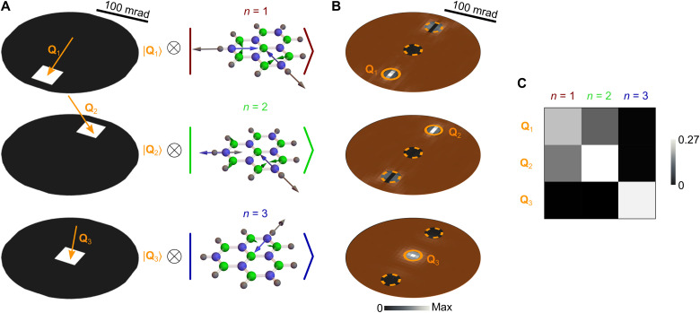
The electron–sample entanglement scheme under consideration can be applied to sample excitations of different nature. We illustrate this versatility by considering atomic vibrations in a hexagonal boron nitride (hBN) molecule (Fig. 4), which we simulate from first principles (see Methods) (63) assuming passivation of the edges with hydrogen atoms. This structure supports a number of vibrational excitations up to energies ∼450 meV, including a set of triply degenerate N─H bond-stretching modes at 440 meV (see EELS spectrum in fig. S4), on which we focus our analysis. We again optimize the incident electron wave function to achieve entanglement between final electron states and vibrational modes of the molecule [see the resulting and profiles in figs. S5 and S6C]. Because of the strong spatial confinement of vibrational modes, the angular ranges that need to be used for the incident and scattered electron wave functions are now considerably larger than for plasmons (cf. angle scales in Figs. 3 and 4). The achieved electron–sample state, illustrated in Fig. 4B, exhibits a decent degree of entanglement when selecting electrons scattered along the colored circles in Qf space, also revealed through the partial probabilities contributed by each of the three vibrational modes to each of the regions enclosed by those circles (see table in Fig. 4C).
Fig. 4. Entanglement of free electrons and atomic vibrations.
(A) Pursued electron–sample entangled state, consisting of the superposition of selected (by means of three apertures) electron-momentum states within the white pixels in Qf space (left) and correlated triply degenerate 440-meV vibrational modes of an hBN molecule (right). (B) Resulting probability distributions ∣〈Qf, n∣Ψf〉∣2 with n = 1 (top), n = 2 (middle), and n = 3 (bottom) in Qf space. The area outside the apertures (small circles) is colored to emphasize the selected momentum regions of interest. (C) Probability matrix showing the fractional contribution associated with the excitation of each of the three vibrational modes n = 1 to 3 to the energy-filtered electron signal contained within the three selected circular areas around final transverse wave vectors Q1, Q2, and Q3 in (A). The sum of the nine matrix elements is normalized to one. We consider 60 keV electrons, 29 detector pixels, and φi = φf = 100 mrad.
DISCUSSION
By entangling the transverse momenta of free electrons with localized optical excitations in a nanostructure, we could selectively measure one of the corresponding outgoing electron directions, thus providing a way to herald the creation of single designated excitations in the studied specimen. This should allow us to follow the dynamics of the latter and gain insight into the state-dependent decay pathways, for example, by subsequently probing the evolution of the specimen through scattering of laser pulses that are synchronized with the electron in an electron-pump/photon-probe approach. An additional possibility is offered by correlating the angle-resolved electron signal with traces originating in the decay of excited states of the specimen (e.g., an electrical signal produced by coupling to electron-hole pairs in a proximal semiconductor or also the polarization- and angle-resolved cathodoluminescence emission associated with radiative decay). The present scheme could further be extended to incorporate gain processes similar to those in photon-induced near-field electron microscopy (PINEM) (54) upon illumination of the sample with symmetry-matched optical pulses that can simultaneously excite a subset of its supported excitations.
We remark that the proposed approach holds elements of novelty with respect to traditional quantum optics methods because one of the entangled particles (the free electron) can be highly energetic and, therefore, capable of undergoing subsequent strong collisions with other objects. These collisions could, for instance, trigger chemical reactions that would then be entangled with optical modes in the specimen with which the electron has previously interacted.
Although we have illustrated some possibilities based on heuristic electron wave function designs and a straightforward application of the steepest-descent maximization method, improved solutions to the problem of entanglement optimization could be obtained through neural-network training (59), possibly combined with iterative physical improvement of the wave function profile based on currently explored tunable electron phase plates (46–51). As an alternative to the use of aloof interaction with the optical modes of the sampled nanostructure to avoid strong electron collisions with atomic potentials, this type of adaptive improvement could potentially be used to compensate for the effect of these potentials and morphological imperfections. In addition to the investigated examples of plasmons in nanoparticles and atomic vibrations in molecules, we envision the entanglement of free electrons with optical modes in dielectric cavities (30) and photons guided along optical waveguides (64), which together configure a vast range of possibilities for leveraging the quantum nature of free electrons in the design of improved microscopy and metrology schemes.
METHODS
Transfer matrix for inelastic electron–sample scattering
The time-dependent electron–sample system can be generally described by a wave function of the form , where ∣q〉 and ∣n〉 are electron and sample eigenstates of the noninteracting Hamiltonian with energies ħεq and ħωn, respectively. In particular, electron states are labeled by the 3D momentum ħq and satisfy the orthonormality relation 〈q∣q′〉 = δ(q − q′). The expansion coefficients αq,n(t) are determined by solving the Schrödinger equation with an electron–sample interaction Hamiltonian , which is generally weak for the energetic electrons that are typically used in electron microscopes, so we can work within first-order perturbation theory. Then, taking the sample to be initially prepared in its ground state n = 0, the post-interaction wave function has coefficients , where we set ω0 = 0 without loss of generality. We further adopt the nonrecoil approximation (13) ϵq − ϵq′ ≈ (q − q′) · v under the assumption that the transverse electron energy is negligible compared with the longitudinal energy along the e-beam direction defined by the average electron velocity v. This condition is commonly satisfied in electron microscopes. In this approximation, the energy ħωn transferred from the electron to the sample is fully absorbed by a change in the longitudinal electron wave vector given by −ωn/v, so for monochromatic incident electrons, the initial and final longitudinal components of the electron wave function play a trivial role and can be disregarded in the description of the present problem. Consequently, we can expand the final wave function as shown in Eq. 1, with coefficients that only depend on the transverse electron wave vector Qf for each sample excitation n and are determined from the incident electron wave function coefficients through the linear relation (Eq. 3) with
| (7) |
We remark that the transfer matrix elements defined in Eq. 7 involve just the difference between incident and scattered transverse wave vectors. In what follows, we develop a formalism to relate MQf −Qi,n to the EELS probability and obtain specific expressions for plasmonic and atomic-vibration modes.
EELS with shaped electron beams
We consider the configuration of Fig. 1A and assume the electron velocity and sample dimensions to be small enough as to neglect retardation effects and work in the electrostatic regime. Further adopting the aforementioned nonrecoil approximation, we can disregard the longitudinal component of the electron wave function and only consider the dependence on transverse coordinates R = (x, y) (i.e., taking the electron velocity v along z). We can then write a general expression for the EELS probability ΓEELS(ω) in terms of the energy loss ħω, the transverse wave vector of the final (f) electron state (corresponding to a wave function ∝ eiQf · R), and the transverse component of the initial (i) electron wave function, ψi(R). More precisely, using equation 17 of (6), we have ΓEELS(ω) = ∫ d2Qf ΓEELS(Qf, ω), where
| (8) |
is the momentum-resolved probability and
| (9) |
is a transverse screened interaction obtained from the full screened interaction W(r, r′, ω). The latter stands for the Coulomb potential created at r by a point charge of magnitude e−iωt placed at r′, including the effect of screening by the environment. Now, as we show below for plasmonic and phononic structures, the transverse screened interaction in Eq. 9 is separable as
| (10) |
where n runs over excitation modes characterized by spatial profiles wn(R, ω) and dimensionless spectral functions gn(ω). Finally, inserting Eq. 10 into Eq. 8, we readily find Eq. 4 in the main text. Incidentally, the angle-integrated inelastic electron signal (i.e., the integral of Eq. 8 over Qf) reduces to ΓEELS(ω) = (e2/πħv2)∑ngn(ω) ∫ d2R ∣ψi(R)∣2∣wn(R, ω)∣2, which is an average over transverse positions R weighted by both the incident electron probability (6, 61) and the mode spatial profile, and consequently, because the e-beam can generally excite different modes n, the optimization scheme that we pursue here to produce entanglement essentially consists in rearranging the Qf distribution of the scattered electron components associated with the excitation of each of those modes.
We note that the spectral functions in this formalism can be generally approximated by Lorentzians
peaked at the mode energies ħωn and having areas Gn and widths γn (see below) that determine the spectral positions and strengths of the EELS features.
Numerical determination of for creating selected excitations and entangled electron–sample states
Given a desired final state defined through the coefficients , we numerically obtain by inverting Eq. 3 upon discretization of Qi using a finite number of points and pixels at the electron analyzer in the Fourier plane Qf, as noted in the main text. More precisely, we follow a simple procedure consisting in specifying target values of within a region Q < Qf,max (effectively setting it to zero outside it) and obtain for Qi < Qi, max through the aforementioned numerical inversion method. The wave vector ranges are related to the maximum incidence|collection half-angle φi∣f through Qi∣f, max = (mev/ħ) sin φi∣f. In this scheme, to select a single sample excitation n = n0 (Fig. 2), we set , where C is a constant and Θ is the step function. However, to produce electron–sample entanglement involving two (Fig. 3) or three (Fig. 4) sample states nj correlated with final electron wave vectors Qj (see Fig. 1B), we set to a constant at the Qf space pixel that contains Qj and zero elsewhere. We then construct from the obtained coefficients (also setting them to zero for Qi > Qi, max) and insert this input wave function in Eq. 4 to generate the actual final probability distributions, plotted in the figures with a finer discretization in Qf space.
Transfer matrix from the spectral and spatial mode functions
An expression for the EELS probability analogous to Eq. 4 can be readily obtained from Eq. 3 as
| (11) |
The connection between Eqs. 4 and 11 is established by adding finite mode widths γn to the latter and expanding the incident electron wave function in the former as an integral over momentum components, as indicated in Eq. 2. Comparing the two resulting expressions, we find
| (12) |
which provides a prescription to obtain the transfer matrix coefficients defined in Eq. 7 directly from the screened interaction, thus bypassing the need for a detailed specification of the interaction Hamiltonian. Then, the spatial profiles in Eq. 5 are simply given by the inverse Fourier transform of Eq. 12.
Transfer matrix and transverse screened interaction for plasmonic nanoparticles
In the electrostatic limit under consideration, we can recast the response of an arbitrarily shaped homogeneous nanoparticle into an eigenvalue problem (65, 66). We then need to find the real eigenvalues λn and eigenvectors σn(s) of the integral equation 2πλnσn(s) = ∮ ds′F(s, s′)σn(s′), where s and s′ run over particle surface coordinates, F(s, s′) = − · (s − s′)/∣s − s′∣3, and is the outer surface normal. Here, we solve this eigensystem for triangular particles using the MNPBEM toolbox (67) based on a finite boundary element discretization of the particle surface. Then, the spectral functions in Eq. 10 reduce to (65, 66)
whereas the spatial profiles become
with . This expression neglects the contribution of bulk modes, which should be a reasonable approximation at loss energies well below the bulk plasmon. Inserting it into Eq. 12, the transfer matrix elements reduce to
where we have approximated ω ≈ ωn. For silver, we model the dielectric function as (6) with ϵb = 4.0, ħωp = 9.17 eV, and ħγ = 21 meV, yielding mode frequencies , flat widths γn ≈ γ, and spectral weights .
Transfer matrix and transverse screened interaction for atomic vibrations
For molecules or nanoparticles whose mid-infrared response is dominated by atomic vibrations, we find the spectral and spatial dependence of the modes in Eq. 10 to be governed by (63, 68).
| (13) |
and
where n now runs over vibrational modes, ωn and enl are the corresponding real frequencies and normalized atomic displacement vectors (∑l enl · en′l = δnn′), respectively, the l sum extends over the atoms in the structure, Ml is the mass of atom l, denotes the gradient of the charge distribution associated with displacements of that atom, and we have incorporated a phenomenological damping rate γ (here set to ħγ = 1 meV). From Eq. 13, we have γn ≈ γ for all modes and Gn ≈ πωn/2. Following (63), we use density functional theory (DFT) to calculate , ωn, and enl (see below). The prescription is also adopted with to approximately account for a cutoff ∼ħ/Δ in momentum transfer (6) and so avoid the unphysical divergence associated with close electron-atom encounters.
First-principles description of atomic vibrations
We use DFT and the projector augmented wave method (69) as implemented in the Vienna Ab initio Simulation Package (70–72) with the Perdew-Burke-Ernzerhof–generalized gradient approximation for electron exchange and correlation (73). This method is applied to describe hBN flakes with hydrogen-passivated edges using a plane wave cutoff energy of 500 eV and a sufficient amount of vacuum spacing in all directions around the structure to avoid interaction among the periodic images. Atomic equilibrium positions are found by minimizing the total energy using the conjugate gradient method with convergence criteria between consecutive iteration steps set to 10−5 eV for the total energy and 0.02 eV/Å for the atomic forces. Vibrational frequencies and eigenmodes are found by diagonalizing the dynamical matrix, which is calculated for 0.01-Å displacements. The corresponding gradients of the charge distribution are obtained by treating core electrons and nuclei as point particles, while the contribution coming from valence electrons is directly taken from DFT using a dense grid.
Acknowledgments
Funding: This work has been supported in part by the European Research Council (Advanced grant 789104-eNANO), the Spanish MICINN (PID2020-112625GB-I00 and Severo Ochoa CEX2019-000910-S), the Catalan CERCA Program, and Fundaciós Cellex and Mir-Puig. A.K. was supported by the ESF under the project CZ.02.2.69/0.0/0.0/20_079/0017436.
Author contributions: F.J.G.d.A. conceived the project. A.K. and F.J.G.d.A. developed the theoretical formalism. A.K. performed theoretical calculations for all figures using DFT input generated by F.I.
Competing interests: The authors declare that they have no competing interests.
Data and materials availability: All data needed to evaluate the conclusions in the paper are present in the paper and/or the Supplementary Materials.
Supplementary Materials
This PDF file includes:
Figs. S1 to S6
REFERENCES AND NOTES
- 1.Horodecki R., Horodecki P., Horodecki M., Horodecki K., Quantum entanglement. Rev. Mod. Phys. 81, 865–942 (2009). [Google Scholar]
- 2.Togan E., Chu Y., Trifonov A. S., Jiang L., Maze J., Childress L., Dutt M. V. G., Sörensen A. S., Hemmer P. R., Zibrov A. S., Lukin M. D., Quantum entanglement between an optical photon and a solid-state spin qubit. Nature 466, 730–734 (2010). [DOI] [PubMed] [Google Scholar]
- 3.Kfir O., Entanglements of electrons and cavity photons in the strong-coupling regime. Phys. Rev. Lett. 123, 103602 (2019). [DOI] [PubMed] [Google Scholar]
- 4.Di Giulio V., Kociak M., García de Abajo F. J., Probing quantum optical excitations with fast electrons. Optica 6, 1524–1534 (2019). [Google Scholar]
- 5.Reinhardt O., Mechel C., Lynch M., Kaminer I., Free-electron qubits. Ann. Phys. 533, 2000254 (2021). [Google Scholar]
- 6.García de Abajo F. J., Optical excitations in electron microscopy. Rev. Mod. Phys. 82, 209–275 (2010). [Google Scholar]
- 7.R. F. Egerton, Electron Energy-Loss Spectroscopy in the Electron Microscope (Plenum Press, 1996). [Google Scholar]
- 8.Egerton R. F., New techniques in electron energy-loss spectroscopy and eneorgy-filtered imaging. Micron 34, 127–139 (2003). [DOI] [PubMed] [Google Scholar]
- 9.Erni R., Browning N. D., Valence electron energy-loss spectroscopy in monochromated scanning transmission electron microscopy. Ultramicroscopy 104, 176–192 (2005). [DOI] [PubMed] [Google Scholar]
- 10.R. Brydson, Electron Energy Loss Spectroscopy (BIOS Scientific Publishers, 2001). [Google Scholar]
- 11.Krivanek O. L., Lovejoy T. C., Dellby N., Aoki T., Carpenter R. W., Rez P., Soignard E., Zhu J., Batson P. E., Lagos M. J., Egerton R. F., Crozier P. A., Vibrational spectroscopy in the electron microscope. Nature 514, 209–212 (2014). [DOI] [PubMed] [Google Scholar]
- 12.Krivanek O. L., Dellby N., Hachtel J. A., Idrobo J.-C., Hotz M. T., Plotkin-Swing B., Bacon N. J., Bleloch A. L., Corbin G. J., Hoffman M. V., Meyer C. E., Lovejoy T. C., Progress in ultrahigh energy resolution EELS. Ultramicroscopy 203, 60–67 (2019). [DOI] [PubMed] [Google Scholar]
- 13.García de Abajo F. J., Di Giulio V., Optical excitations with electron beams: Challenges and opportunities. ACS Photonics 8, 945–974 (2021). [DOI] [PMC free article] [PubMed] [Google Scholar]
- 14.Hage F. S., Radtke G., Kepaptsoglou D. M., Lazzeri M., Ramasse Q. M., Single-atom vibrational spectroscopy in the scanning transmission electron microscope. Science 367, 1124–1127 (2020). [DOI] [PubMed] [Google Scholar]
- 15.Yan X., Liu C., Gadre C. A., Gu L., Aoki T., Lovejoy T. C., Dellby N., Krivanek O. L., Schlom D. G., Wu R., Pan X., Single-defect phonons imaged by electron microscopy. Nature 589, 65–69 (2021). [DOI] [PubMed] [Google Scholar]
- 16.Rez P., Aoki T., March K., Gur D., Krivanek O. L., Dellby N., Lovejoy T. C., Wolf S. G., Cohen H., Damage-free vibrational spectroscopy of biological materials in the electron microscope. Nat. Commun. 7, 10945 (2016). [DOI] [PMC free article] [PubMed] [Google Scholar]
- 17.Haiber D. M., Crozier P. A., Nanoscale probing of local hydrogen heterogeneity in disordered carbon nitrides with vibrational electron energy-loss spectroscopy. ACS Nano 12, 5463–5472 (2018). [DOI] [PubMed] [Google Scholar]
- 18.Jokisaari J. R., Hachtel J. A., Hu X., Mukherjee A., Wang C., Konečná A., Lovejoy T. C., Dellby N., Aizpurua J., Krivanek O. L., Idrobo J.-C., Klie R. F., Vibrational spectroscopy of water with high spatial resolution. Adv. Mater. 12, 430–436 (2018). [DOI] [PubMed] [Google Scholar]
- 19.Hachtel J. A., Huang J., Popovs I., Jansone-Popova S., Keum J. K., Jakowski J., Lovejoy T. C., Dellby N., Krivanek O. L., Idrobo J. C., Identification of site-specific isotopic labels by vibrational spectroscopy in the electron microscope. Science 363, 525–528 (2019). [DOI] [PubMed] [Google Scholar]
- 20.Bosman M., Keast V. J., Watanabe M., Maaroof A. I., Cortie M. B., Mapping surface plasmons at the nanometre scale with an electron beam. Nanotechnology 18, 165505 (2007). [Google Scholar]
- 21.Nelayah J., Kociak M., Stéphan O., García de Abajo F. J., Tencé M., Henrard L., Taverna D., Pastoriza-Santos I., Liz-Marzán L. M., Colliex C., Mapping surface plasmons on a single metallic nanoparticle. Nat. Phys. 3, 348–353 (2007). [Google Scholar]
- 22.Rossouw D., Botton G. A., Plasmonic response of bent silver nanowires for nanophotonic subwavelength waveguiding. Phys. Rev. Lett. 110, 066801 (2013). [DOI] [PubMed] [Google Scholar]
- 23.Tan S. F., Wu L., Yang J. K. W., Bai P., Bosman M., Nijhuis C. A., Quantum plasmon resonances controlled by molecular tunnel junctions. Science 343, 1496–1499 (2014). [DOI] [PubMed] [Google Scholar]
- 24.Mkhitaryan V., March K., Tseng E., Li X., Scarabelli L., Liz-Marzán L. M., Chen S.-Y., Tizei L. H. G., Stéphan O., Song J.-M., Kociak M., García de Abajo F. J., Gloter A., Can copper nanostructures sustain high-quality plasmons? Nano Lett. 21, 2444–2452 (2021). [DOI] [PubMed] [Google Scholar]
- 25.Lagos M. J., Trügler A., Hohenester U., Batson P. E., Mapping vibrational surface and bulk modes in a single nanocube. Nature 543, 529–532 (2017). [DOI] [PubMed] [Google Scholar]
- 26.Govyadinov A. A., Konečná A., Chuvilin A., Vélez S., Dolado I., Nikitin A. Y., Lopatin S., Casanova F., Hueso L. E., Aizpurua J., Hillenbrand R., Probing low-energy hyperbolic polaritons in van der Waals crystals with an electron microscope. Nat. Commun. 8, 95 (2017). [DOI] [PMC free article] [PubMed] [Google Scholar]
- 27.Li N., Guo X., Yang X., Qi R., Qiao T., Li Y., Shi R., Li Y., Liu K., Xu Z., Liu L., García de Abajo F. J., Dai Q., Wang E.-G., Gao P., Direct observation of highly confined phonon polaritons in suspended monolayer hexagonal boron nitride. Nat. Mater. 20, 43–48 (2020). [DOI] [PubMed] [Google Scholar]
- 28.Kfir O., Lourenço-Martins H., Storeck G., Sivis M., Harvey T. R., Kippenberg T. J., Feist A., Ropers C., Controlling free electrons with optical whispering-gallery modes. Nature 582, 46–49 (2020). [DOI] [PubMed] [Google Scholar]
- 29.Wang K., Dahan R., Shentcis M., Kauffmann Y., Ben Hayun A., Reinhardt O., Tsesses S., Kaminer I., Coherent interaction between free electrons and a photonic cavity. Nature 582, 50–54 (2020). [DOI] [PubMed] [Google Scholar]
- 30.Auad Y., Hamon C., Tencé M., Lourenço-Martins H., Mkhitaryan V., Stéphan O., García de Abajo F. J., Tizei L. H. G., Kociak M., Unveiling the coupling of single metallic nanoparticles to whispering-gallery microcavities. Nano Lett. 22, 319–327 (2022). [DOI] [PubMed] [Google Scholar]
- 31.Pettit R. B., Silcox J., Vincent R., Measurement of surface-plasmon dispersion in oxidized aluminum films. Phys. Rev. B 11, 3116–3123 (1975). [Google Scholar]
- 32.Krivanek O. L., Tanishiro Y., Takayanagi K., Yagi K., Electron energy loss spectroscopy in glancing reflection from bulk crystals. Ultramicroscopy 11, 215–222 (1983). [Google Scholar]
- 33.Hage F. S., Nicholls R. J., Yates J. R., McCulloch D. G., Lovejoy T. C., Dellby N., Krivanek O. L., Refson K., Ramasse Q. M., Nanoscale momentum-resolved vibrational spectroscopy. Sci. Adv. 4, eaar7495 (2018). [DOI] [PMC free article] [PubMed] [Google Scholar]
- 34.Shekhar P., Malac M., Gaind V., Dalili N., Meldrum A., Jacob Z., Momentum-resolved electron energy loss spectroscopy for mapping the photonic density of states. ACS Photonics 4, 1009–1014 (2017). [Google Scholar]
- 35.Mechel C., Kurman Y., Karnieli A., Rivera N., Arie A., Kaminer I., Quantum correlations in electron microscopy. Optica 8, 70–78 (2021). [Google Scholar]
- 36.Y. Adiv, H. Hu, S. Tsesses, R. Dahan, K. Wang, Y. Kurman, A. Gorlach, H. Chen, X. Lin, G. Bartal, I. Kaminer, Observation of 2D Cherenkov radiation, https://arxiv.org/abs/2203.01698 (2022).
- 37.Powell C. J., Swan J. B., Origin of the characteristic electron energy losses in aluminum. Phys. Rev. 115, 869–875 (1959). [Google Scholar]
- 38.Schattschneider P., Födermayr F., Su D. S., Coherent double-plasmon excitation in aluminum. Phys. Rev. Lett. 59, 724–727 (1987). [DOI] [PubMed] [Google Scholar]
- 39.Batson P. E., Dellby N., Krivanek O. L., Sub-ångstrom resolution using aberration corrected electron optics. Nature 418, 617–620 (2002). [DOI] [PubMed] [Google Scholar]
- 40.Clark L., Béché A., Guzzinati G., Lubk A., Mazilu M., Van Boxem R., Verbeeck J., Exploiting lens aberrations to create electron-vortex beams. Phys. Rev. Lett. 111, 064801 (2013). [DOI] [PubMed] [Google Scholar]
- 41.Möllenstedt G., Düker H., Beobachtungen und Messungen an Biprisma-Interferenzen mit Elektronenwellen. Z. Phys. 145, 377–397 (1956). [Google Scholar]
- 42.Guzzinati G., Beche A., Lourenço-Martins H., Martin J., Kociak M., Verbeeck J., Probing the symmetry of the potential of localized surface plasmon resonances with phase-shaped electron beams. Nat. Commun. 8, 14999 (2017). [DOI] [PMC free article] [PubMed] [Google Scholar]
- 43.Johnson C. W., Turner A. E., McMorran B. J., Scanning two-grating free electron Mach-Zehnder interferometer. Phys. Rev. Res. 3, 043009 (2021). [Google Scholar]
- 44.Verbeeck J., Tian H., Schattschneider P., Production and application of electron vortex beams. Nature 467, 301–304 (2010). [DOI] [PubMed] [Google Scholar]
- 45.McMorran B. J., Agrawal A., Anderson I. M., Herzing A. A., Lezec H. J., McClelland J. J., Unguris J., Electron vortex beams with high quanta of orbital angular momentum. Science 331, 192–195 (2011). [DOI] [PubMed] [Google Scholar]
- 46.Verbeeck J., Béché A., Müller-Caspary K., Guzzinati G., Luong M. A., Hertog M. D., Demonstration of a 2 × 2 programmable phase plate for electrons. Ultramicroscopy 190, 58–65 (2018). [DOI] [PubMed] [Google Scholar]
- 47.Konečná A., García de Abajo F. J., Electron beam aberration correction using optical near fields. Phys. Rev. Lett. 125, 030801 (2020). [DOI] [PubMed] [Google Scholar]
- 48.Schwartz O., Axelrod J. J., Campbell S. L., Turnbaugh C., Glaeser R. M., Müller H., Laser phase plate for transmission electron microscopy. Nat. Methods 16, 1016–1020 (2019). [DOI] [PMC free article] [PubMed] [Google Scholar]
- 49.García de Abajo F. J., Konečná A., Optical modulation of electron beams in free space. Phys. Rev. Lett. 126, 123901 (2021). [DOI] [PubMed] [Google Scholar]
- 50.Mihaila M. C. C., Weber P., Schneller M., Grandits L., Nimmrichter S., Juffmann T., Transverse electron-beam shaping with light. Phys. Rev. X 12, 031043 (2022). [Google Scholar]
- 51.Madan I., Leccese V., Mazur A., Barantani F., LaGrange T., Sapozhnik A., Tengdin P. M., Gargiulo S., Rotunno E., Olaya J.-C., Kaminer I., Grillo V., García de Abajo F. J., Carbone F., Vanacore G. M., Ultrafast transverse modulation of free electrons by interaction with shaped optical fields. ACS Photonics 10, 3215–3224 (2022). [DOI] [PMC free article] [PubMed] [Google Scholar]
- 52.Aseyev S. A., Ryabov E. A., Mironov B. N., Ischenko A. A., The development of ultrafast electron microscopy. Crystals 10, 452 (2020). [Google Scholar]
- 53.Baum P., Zewail A. H., Attosecond electron pulses for 4D diffraction and microscopy. Proc. Natl. Acad. Sci. U.S.A. 104, 18409–18414 (2007). [DOI] [PMC free article] [PubMed] [Google Scholar]
- 54.Priebe K. E., Rathje C., Yalunin S. V., Hohage T., Feist A., Schäfer S., Ropers C., Attosecond electron pulse trains and quantum state reconstruction in ultrafast transmission electron microscopy. Nat. Photonics 11, 793–797 (2017). [Google Scholar]
- 55.Morimoto Y., Baum P., Diffraction and microscopy with attosecond electron pulse trains. Nat. Phys. 14, 252–256 (2018). [Google Scholar]
- 56.Kfir O., Di Giulio V., García de Abajo F. J., Ropers C., Optical coherence transfer mediated by free electrons. Sci. Adv. 7, eabf6380 (2021). [DOI] [PMC free article] [PubMed] [Google Scholar]
- 57.Di Giulio V., Kfir O., Ropers C., García de Abajo F. J., Modulation of cathodoluminescence emission by interference with external light. ACS Nano 15, 7290–7304 (2021). [DOI] [PMC free article] [PubMed] [Google Scholar]
- 58.Uchida M., Tonomura A., Generation of electron beams carrying orbital angular momentum. Nature 464, 737–739 (2010). [DOI] [PubMed] [Google Scholar]
- 59.Spurgeon S. R., Ophus C., Jones L., Petford-Long A., Kalinin S. V., Olszta M. J., Dunin-Borkowski R. E., Salmon N., Hattar K., Yang W.-C. D., Sharma R., Du Y., Chiaramonti A., Zheng H., Buck E. C., Kovarik L., Penn R. L., Li D., Zhang X., Murayama M., Taheri M. L., Towards data-driven next-generation transmission electron microscopy. Nat. Mater. 20, 274–279 (2021). [DOI] [PMC free article] [PubMed] [Google Scholar]
- 60.Schmidt F. P., Ditlbacher H., Hofer F., Krenn J. R., Hohenester U., Morphing a plasmonic nanodisk into a nanotriangle. Nano Lett. 14, 4810–4815 (2014). [DOI] [PMC free article] [PubMed] [Google Scholar]
- 61.Ritchie R. H., Howie A., Electron excitation and the optical potential in electron microscopy. Philos. Mag. 36, 463–481 (1977). [Google Scholar]
- 62.Abouraddy A. F., Saleh B. E. A., Sergienko A. V., Teich M. C., Degree of entanglement for two qubits. Phys. Rev. A 64, 050101 (2001). [Google Scholar]
- 63.Konečná A., Iyikanat F., García de Abajo F. J., Theory of atomic-scale vibrational mapping and isotope identification with electron beams. ACS Nano 15, 9890–9899 (2021). [DOI] [PubMed] [Google Scholar]
- 64.Bendaña X. M., Polman A., García de Abajo F. J., Single-photon generation by electron beams. Nano Lett. 11, 5099–5103 (2011). [DOI] [PubMed] [Google Scholar]
- 65.García de Abajo F. J., Aizpurua J., Numerical simulation of electron energy loss near inhomogeneous dielectrics. Phys. Rev. B 56, 15873–15884 (1997). [Google Scholar]
- 66.Boudarham G., Kociak M., Modal decompositions of the local electromagnetic density of states and spatially resolved electron energy loss probability in terms of geometric modes. Phys. Rev. B 85, 245447 (2012). [Google Scholar]
- 67.Hohenester U., Trügler A., MNPBEM - A matlab toolbox for the simulation of plasmonic nanoparticles. Comput. Phys. Commun. 183, 370–381 (2012). [Google Scholar]
- 68.Saavedra J. R. M., García de Abajo F. J., Phonon excitation by electron beams in nanographenes. Phys. Rev. B 92, 115449 (2015). [Google Scholar]
- 69.Blöchl P. E., Projector augmented-wave method. Phys. Rev. B 50, 17953–17979 (1994). [DOI] [PubMed] [Google Scholar]
- 70.Kresse G., Furthmüller J., Efficient iterative schemes for ab initio total-energy calculations using a plane-wave basis set. Phys. Rev. B 54, 11169–11186 (1996a). [DOI] [PubMed] [Google Scholar]
- 71.Kresse G., Hafner J., Ab initio molecular dynamics for liquid metals. Phys. Rev. B 47, 558–561 (1993). [DOI] [PubMed] [Google Scholar]
- 72.Kresse G., Furthmüller J., Efficiency of ab-initio total energy calculations for metals and semiconductors using a plane-wave basis set. Comput. Mater. Sci. 6, 15–50 (1996). [Google Scholar]
- 73.Perdew J. P., Burke K., Ernzerhof M., Generalized gradient approximation made simple. Phys. Rev. Lett. 77, 3865–3868 (1996). [DOI] [PubMed] [Google Scholar]
Associated Data
This section collects any data citations, data availability statements, or supplementary materials included in this article.
Supplementary Materials
Figs. S1 to S6


