

Case Report
The mother, a 26-year-old gravida2 para1 living1, with no significant medical or obstetric history, presented at 32 weeks of gestation with ultrasound suggestive of cystic lesion on the umbilical cord at the fetal end, with dimensions of 3.6 * 1.9 cm, which increased in size to 4.3 * 5.7 cm at 34 weeks. The Color Doppler confirmed normal flow through the umbilical vessels with no vascularity around the cyst and no fetal abdominal herniation. Subsequent scan done at 35 weeks revealed a further increase in size of the cyst to 5.5 * 6.0 cm with good fetal condition. Since the presentation was at 32 weeks of gestation, the fetal genetic testing was offered but refused by the patient. On account of the increasing size of the cord lesion close monitoring was kept and patient presented with decrease fetal monitoring at 36 weeks for which Cardiac tocogram was done. CTG was a non reassuring one for which she was taken for emergency LSCS and delivered a male child with APGAR score 10 at 1 and 5 min.
The placenta was discoid in shape with normal maternal and fetal sides. It weighed 500 gm and measured 17 cm * 10 cm * 2.5 cm. The cut section showed no gross abnormality. The free membranes were translucent, marginally attached, and not discoloured. On cut section, the placenta was of normal colour and consistency with no gross abnormalities. The umbilical cord was 53 cm in length with paracentral insertion to the placenta. There was a collapsed cyst of sized 3 * 4 cm (Fig. 1) seen at the fetal end with thickened umbilical cord 4.0 cm diameter. Though there was no poor outcome in the pregnancy with umbilical cyst in the first trimester [1], in our case the cyst was diagnosed at 28 weeks of gestation. The thickened umbilical cord continued for around 3 cm from fetal end in continuation to normal looking umbilical cord till the placental attachment. Cut section of the enlarged portion of the umbilical cord exuded copious mucoid, slimy substance with three vessels well identified within. Histologic sections from the dilated portion of the cord showed extensive edema and myxomatous degeneration of the substance of the cord with formation of pseudocysts containing mucoid fluid. The vessels of the cord (2 arteries and 1 vein) were normal in appearance. Histologic sections of the placenta and the membranes were unremarkable.
Fig. 1.
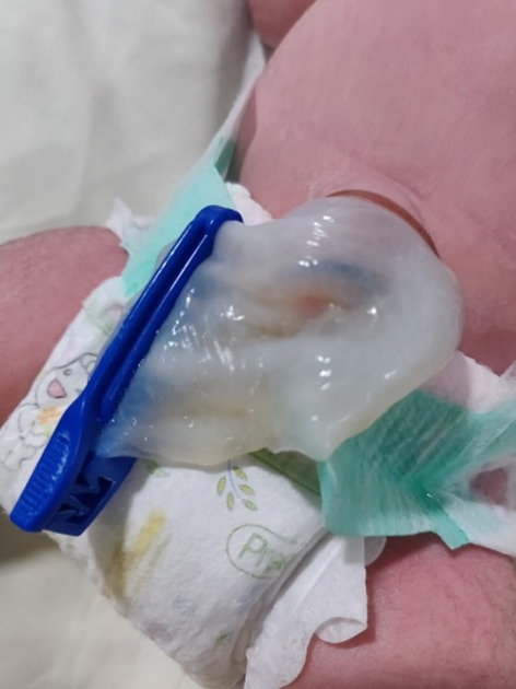
Thickened umbilical cord with collapsed cyst at fetal end
The baby was kept in the ICU for observation and then discharged with mother on the third day. During subsequent visit, the mother gave history of persistent umbilical wetness. On examination, umbilical wetness was observed and a micturating cystourethrogram (Fig. 2) was performed on the baby via an umbilical catheter which confirmed patent urachus. There was no fever or excessive crying, and the urine microscopy was negative for infection. A thorough review of all the systems did not reveal any other congenital anomaly. On Day7, of birth the baby was scheduled for laparotomy and extraperitoneal excision of tract of the patent urachus. Catheter was kept for 4 days and covered with single antibiotic. Gradual feeds were started under observation and he was discharged on day 7 of the surgery and was followed up in the OPD.
Fig. 2.

Micturating cystourethrogram from urachus catheter shows patent tract
Discussion
Ours is a case of umbilical cord cyst detected at 32-week prenatal ultrasound scan which was associated with patent urachus. The pseudocyst in our case could have been formed due to the back flow of urine through the patent urachus leading to dilatation of the umbilical cord. The suspected reasoning aroused when the baby was delivered and a collapsed cyst was seen. The antenatal presentation of a progressively increasing cyst was the only presentation to the patent urachus. The urachus is a duct forming the connection between the umbilicus and fetal bladder which obliterates on its own near term forming the median umbilical ligament. The urachus is an embryological ductal remnant that extends between the bladder dome and the umbilicus [2]. Failure of the tube to obliterate leads to patent urachus, which may present in isolation or with other fetal anomalies as in genetic defects. The occurrence for posterior urethral valves in an infant with patent urachus should not be overlooked [3]. This was ruled out in our case by performing a micturating cystourethrogram. (Fig. 2). Also, this embryonic defect is seen in one third cases of prune belly syndrome (also called as Eagle Barette syndrome or triad syndrome) which was ruled out.
Pathologically cord cysts are classified as either true cysts or pseudocysts. True cysts can derive either from the embryologic remaining part of the extra embryonic allantois or from the omphalomesenteric duct, and they are typically located near the foetal cord insertion. They are covered with epithelium. Allantoid cysts resolve themselves but may be associated with omphalocele, persistent urachus and obstructive uropathy. Cysts in the omphalomesenteric duct can be associated with defects in the abdominal wall and with Meckel's diverticulum. Cysts covered with amniotic epithelium (amniotic inclusion cysts) which are produced by amnion entrapment in the cord can be found inside true cysts.
Hannaford et al. [1] studied the ultrasonographic characteristics and natural history of umbilic cord cysts detected in the first trimester of pregnancy. They agree that the detection of an umbilical cord cyst in the second trimester should be an indication for foetal karyotyping. Routine karyotyping may not be necessary if the cyst disappears after first trimester. Regarding the mode of delivery, if the gestational course has run normally without associated structural anomalies, there is no contraindication to vaginal delivery [3]. However, in our case an emergency caesarean section was done in view of fetal distress.
Conclusion
Of all the umbilical cord cysts which persist in the second and third trimester, almost half have no other ultrasonographic or chromosomal anomalies and carry a good fetal prognosis. In the above, case timely antenatal follow up and prompt postnatal intervention led to successful feto-maternal outcome. Due to lack of insufficient data, proper management guidelines are deficient, hence larger case series would prove useful.
Acknowledgements
The authors received no financial support for the research, authorship and/or publication of this article.
Mukti S. Harne
is a junior consultant at Apollo Cradle hospital. She did her under graduation from DMIMSU, Wardha and pursued Postgraduation from MUHS University. Post her experience in ESIC as a senior resident she did fellowships in infertility and laparoscopy and has 8 publications to her credits. Her interest is more towards being a laparoscopic surgeon.
Declarations
Conflict of interest
The authors declare that they have no conflict of interest.
Ethical Approval
This article does not contain any studies with human participants or animals performed by any of the authors.
Informed Consent
Informed consent was obtained from the patient mentioned in the case report. Consent was obtained from the patient for publication of this case report.
Footnotes
Dr. Mukti Harne is a junior consultant at Apollo Cradle Gurugram, Gurugram, India; Dr Amita Shah is in Apollo Cradle Gurugram, Gurugram, India.
Publisher's Note
Springer Nature remains neutral with regard to jurisdictional claims in published maps and institutional affiliations.
References
- 1.Hannaford K, Reeves SE. Wegner Umbilical cord cysts in the first trimester: are they associated with pregnancy complications? J Ultrasound Med. 2013;32(5):801–806. doi: 10.7863/ultra.32.5.801. [DOI] [PubMed] [Google Scholar]
- 2.Severson CR. Enhancing nurse practitioner understanding of urachal anomalies. J Am Acad Nurse Pract. 2011;23(1):2–7. doi: 10.1111/j.1745-7599.2010.00580.x. [DOI] [PubMed] [Google Scholar]
- 3.Ruiz Campo L, SavirónCornudella R, GámezAlderete F, et al. Prenatal diagnosis of umbilical cord cyst: Clinical significance and prognosis. Taiwan J Obstet Gynecol. 2017;56(5):622–627. doi: 10.1016/j.tjog.2017.08.008. [DOI] [PubMed] [Google Scholar]