

Introduction
Surgical site infections (SSI) are frequently encountered in our practice. However, when a case of SSI does not respond to routine treatment with daily dressings and antibiotics, we start looking for underlying causes of poor wound healing, like diabetes and immunocompromised state. In this case report, we will be sharing our experience of managing a surgical wound complication resulting from a rare condition called Pyoderma Gangrenosum (PG).
Case Report
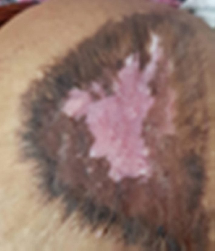
A 25-year-old primigravida with no associated co-morbidities underwent an emergency Caesarean section for abruptio placenta. She started having persistent fever from post-operative day (POD) 3, followed by serosanguinous discharge from the incision wound. CBC showed neutrophilic leucocytosis, other investigations were normal, wound discharge swab was culture negative, and fever persisted, reaching a maximum of 103°F. Suture removal on POD 7 revealed non-union of the skin and subcutaneous tissue. Patient was managed with wound dressing and appropriate antibiotics. Clinically, there were no other localizing signs. In spite of antibiotics and daily dressings, the wound showed no signs of healing. Instead, its margins were spreading as if something was burrowing into surrounding healthy tissue (Fig. 2a). Suspecting a staphylococcal skin infection due to the nature of the wound, antibiotic cover was stepped up after sending a second wound swab culture. The patient experienced unusual pain during the daily dressings. Repeat wound culture was again negative.
Fig. 2.

a Initial state of the wound with claw like margins extending into healthy tissue with a violaceous hue, as seen on POD9 LSCS b Collagen dressing applied over the wound c Wound as seen on POD20 LSCS; significant reduction in margins and showing signs of healing d Completely healed wound
Around this time, during a literature search on non-healing SSI wounds, we chanced upon a case report. The clinical picture of this case was quite similar to our patient and it was diagnosed as a case of PG. This was a novel revelation which drove us to consult our dermatologist. He carried out a punch biopsy from the wound margin. The biopsy revealed massive leucocytic infiltration.
On further reading, we came to know of a phenomenon called ‘Pathergy’ which is commonly associated with PG. Pathergy is described as an exaggerated skin reaction after minor trauma or surgery, resulting in the formation of a pustule, papule or ulceration and can recur in any future skin trauma or surgery. After this newfound information we enquired from the patient and her husband about any past wound which specifically took a long time to heal. A corroborative history surfaced that she had an injection abscess over the right gluteal region almost two years ago which took more than a month to heal and left behind a large residual scar (Fig. 1). This history confirmed the ‘Pathergy’ phenomenon in our patient.
Fig. 1.
Broad scar resulting from an injection abscess
Based on the history, clinical features and biopsy finding, the diagnosis of PG was confirmed and the patient was started on prednisolone 40 mg OD. Here onwards, the case was jointly managed by us and the dermatologist. From POD12 gauze dressing was switched over to collagen dressing (Fig. 2b). The patient, however, continued experiencing significant pain during the dressings. Oral steroids were continued and dapsone 25 mg OD was added on POD 40 when the healing appeared to be delayed. Subsequently, as the wound started healing prednisolone and dapsone were gradually tapered off. By POD 90, the wound had completely healed. The progression of wound healing is depicted in Fig. 2c, d.
Discussion
PG is an ulcerative, cutaneous condition with distinctive clinical characteristics first described by Brunsting et al. in 1930. The term Pyoderma Gangrenosum is more historical as there is neither any pus forming infection, nor any gangrene. It is basically a neutrophilic dermatosis. The peak incidence occurs between the ages of 40 and 50 years. The incidence has been estimated to be 3–10 per million population per year. In approximately 50% cases, it is seen associated with systemic diseases such as IBD, collagen vascular diseases, leukaemia and malignancy [1].
The prevalence of PG after Caesarean section is not very high. As per a study, only 17 cases have been reported worldwide between 1996 and 2019 [2]. Among other surgeries, it is more often seen after breast, cardio-thoracic, and limb surgeries.
PG is mostly a diagnosis of exclusion. Clinical features include a non-healing ulcer seen typically over the surgical site with excruciating pain over the wound. Constitutional symptoms of fever and malaise may be there and wound margins show a violaceous hue with undermined borders with rapidly widening margins.
The Maverakis diagnostic criteria [3] for Pyoderma Gangrenosum which was put forward following a consensus in 2018 cited a single major criterion of histopathology of ulcer edge showing neutrophilic infiltrate for the diagnosis and the addition of four or more of the eight minor criteria yielded a sensitivity of 86% and a specificity of 90%.
The suspicion of this auto-immune condition usually arises after excluding the common infective aetiology of so-called SSI. The cornerstones of clinching the diagnosis are the following:
Presence of violaceous undermined wound margin
Excruciating pain over the wound
A non-specific histological picture of massive dermal neutrophilic infiltration
Pathergy phenomenon
Dramatic clinical recovery after a course of steroids with or without immuno-modulators.
Pathergy can be demonstrated by a Pathergy test which involves a skin prick to any site leading to the formation of either an ulcer, papule or pustule.
Post-operative PG is often mistaken for bacterial wound infection. In a series of 36 PG patients, 29 were diagnosed as wound infection and 13 of them even underwent debridement [4]. This misdiagnosis by surgeons of any discipline could be due to their infrequent exposure and thereby inadequate knowledge of this rare entity. Thereby the treatment is often delayed.
The various systemic therapies include cyclosporine, azathioprine, cyclophosphamide and TNF-alpha inhibitors. Some studies also show beneficial action of biological agents like infliximab and adalimumab. Steroid, which may be a contraindication in infective wounds, is the backbone of treatment of this auto-immune condition and the choice of steroid is prednisolone. Immuno-modulators like cyclosporine and dapsone are also used. The role of surgical intervention in such cases remains controversial as it may further aggravate the condition [1]. However, skin grafting for extensive skin loss has been reported.
Collagen dressings play an important role in wound healing. It helps to promote fibroblast production, deposition of organized collagen fibres and also maintain an optimal chemical and thermal micro-environment of the wound. Moreover, all chronic wounds have elevated levels of matrix metalloproteinases leading to delayed healing of the wound. Collagen dressings inhibit metalloproteinases.
Long-term outcome of patients with Pyoderma Gangrenosum remains unpredictable, even after effective treatment. Recurrence rates up to 30% have been described [1].
Conclusion
This case report is focussed on the management of a relatively rare cause of a non-healing surgical wound. It reminded us of an old adage “What the mind does not know, the eyes cannot see”. Early diagnosis of this debilitating condition requires a high degree of clinical suspicion along with background knowledge of this rare entity.
The question hovering in our mind about this lady, a primipara who had undergone a Caesarean section, is:
“Should she plan a second pregnancy where she may land up with a repeat Caesarean section with a repeat Pyoderma Gangrenosum with its accompanying suffering?”
This will remain a food for thought.
Acknowledgements
We would like to acknowledge the support from the Department of Dermatology in the management of this case.
Abbreviations
- SSI
Surgical Site Infection
- PG
Pyoderma Gangrenosum
- POD
Post-Operative Day
- CBC
Complete Blood Count
- OD
Once Daily
- LSCS
Lower Segment Caesarean Section
- IBD
Inflammatory Bowel Disease
- SLE
Systemic Lupus Erythematosus
- TNF
Tumour Necrosis Factor
Himadri Bal
graduated from Nilratan Sircar Medical College, Kolkata and postgraduate from Armed Forces Medical College followed by DNB from NBE. He is presently working as Professor & HOU at Dr DY Patil Medical College, Pune. Before this he had served in various hospitals of the Armed Forces and finally retired from the Armed Forces Medical College where he was appointed as Professor in Obstetrics & Gynaecology. Currently besides his professional work he is focused on medical education..
Funding
No funding was obtained from any source for this case report.
Declarations
Conflict of interest
The authors report no conflict of interest.
Informed consent
Informed consent was obtained from the patient to prepare this manuscript. No personal information is shared in the description of this case, and no experiments were carried out on the patient.
Human and Animal Rights
No research was conducted on human participants or animals.
Footnotes
Himadri Bal is an MD, DNB, Department of Obstetrics and Gynaecology, Dr. D. Y. Patil Medical College, Hospital and Research Centre, Pimpri, Pune, 411,018, India; Supraja Subramanian is a MBBS, Department of Obstetrics and Gynaecology, Dr. D. Y. Patil Medical College, Hospital and Research Centre, Pimpri, Pune, 411,018, India; Y.K Sharma is an MD, Department of Dermatology, Dr. D. Y. Patil Medical College, Hospital and Research Centre, Pimpri, Pune, 411,018, India; Swati Lal is an MBBS, Department of Obstetrics and Gynaecology, Dr. D. Y. Patil Medical College, Hospital and Research Centre, Pimpri, Pune, 411,018 India.
Publisher's Note
Springer Nature remains neutral with regard to jurisdictional claims in published maps and institutional affiliations.
Contributor Information
Himadri Bal, Email: himadri.bal@gmail.com.
Supraja Subramanian, Email: supraja94@yahoo.com.
Y. K. Sharma, Email: yugalsharmadr@gmail.com
Swati Lal, Email: swatilal21@gmail.com.
References
- 1.Bhat RM. Pyoderma gangrenosum: an update. Indian Dermatol Online J. 2012;3(1):7. doi: 10.4103/2229-5178.93482. [DOI] [PMC free article] [PubMed] [Google Scholar]
- 2.Shen J, Zhang W, Jiang X. Pyoderma Gangrenosum after Caesarean section treated with skin graft: a case report. Medicine. 2019;98(18):e15380. doi: 10.1097/MD.0000000000015380. [DOI] [PMC free article] [PubMed] [Google Scholar]
- 3.Maverakis E, Ma C, Shinkai K, Fiorentino D, Pcallen J, Wollina U. Diagnostic criteria of ulcerative Pyoderma Gangrenosum a delphi consensus of international experts. JAMA Dermatol. 2018;154(4):461–466. doi: 10.1001/jamadermatol.2017.5980. [DOI] [PubMed] [Google Scholar]
- 4.Wong WW, Machado GR, Hill ME. Pyoderma gangrenosum: the great pretender and a challenging diagnosis. J Cutaneous Med. Surg. 2011;15(6):322–328. doi: 10.2310/7750.2011.10119. [DOI] [PubMed] [Google Scholar]