

Abstract
Mucormycosis is a potentially life-threatening fungal infection with a high mortality rate. The difficulty and delay in diagnosis due to its rarity usually results in a poor prognosis. Most common site in head and neck region is the nose and paranasal sinuses. However there are other very unusual areas in head and neck region where mucormycosis is encountered. Knowledge of these unusual areas is must and can save a patient’s life. To elaborate and highlight the unusual areas in head and neck region where mucormycosis can mimic other common diseases. This retrospective study was done from May 2010 to May 2019 over a period of 9 years. All histopathologically confirmed cases of Head and Neck mucormycosis were evaluated and data analyzed. Total 35 cases of head neck mucormycosis were encountered from May 2010 to May 2019 over a period of 9 years. Out of which 30 cases (85.72%) were of rhino-orbito-cerebral mucormycosis and 5 cases (14.28%) were EXTRA rhino-orbito-cerebral mucormycosis in head neck region. Mucormycosis at such unusual sites can cause diagnostic dilemma for the treating doctor. Clinical knowledge with anticipation is a must for success in treatment of mucormycosis in unusual places in head neck region especially when normal looking diseases does not respond to usual treatment.
Keywords: Mucormycosis, Rhizopus oryzae, Invasive fungal sinusitis, Amphotericin
Introduction
Mucormycosis is an acute, invasive and potentially fatal fungal infection. [1] It is the third commonest fungal infection after candidiasis and aspergillosis in immunocompromised patients. Mucormycosis can be classified based on location into (1) Rhinoorbitocerebral (2) Pulmonary (3) Cutaneous (4) Gastrointestinal (5) Disseminated and (6) Uncommon type [2]. In head and neck region rhinoorbitocerebral type is the most frequent form seen with extensive published work.
However there are other sites in head & neck region as well where very rarely mucormycosis can be encountered and can then become a diagnostic dilemma. This article is an attempt to highlight extra rhinocerebral mucormycosis areas in head neck region which we have encountered in our practice.
Materials and Methods
The retrospective study was done in a tertiary care hospital from May 2010 to May 2019 over a period of 9 years. All histopathologically confirmed cases of Head and Neck mucormycosis were evaluated and data analyzed.
Inclusion Criteria
Specific cases of Head and Neck mucormycosis only, EXCLUDING Rhino-orbito-cerebral mucormycosis.
Histopathologically confirmed cases
Follow up of atleast 1 year
Exclusion Criteria
All cases of Rhino-orbito-cerebral mucormycosis.
All cases of mucormycosis involving other parts of the body except head & neck region
Histopathologically unconfirmed cases
Follow up of less then 1 year or patients lost to follow up
Detailed history, Dental, ENT & neurological examination was done in all patients. Routine blood investigations were done. Diagnostic nasal endoscopy was done to rule out any cases of sinonasal mucormycosis. Biopsy was taken from the lesion. CT scan skull base to clavicle was done to assess the extent of the disease. MRI was done to evaluate/rule out intraorbital or intracranial extent if any. Treatment was started for any underlying metabolic disorder. Surgical debridement was done within 24 h and a second look surgery done after 48 h if required. Once a histopathological diagnosis of mucormycosis was established, lyophilized amphotericin-B was started according to weight and severity of each case. First a test dose of 1 mg Amphotericin-B in 200 ml of 5% dextrose was given over 4–6 h. Therapy is initiated at 0.25 mg/kg to 0.30 mg/kg, then the dose is gradually increased by 0.25 mg per day. Every alternate day renal function test and serum electrolytes were monitored. Follow-up was done every week for first 3 weeks and then every month for 6 months and then after 1 year.
Results
Total 5 cases of confirmed EXTRA Rhino-orbito-cerebral mucormycosis in head & neck region were reported in a tertiary care hospital from May 2010 to May 2019 over a period of 9 years. There were 3 males and 2 female cases. Average age of presentation was 43 years.
Incidence
Total 35 cases of head neck mucormycosis were encountered from May 2010 to May 2019 over a period of 9 years. Out of which 30 cases (85.72%) were of rhino-orbito-cerebral mucormycosis and 5 cases (14.28%) were EXTRA rhino-orbito-cerebral mucormycosis in head neck region (Tables 1 and 2).
Table 1.
Incidence
| Region involved | Number of cases |
|---|---|
| Rhino-orbito-cerebral mucormycosis | 30 (85.72%) |
| EXTRA rhino-orbito-cerebral mucormycosis in head neck region | 5 (14.28%) |
| Head and neck mucormycosis (TOTAL) | 35 (100%) |
Table 2.
Patients data
| S. no | Area involved | No of cases | Chief Presentation of patient | Comorbid condition | Immunocompromised status | Treatment given | Outcome |
|---|---|---|---|---|---|---|---|
| 1 | Right submandibular region in neck | 1 | Abscess formation with loss of overlying skin | Diabetes Mellitus (DM) | Reversed | I and D with Debridement with IV Amphotericin B | Recovered |
| 2 | Oral cavity–left mandibular gingiva | 1 | Ulcerative lesion | Diabetes Mellitus (DM) | Reversed | Wide Excision with IV Amphotericin B | Recovered |
| 3 | Palatal region | 1 | Black necrotic eschar lesions | DM with Diabetic Ketoacidosis | Could not be reversed | Debridement with IV Amphotericin B | Mortality |
| 4 | Temporal bone | 1 | Left ear discharge with left Lower Motor Neuron Facial palsy | Diabetes Mellitus (DM) | Reversed | Modified Radical Mastoidectomy with Facial nerve decompression with IV Amphotericin B with facial physiotherapy | Recovered |
| 5 | Cutaneous area of Forehead | 1 | Nodular lesions | Prolonged steroid use | Steroids stopped | IV Amphotericin B | Recovered |
Patient Data
Data of 5 cases of EXTRA Rhino-orbito-cerebral mucormycosis encountered in head and neck region is as follow-
Case with involvement of right submandibular region in neck presented as any other case of neck abscess. An incision and drainage of the abscess was done and the removed tissue was sent for histopathology. It was indicative of fungal disease. After special stains for fungus diagnosis of mucormycosis was confirmed. Debridement with local application of amphotericin B as well as IV amphotericin B was started and the patient could be saved.
Case with involvement of Oral cavity was referred from dental department with complaints of ulcerative lesion of the left mandibular gingiva. Patient also had past history of treatment from outside for the same problems which was not relieved. Initial suspicion was of a neoplastic etiology and a biopsy was taken to prove the same. But again it was indicative of fungal disease. After special stains for fungus diagnosis of mucormycosis was established. Wide excision with local application of amphotericin B as well as IV amphotericin B was started and the patient responded well to the treatment.
Another case presented with involvement of Palatal region. It was clinically suggestive of mucormycosis and the histopathology confirmed our diagnosis. But the patient’s immunocompromised status could not be treated and eventually dissemination of the disease occurred leading to failure of treatment and eventual mortality.
One case presented with classical features of chronic otitis media squamous disease with facial nerve palsy. Modified Radical Mastoidectomy with Facial nerve decompression was done. Intraoperatively necrosed bone and slough like tissue was encountered which raised our suspicion and biopsy was sent, which came out to be mucormycosis. IV amphotericin B was started and the patient could be saved.
Another case was referred from skin department with complaints of persistent nodular lesions over cutaneous area of forehead to rule out any underlying pathology in sinuses. On evaluation paranasal sinuses were clear and tissue diagnosis came out to be mucormycosis. Disease was controlled with IV amphotericin B.
Extent of Disease
All the cases of extra rhino-orbito-cerebral mucormycosis in head neck region were primary in origin [3].
Extra rhinocerebral mucormycosis in head neck region can be divided according to following two categories [3]:
-
A.
Primary or Secondary: based on source of infection
-
B.
Localized, deep or disseminated: based on extent of disease
In primary disease [3] the infection is by direct inoculation of the involved region whereas in secondary disease [3] the infection spreads from other locations like from nose or paranasal sinus disease in case of palatal mucormycosis.
In localized disease [3], there is only involvement of skin or mucosa of the involved area with no underlying spread. In Deep disease [3] there is involvement of skin or mucosa of the involved area with involvement of underlying structures like muscle or bone. In Disseminated disease [3] along with these there is hematogenous dissemination to other anatomical sites as well (Table 3).
Table 3.
Extent of disease
| S. no | Area involved | Primary or secondary | Localized, deep or disseminated |
|---|---|---|---|
| 1 | Right submandibular region in neck | Primary | Deep |
| 2 | Oral cavity—left mandibular gingiva | Primary | Localized |
| 3 | Palatal region | Primary | Disseminated |
| 4 | Left Temporal bone | Primary | Deep |
| 5 | Cutaneous area of Forehead | Primary | Localized |
Discussion
Mucormycosis is an opportunistic fungal infection attacking the immunocompromised patients [1, 2] i.e., patients with uncontrolled diabetes mellitus, prolonged steroid use, with allogenic hematopoietic stem cell transplantations and with other hematologic malignancies etc.
These fungi are ubiquitous in nature and are commonly found in decaying organic matter, water, soil, human body and waste products [1]. They attack the immunocompromised host by direct inoculation. Their growth is favored by low oxygen concentration, acidic medium and high iron levels [1]. They germinate into hyphae. Due to the metabolic hypoxic conditions, the polymorphonuclear cells are less effective at removing these hyphae, as it is often found in patients with mucormycosis associated with diabetes mellitus, thereby favoring the establishment of infection. Fungus then invades the arteries and form thrombus within the blood vessels and cause necrosis of the involved area. This necrosis leads to characteristic brownish black eschar tissue in mucormycosis [1].
Main presenting symptoms in head neck region are the formation of ulcerative lesion in oral cavity or an abscess formation in the neck region [3, 4]. Mucormycosis of the temporal bone presents as any other chronic otitis media case with ear discharge and with complaints of decreased hearing. However cutaneous mucormycosis of the temporal bone starts as necrosis of the pinna and spreads towards the middle ear and mastoid cavity. Symptoms vary with the anatomic site involved, however, anesthesia of the involved area in an immunocompromised case should be dealt carefully with the possibility of mucormycosis that must always be kept in mind (Figs. 1, 2 and 3).
Fig. 1.

Abscess formation in right submandibular region in neck with loss of skin
Fig. 2.
Ulcerative lesion in left mandibular gingiva in oral cavity
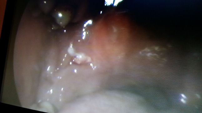
Fig. 3.

Pale mucosa over hard palate
Epstein [4] et al. in their study found out that the most common oral sign of mucormycosis is ulceration of the palate. They also documented a case of gingival mucormycosis following a hematopoietic stem cell transfer for chronic myeloid leukemia.
Sathish [5] et al. in their study described the only case of mucormycosis of temporal bone with facial nerve palsy. They also emphasized that the spread of infection to the mastoid region can be either direct spread through the Eustachian tube or through the vascular channels.
Varghese [6] et al. documented 7 cases of fungal otomastoiditis in their study, out of which 2 cases were of mucormycosis. They described the route of infection into the mastoid cavity may be either tympanogenic, meningogenic, haematogenic or nasopharyngeal. The route of entry to tympanic cavity and middle ear could be through the perforated tympanic membrane. Comorbid condition associated was prolonged use of topical antibiotic-steroid medication which might have hampered the innate immunity and led to the spread of fungal infection. Treatment consists of three parts, i.e., control of underlying immunocompromised condition if present, surgical debridement and antifungal chemotherapy (Figs. 4, 5 and 6).
Fig. 4.
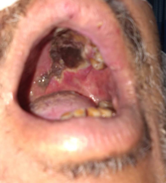
Black necrotic eschar lesion at Palatal region
Fig. 5.

Nodular lesions over cutaneous area of forehead
Fig. 6.

Broad, ribbon like, irregular and aseptate fungal hyphae with branching at right angle on histopathological examination
Hazarika [7] et al. in their study highlighted the point that mucormycosis can also involve middle ear and mastoid in non-diabetic patients. In their study they documented that the involvement of the middle ear and mastoid in a nondiabetic patient is very rare and the early symptom is otalgia, unlike the foul smelling ear discharge as seen in the attico antral disease. Radical debridement in the form of modified radical mastoidectomy with or without the use of Amphotericin B may suffice in non-diabetic patients. They also suggested that it is difficult to predict preoperative or intraoperatively about mucormycosis as the case is almost indistinguishable from unsafe Chronic Otitis Media and fungal culture is the only method to diagnose mucormycosis. Similar situation was encountered by the authors in this study during the treatment of mucormycosis of the temporal bone. Thus the possibility of mucormycosis infecting the middle ear and mastoid region must be kept in mind at all times and wherever suspected a separate biopsy specimen must be sent in plain saline solution for fungal culture specifically.
Mc Dermott [8] et al. in their study described 6 cases of periodontal mucormycosis. In this study they found out that there was periodontal ulceration along with involvement of underlying bone in patients undergoing treatment for AML. Diagnosis was made by culture and treatment was done by local debridement with antifungal amphotericin C.
Bakathir [9] et al. in their study reported two cases of mucormycosis of the mandible region post dental extractions. First case was undergoing chemotherapy for AML and second case was in diabetic ketoacidosis. With treatment good loco regional control over the disease was attained.
Gold standard for diagnosis is the clinical presentation of black necrosed eschar tissue in an immunocompromised case with prompt immediate biopsy for histopathological confirmation [1]. On biopsy the fungal hyphae seen are broad, ribbon like, irregular and aseptate with branching at right angle with submucosal infiltration and angioinvasion [1].
In differential diagnosis it should be differentiated from aspergillosis and gangrene caused by other causes. Again biopsy is crucial and a timely done biopsy can save a patient’s life. Recently Bernal Martinez [10] et al. developed a single tube multiplex real time Polymerase Chain Reaction to detect the genus mucor and rhizopus. This technique has high specificity and most importantly provides the results in upto 3 h. There is need for similar studies in today’s time to get an early diagnosis, however such techniques are not available at all the places and most importantly not affordable to all.
Treatment mainly consists of [1].
Early prompt diagnosis
Reversal of the Immunocompromised status
Appropriate aggressive surgical debridement
Rapid antifungal therapy
High clinical suspicion based on clinical diagnosis and immediate biopsy helps in early diagnosis of disease which has a direct impact on prognosis. Underlying immunocompromised comorbidity should be treated. Immediate debridement of the involved area is a must till the healthy area with bleeding is encountered. This reduces the fungal load and also acts as local control because anti fungal treatment is non effective at this area due to thrombosis. Once diagnosis of mucormycosis is established, Antifungal therapy should be immediately started with the help of physician colleagues. Amphotericin B and Isavoconazole are the two agents approved for primary therapy of mucormycosis [1]. Posaconazole can be used for salvage treatment in patients who are intolerant to Amphotericin B [1]. It can also be used as a step down therapy after initial control of the disease with Amphotericin B. However both posaconazole and specially isavoconazole can have an availability problem. Initiation of the polyene therapy within 5 days of diagnosis of mucormycosis was associated with improvement in survival, when compared with initiation of polyene therapy at 6 days or more after diagnosis (83% vs 49% survival) [1]. Thus treatment requires a joint and team effort of various specialties in saving a patient’s life. If the disease is detected early and treatment started immediately i.e. within 5 days of presentation then the outcome is favorable [1].
It is very important to keep mucormycosis in mind as a differential diagnosis while treating such cases as an ENT specialist because both the morbidity and mortality in mucormycosis is very high and knowledge of this rare entity can not only save a patient’s life but can also save a doctor from a potential lawsuit. Also mucormycosis in such unusual regions can come as a sudden surprise for the treating doctor at times and can cause problems in patient counselling, especially when it mimics other common diseases. Thus anticipation and knowledge with high suspicion plays a key role in diagnosis of such cases.
Conclusion
Clinical knowledge with anticipation is a must for success in treatment of mucormycosis especially in unusual places in head neck region as discussed. Mucormycosis at such rare sites can cause diagnostic dilemma for the treating doctor if not anticipated in non-healing or non-responding cases increasing patient’s morbidity.
Funding
There are no financial interests the authors may have in companies or other entities that have an interest in the information in the Contribution (e.g., grants, advisory boards, employment, consultancies, contracts, honoraria, royalties, expert testimony, partnerships, or stock ownership in medically related fields). The authors have no financial interest.
Footnotes
Publisher's Note
Springer Nature remains neutral with regard to jurisdictional claims in published maps and institutional affiliations.
References
- 1.Singh VP, Bansal C, Kaintura M. Sinonasal mucormycosis: A–Z. Indian J Otolaryngol Head Neck Surg. 2019;71(Suppl 3):1962–1971. doi: 10.1007/s12070-018-1384-6.PMID:31763277;PMCID:PMC6848679. [DOI] [PMC free article] [PubMed] [Google Scholar]
- 2.Desai V, Pratik P. Mucormycosis Of Oral Mucosa: A Rare Case Report. Int J Pharm Chem Biol Sci. 2014;4(3):509–511. [Google Scholar]
- 3.Castrejon-Perez A, Welsh EC, Miranda I, Ocampo-Candiani J, Welsh O. Cutaneous mucormycosis. An Bras Dermatol. 2017;92(3):304–311. doi: 10.1590/abd1806-4841.20176614. [DOI] [PMC free article] [PubMed] [Google Scholar]
- 4.Joel B. Epstein, Steven B. Kupferman , Rachel Zabner, Ali Rejali, Martin L. Hopp, Michael Lill, Dimitrios Tzachanis (2016) Early diagnosis and successful management of oral mucormycosis in a hematopoietic stem cell transplant recipient: case report and literature review. 24(8):3343–3346 [DOI] [PubMed]
- 5.Sathish Kumar KN, Nishan N. Mucormycosis of the temporal bone with facial nerve palsy: A rare case report. Indian J Otol. 2015;21:61–63. doi: 10.4103/0971-7749.152870. [DOI] [Google Scholar]
- 6.Varghese R, Nair RM, Kavalakkat FJ. Fungal otomastoiditis: A case series in immunocompetent Adults. Indian J Otolaryngol Head Neck Surg. 2014;66(1):110–113. doi: 10.1007/s12070-012-0527-4. [DOI] [PMC free article] [PubMed] [Google Scholar]
- 7.Hazarika P, Zachariah J, Victor J, John M, Devi C, Abraham P. Mucormycosis of the Middle Ear: A Case Report with Review of Literature. Indian J Otolaryngol Head Neck Surg. 2012;64(1):90–94. doi: 10.1007/s12070-011-0156-3. [DOI] [PMC free article] [PubMed] [Google Scholar]
- 8.McDermott NE, Barrett J, Hipp J. Successful treatment of periodontal mucormycosis: Report of a case and literature review. Oral Surg Oral Med Oral Pathol Oral Radiol Endodont. 2010;109:e64–e69. doi: 10.1016/j.tripleo.2009.11.012. [DOI] [PubMed] [Google Scholar]
- 9.Bakathir AA. Mucormycosis of the Jaw after dental extractions: two case reports. Sultan Qaboos Univ Med. 2006;6(2):77–82. [PMC free article] [PubMed] [Google Scholar]
- 10.Bernal-Martínez L, Buitrago MJ, Castelli MV, Rodriguez-Tudela JL, Cuenca-Estrella M. Development of a single tube multiplex real-time PCR to detect the most clinically relevant Mucormycetes species. Clin Microbiol Infect. 2013;19:E1–7. doi: 10.1111/j.1469-0691.2012.03976.x. [DOI] [PubMed] [Google Scholar]