Abstract
During neck surgery; Zuckerkandl’s tubercle, Berry’s ligament, the inferior horn of thyroid cartilages have become crucial anatomical landmarks in order to protect the integrity of the recurrent laryngeal nerve. Forty-two male postmortem human cadavers were used. The proximal part of the recurrent laryngeal nerve, before the inferior thyroid artery arises from its source has been observed in 87% inside the tracheoesophageal groove and in 13% running laterally to the trachea. The recurrent laryngeal nerve was encountered passing behind and through the branches of the inferior thyroid artery in 92% and 8% respectively. At all sides; the nerve was piercing the larynx 0.6 ± 0.1 mm below the inferior horn of thyroid cartilage, passing next to the inner-lower side of Berry’s ligament and running under the lower middle part of Zuckerkandl’s tubercle. These landmarks and their upper mentioned distances to the laryngeal nerve can be taken into consideration as important surgical guides.
Keywords: Recurrent laryngeal nerve, Inferior laryngeal nerve, Thyroid cartilage, Berry’s ligament, Zuckerkandl’s tubercle
Introduction
The recurrent laryngeal nerve injury is the most feared complication of thyroid surgeries. The recurrent laryngeal nerves may be injured as a result of the complete or partial cut, contusion, burning, failure of arterial supply, etc. [1]. Unilateral paralysis of the recurrent laryngeal nerve can result in hoarseness and bilateral paralysis of the recurrent laryngeal nerve can result in life-threatening glottal closure. There are different landmarks to expose the recurrent laryngeal nerve during the surgery. These are; inferior thyroid artery, Berry’s ligament (suspensory ligament of the thyroid gland, posterior suspensory ligament) and Zuckerkandl's tubercle. In conclusion, nowadays finding and tracing the recurrent laryngeal nerve during the thyroidectomy is very important. The anatomical structure shouldn’t be cut before the exposure of the recurrent laryngeal nerve and the surgeon shouldn’t trust only one landmark, to avoid the risk of damage to the nerve [2, 3].
The aim of this study to investigate the relationship between the recurrent laryngeal nerve and the inferior thyroid artery, Berry’s ligament, Zuckerkandl's tubercle.
The recurrent laryngeal nerve is a branch of the vagus nerve and terminal branch of the recurrent laryngeal nerve is called the inferior laryngeal nerve [4]. There are two recurrent laryngeal nerves, right and left recurrent laryngeal nerves are in intimate proximity to the thyroid gland. The right recurrent laryngeal nerve originates from the vagus nerve and surrounds the subclavian artery then ascends behind the common carotid artery and lateral to the trachea. It passes behind the right lobe of the thyroid gland and enters the larynx behind the cricothyroid cartilage. Mostly (64%) it lies in the tracheoesophageal groove in an oblique and lateral position [5–8]. Till it arrives at the inferior laryngeal artery it may lie in the tracheoesophageal groove approximately 10% it passes between the branches of the inferior laryngeal artery, 50% behind and 40% in front of the artery [9, 10]. On the left side, the recurrent laryngeal nerve originates from the vagus nerve in the thoracic cavity lateral to the aortic arch and surrounds the aortic arch then ascends to the neck. (77%), lateral to the trachea (17%) or close to in front of the trachea (6%). When it arrives the inferior laryngeal artery mostly passes behind the artery (69%), sometimes in front of the artery (24%) and rarely between the branches of the artery (5–6%) and then arises to the neck [10, 11]. Both nerves arrive at the larynx from the internal surface of the thyroid gland lobes [5]. The inferior thyroid artery is the primary artery of the thyroid gland. The recurrent laryngeal nerves lie deep to the thyroid gland and come very close to the inferior thyroid artery. Relations between the terminal branches of the artery and recurrent laryngeal nerve are very variable. But it is certain that these two structures are crossing structures. The left nerve is usually posterior to the artery and the right nerve is equally likely to be anterior, posterior or amongst, the branches of the artery [10, 11]. The inferior cornu of the thyroid cartilage is palpable lower behind the thyroid cartilage. It is reported that; because of the relations between the inferior thyroid artery and the recurrent laryngeal nerve are very variable the inferior cornu of the thyroid cartilage is a more reliable landmark. The reccurent laryngeal nerve enters to the larynx 0.5–0.8 mm below to the inferior cornu [12–14]. Berry ligament is the main ligament that binding the thyroid gland to the laryngotracheal complex. Berry’s ligament is a strong and vascular structure. Branches of the inferior thyroid artery lies through the inferior margin of the Berry’s ligament and could cause to bleeding during the thyroidectomy. Besides Berry’s ligament has a close relationship with the thyroid parenchyma. The recurrent laryngeal nerve also is known close to the Berry’s ligament, moreover some studies showed that in 10–38% it passes through the ligament [15–17]. Because of the strong and vascular structure of the Berry’s ligament, the close relationship between the branches of the recurrent laryngeal nerve and the thyroid parenchyma with the Berry’s ligament this area became the most difficult dissection area during the thyroidectomy [18].
The posterolateral lobe of the thyroid gland which locates caudally to the Berry’s ligament named as the Zuckerkandl’s tubercle. This tubercle and the recurrent laryngeal nerve have a variable relationship and it makes difficult to dissect the distal part of the nerve. The Zuckerkandl tubercle is described as an important and reliable landmark for the recurrent laryngeal nerve during thyroid surgeries [18–23]. Pelizzo et al. described 4 grades for the Zuckerkandl tubercle. According to this classification Grade 0 is the absence of the tubercle, Grade 1 is a little thickening of the thyroid lobe, Grade 2 is a thickening < 1 cm, Grade 3 is a thickening > 1 cm. The Zuckerkandl tubercle, which described as an important landmark for the recurrent laryngeal nerve, is found 14–62% during the thyroidectomy with different levels [24]. The knowledge of the anatomy of the Zuckerkandl tubercle is very important for a safe surgical dissection.
Materials and Methods
This study is performed with forty-two formalin-fixed adult male cadavers at the Akdeniz University Faculty of Medicine, Department of Anatomy. After the dissection the relation between the recurrent laryngeal nerve and it’s acknowledged landmarks that the inferior thyroid artery, the inferior cornu of the thyroid cartilage, Berry’s ligament, Zuckerkandl tubercle, is investigated.
Results
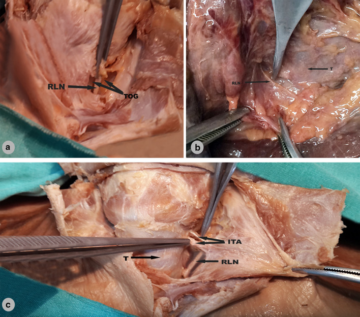
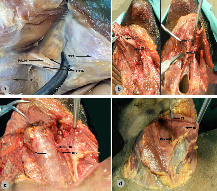
This study performed with forty-two cadavers (right and left totally 84 sides). After meticulous exposure of the recurrent laryngeal nerve we found that the nerve lies in the tracheoesophageal groove 87% (73 sides) (Fig. 1a). 13% (11 sides) it lies lateral to the trachea (Fig. 1b). We haven’t found any recurrent laryngeal nerve which lies anteriorly to the trachea in our study. The recurrent laryngeal nerve was passing behind the inferior thyroid artery in 77 sides (92%) (Fig. 1c). In 7 sides (8%) it was passing between the branches of the inferior thyroid artery (Fig. 2a). In all cadavers the recurrent laryngeal nerve was entering the larynx 0.6 ± 0.1 mm below to the inferior cornu of the thyroid cartilage (Fig. 2b) and it was also passing inferolaterally to the Berry’s ligament (Fig. 2c). In all cadavers the recurrent laryngeal nerve was located beneath the Zuckerland tubercle (Fig. 2d).
Fig. 1.
a Recurrent laryngeal nerve (RLN) lying in the tracheoesophageal groove (TOG). b Recurrent laryngeal nerve (RLN) lying laterally of the trachea (T). c Recurrent laryngeal nerve (RLN) passing behind the inferior thyroid artery (ITA), (T: Trachea)
Fig. 2.
a Recurrent laryngeal nerve (RLN) passing between the branches of inferior thyroid artery (ITA). (T: Trachea, TG: Thyroid gland), b Recurrent laryngeal nerve (RLN) in related to inferior corn of the thyroid cartilage (ICTC), c Recurrent laryngeal nerve (RLN) passing inferomedially to the Berry’s ligament (BL), (T:Trachea), d Elevation of Zuckerkandl tubercle ( ZT) laterally and exposed to recurrent laryngeal nerve (RLN), (TG: Thyroid gland)
Discussion
The recurrent laryngeal nerve is one of the most important structures which needs attention during the thyroid surgeries. The recurrent laryngeal nerves may be injured as a result of neck trauma, thyroid surgery, aortic aneurysm, oesophageal cancer, mediastinal tumor, lung cancer, etc. Injury of the recurrent laryngeal nerve is clinically important. Some authors indicated that certain landmarks should use during the surgery to avoid the recurrent laryngeal nerve injury. One of the landmarks is the inferior thyroid artery. The relation between the recurrent laryngeal nerve and the inferior thyroid artery is described well. Campos and Heriques [9] studied the anatomical relationship between found in both sides the recurrent laryngeal nerve lay between the branches of the inferior laryngeal artery in 45.8%, anterior to the artery in 16.7% and posterior to the artery in 37.5%. in 76 cadavers, 8 females and 68 males. They found the recurrent laryngeal nerve lay between the branches of the inferior laryngeal artery in 48.86%, anterior to the artery in 27.97% and posterir to the artery in 24.47%, in both sexes. Kulekci et al. [11] studied the relationship between the recurrent laryngeal nerve and the inferior thyroid artery in 100 cadavers and they described 6 types. Poyraz ve Calguner studies 48 recurrent laryngeal nerve and they found in both sides the recurrent laryngeal nerve lay between the branches of the inferior thyroid artery in 45.8%, anterior to the artery in 16.7% and posterir to the artery in 37.5%. Lee et al. [6] found in both sides the recurrent laryngeal nerve lay between the branches of the inferior thyroid artery in 23.5%, anterior to the artery in 31.8% and posterior to the artery in 45.5%. In our study in 77 sides (92%) the recurrent laryngeal nerve was layin posterior to the artery, in 7 sides (8%) between the branches of the artery. Because the relation between the recurrent laryngeal nerve and inferior thyroid artery is very variable the inferior cornu of the thyroid cartilage is a more reliable landmark [7, 25–27]. The recurrent laryngeal nerve enters larynx from inferolateral margin of the inferior cornu of the thyroid cartilage [7, 12, 14]. In our study we found that in all cadavers both sides the recurrent laryngeal nerve was entering the larynx 0.6 ± 0.1 mm inferolateral to the inferior cornu of the thyroid cartilage. Another important landmark is Berry’s ligament (posterior suspansory ligament). Berry’s ligament has a close relationship with the motor branches of the recurrent laryngeal nerve. And this relation is an important risk factor during thyroid surgeries (98.2%) [28, 29]. Some studies showed that in 10–38% recurrent laryngeal nerve passes through the Berry’s ligament [15–17].Yalcın et al. [29] found that the nerve could be located posteromedial or posterolateral to the ligament. The thyroid gland is fixed to the trachea by the Berry’s ligament. The recurrent laryngeal nerve is located lateral to the trachea and runs beneath the inferior pharyngeal constrictor muscle just cranial to the Berry’s ligament and enters to the larynx. Because of this close relation lateral mobilization of the recurrent laryngeal nerve facilitates the preservation of the nerve during tracheal surgery [30]. Botelho et al. [15] investigated the relationship between the recurrent laryngeal nerve and Berry’s ligament and classification the nerve to three types; lateral to the ligament, the recurrent laryngeal nerve passes the surface of the Berry’s ligament in 66.9%, passes beneath the ligament in 25.6% and penetrates the ligament in 7.4% [17]. Pradeep et al. [31] clasificated the recurrent laryngeal nerve as deep to the ligament, superficial to the ligament, around the ligament and inside the ligament. They found the nerve lay medial to the ligament in 86.3%. Henry B.M. et al. investigated twenty-three studies (n = 5.970 nerves) examined the recurrent laryngeal nerve/tracheoesophageal groove relationship. The recurrent laryngeal nerve was found most often located superficial to the the Berry’s ligament with a pooled prevalence estimate of 78.2% of nerves, followed by deep to the Berry’s ligament in 14.8% [32]. In our study in all cadavers the recurrent laryngeal nerve was passing inferomedially to the Berry’s ligament. Another landmark is Zuckerkandl tubercle. In diseased gland the tubercle measuring more than 1 cm, is found in 14–55% of thyroidectomies [33]. Zuckerkandl is classified by the size of the gland [34]. This tubercle and the recurrent laryngeal nerve have a variable relationship. The recurrent laryngeal nerve passes mostly posterior, rarely anterior to the tubercle and it makes difficult to dissect the distal part of the nerve [35, 36]. Yalcın et al. [21] classified the tubercle according to localization and relation with the recurrent laryngeal nerve. They pointed out the importance of the tubercle during the surgeries. The Zuckerkandl tubercle has a close relationship not only the nerve also the vascular and glandular structures. Because of that, the tubercle is an important landmark [37]. Gravente et al. [38] showed that the recurrent laryngeal nerve was passing posteromedially to the tubercle in 93%, posterolaterally in 7%. Yalçın B et al. [21] observed that some tubercles pass through the inferior laryngeal nerve, some pass through the laryngeal branches, and some pass only through the anterior laryngeal branch. During the surgical operations the nerve must be found attentively, because of this the tubercle should be deviated laterally gently [33]. Total thyroidectomies should be performed very carefully [39]. In our study the Zuckerkandl tubercle was approximately 8 mm (transverse). In all cadavers, recurrent laryngeal nerve was passing beneath the tubercle. We found all tubercles grade 3 according to Pelizzio [24]. Recurrent laryngeal nerve mostly lies diagonally in the tracheoesophageal groove between the inferior thyroid artery and Berry’s ligament [36]. We found that the recurrent laryngeal nerve lies in the tracheoesophageal groove 87% (52 sides) and 13% (8 sides) it lies lateral to the trachea.
During the neck surgeries finding and tracing the recurrent laryngeal nerve, to avoid paralysis is became a sine qua non rule. Anatomical structures shouldn’t be cut before the exposure of nerve and surgeons shouldn’t trust only one landmark, to avoid risk of damage the nerve. The recurrent laryngeal nerve and inferior thyroid artery have a close relationship. The Zuckerkandl tubercle is an important landmark for the recurrent laryngeal nerve during thyroid surgeries. Berry’s ligament has a close relationship with the recurrent laryngeal nerve. Also, inferior cornu of thyroid cartilage has a relation with the nerve. During the surgeries like thyroidectomy to protect the recurrent laryngeal nerve is essential. Therefore surgeries should be done by taking into consideration all these classical landmarks. We concluded that knowledge of the detailed anatomy of this area and these landmarks is provided more safety and successful operations.
Author Contributions
Özlem Zümre Kaştan and Serra Öztürk were made dissection and making measurements, analysis of data, literature review and revising it critically for important intellectual content Muzaffer Sindel and Engin Çalgüner made drafting the work and substantial contributions to the conception of the work and interpretation of data for the work. Bülent Veli Ağırdır made dissection.
Funding
This research received no specific grant from any funding agency in the public, commercial, or nonprofit sectors.
Data Availability
Cadavers used in department of anatomy Akdeniz University.
Compliance with Ethical Standards
Conflict of interest
The authors have no conflicts of interest to declare.
Ethical Approval
The study was approved by the Clinical Research Ethics Committee of Akdeniz University, Faculty of Medicine (Date: 03.09.2014 & No: 347).
Footnotes
Publisher's Note
Springer Nature remains neutral with regard to jurisdictional claims in published maps and institutional affiliations.
References
- 1.Bergamaschi R, Becouarn G, Ronceray J, Arnaud JP. Morbidity of thyroid surgery. Am J Surg. 1998;176(1):71–75. doi: 10.1016/s0002-9610(98)00099-3. [DOI] [PubMed] [Google Scholar]
- 2.Benouaich V, Porterie J, Bouali O, Moscovici J, Lopez R. Anatomical basis of the risk of injury to the right laryngeal recurrent nerve during thoracic surgery. Surg Radiol Anat. 2012;34(6):509–512. doi: 10.1007/s00276-012-0946-7. [DOI] [PubMed] [Google Scholar]
- 3.Hisham AN, Aina EN. Zuckerkandl's tubercle of the thyroid gland in association with pressure symptoms: a coincidence or consequence? Aust N Z J Surg. 2000;70(4):251–253. doi: 10.1046/j.1440-1622.2000.01800.x. [DOI] [PubMed] [Google Scholar]
- 4.Arıncı K, Elhan A. Anatomi 1. 6. Ankara: Güneş Kitabevi; 2016. [Google Scholar]
- 5.Ardito G, Revelli L, D'Alatri L, Lerro V, Guidi ML, Ardito F. Revisited anatomy of the recurrent laryngeal nerves. Am J Surg. 2004;187(2):249–253. doi: 10.1016/j.amjsurg.2003.11.001. [DOI] [PubMed] [Google Scholar]
- 6.Lee MS, Lee UY, Lee JH, Han SH. Relative direction and position of recurrent laryngeal nerve for anatomical configuration. Surg Radiol Anat. 2009;31(9):649–655. doi: 10.1007/s00276-009-0494-y. [DOI] [PubMed] [Google Scholar]
- 7.Richer SL, Randolph GW. Management of the recurrent laryngeal nerve in thyroid surgery. Operative Tech Otolaryngol Head Neck Surg. 2009;20(1):29–34. doi: 10.1016/j.otot.2009.02.006. [DOI] [Google Scholar]
- 8.Sunanda H, Tilakeratne S, Silva K. Surgical anatomy of the recurrent laryngeal nerve; a cross-sectional descriptive study. Galle Med J. 2010;15(1):14–16. doi: 10.4038/gmj.v15i1.2390. [DOI] [Google Scholar]
- 9.Campos BA, Henriques PR. Relationship between the recurrent laryngeal nerve and the inferior thyroid artery: a study in corpses. Rev Hosp Clin Fac Med Sao Paulo. 2000;55(6):195–200. doi: 10.1590/S0041-87812000000600001. [DOI] [PubMed] [Google Scholar]
- 10.Poyraz M, Calguner E. Bilateral investigation of the anatomical relationships of the external branch of the superior laryngeal nerve and superior thyroid artery, and also the recurrent laryngeal nerve and inferior thyroid artery. Okajimas Folia Anat Jpn. 2001;78(2–3):65–74. doi: 10.2535/ofaj1936.78.2-3_65. [DOI] [PubMed] [Google Scholar]
- 11.Kulekci M, Batioglu-Karaaltin A, Saatci O, Uzun I. Relationship between the branches of the recurrent laryngeal nerve and the inferior thyroid artery. Ann Oto Rhinol Laryn. 2012;121(10):650–656. doi: 10.1177/000348941212101005. [DOI] [PubMed] [Google Scholar]
- 12.Armstrong WG, Hinton JW. Multiple divisions of the recurrent laryngeal nerve. An anatomic study. AMA Arch Surg. 1951;62(4):532–539. doi: 10.1001/archsurg.1951.01250030540011. [DOI] [PubMed] [Google Scholar]
- 13.Weeks C, Hinton JW. Extralaryngeal division of the recurrent laryngeal nerve: its significance in vocal cord paralysis. Ann Surg. 1942;116(2):251–258. doi: 10.1097/00000658-194208000-00009. [DOI] [PMC free article] [PubMed] [Google Scholar]
- 14.Yalcin B, Ozan H. Anatomic configurations of the recurrent laryngeal nerve and inferior thyroid artery. Surg Today. 2008;38(5):478. doi: 10.1007/s00595-006-3640-8. [DOI] [PubMed] [Google Scholar]
- 15.Botelho JB, Vieira D, Monteiro de Carvalho D, Batista MB. Anatomic and surgical study of the recurrent laryngeal nerve and its involvement with the ligament of Berry. Rev Col Bras Cir. 2012;39(5):364–367. doi: 10.1590/s0100-69912012000500004. [DOI] [PubMed] [Google Scholar]
- 16.John A, Etienne D, Klaassen Z, Shoja MM, Tubbs RS, Loukas M. Variations in the locations of the recurrent laryngeal nerve in relation to the ligament of Berry. Am Surg. 2012;78(9):947–951. doi: 10.1177/000313481207800933. [DOI] [PubMed] [Google Scholar]
- 17.Kaisha W, Wobenjo A, Saidi H. Topography of the recurrent laryngeal nerve in relation to the thyroid artery, Zuckerkandl tubercle, and Berry ligament in Kenyans. Clin Anat. 2011;24(7):853–857. doi: 10.1002/ca.21192. [DOI] [PubMed] [Google Scholar]
- 18.Sheahan P, Murphy MS. Thyroid tubercle of Zuckerkandl: importance in thyroid surgery. Laryngoscope. 2011;121(11):2335–2337. doi: 10.1002/lary.22188. [DOI] [PubMed] [Google Scholar]
- 19.Mehmood Z, Khan U, Bokhari I, Hussain A, Subhan A, Nazeer M. Zuckerkandl tubercle: an important landmark in thyroid surgery. J Coll Physicians Surg Pak. 2015;25(7):495–497. [PubMed] [Google Scholar]
- 20.Musajo FG, Mangiante G, Ischia A, Marchiori L, Benati G, Mainente M, Tenci A, Costa V, Asnicar A, Nicoli N. Zuckerkandl tubercle of the thyroid gland (anatomo-surgical study: preliminary considerations) Chir Ital. 1989;41(2–3):129–136. [PubMed] [Google Scholar]
- 21.Yalcin B, Poyrazoglu Y, Ozan H. Relationship between Zuckerkandl's tubercle and the inferior laryngeal nerve including the laryngeal branches. Surg Today. 2007;37(10):919–920. doi: 10.1007/s00595-007-3538-0. [DOI] [PubMed] [Google Scholar]
- 22.Irawati N, Vaish R, Chaukar D, Deshmukh A, D'Cruz A. The tubercle of Zuckerkandl: an important landmark revisited. Indian J Surg Oncol. 2016;7(3):312–315. doi: 10.1007/s13193-015-0482-0. [DOI] [PMC free article] [PubMed] [Google Scholar]
- 23.Gauger PG, Delbridge LW, Thompson NW, Crummer P, Reeve TS. Incidence and importance of the tubercle of Zuckerkandl in thyroid surgery. Eur J Surg. 2001;167(4):249–254. doi: 10.1080/110241501300091363. [DOI] [PubMed] [Google Scholar]
- 24.Pelizzo MR, Toniato A, Gemo G. Zuckerkandl's tuberculum: an arrow pointing to the recurrent laryngeal nerve (constant anatomical landmark) J Am Coll Surg. 1998;187(3):333–336. doi: 10.1016/s1072-7515(98)00160-4. [DOI] [PubMed] [Google Scholar]
- 25.Butskiy O, Chang BA, Luu K, McKenzie RM, Anderson DW. A systematic approach to the recurrent laryngeal nerve dissection at the cricothyroid junction. J Otolaryngol Head Neck Surg. 2018;47(1):57. doi: 10.1186/s40463-018-0306-7. [DOI] [PMC free article] [PubMed] [Google Scholar]
- 26.Cakir BO, Ercan I, Sam B, Turgut S. Reliable surgical landmarks for the identification of the recurrent laryngeal nerve. Otolaryngol Head Neck Surg. 2006;135(2):299–302. doi: 10.1016/j.otohns.2006.03.026. [DOI] [PubMed] [Google Scholar]
- 27.Fatogoma Issa K, Sidiki D, Naouma C, Kassim D, N'Faly K, Djibril S, Neuilly T, Abdoul Wahab H, Boubacary G, Siaka S, Kadidiatou S, Samba Karim T, Mohamed Amadou K. Superior approach of recurrent laryngeal nerve: review of the literature. J Thyroid Res. 2019;2019:5671816. doi: 10.1155/2019/5671816. [DOI] [PMC free article] [PubMed] [Google Scholar]
- 28.Snyder SK, Lairmore TC, Hendricks JC, Roberts JW. Elucidating mechanisms of recurrent laryngeal nerve injury during thyroidectomy and parathyroidectomy. J Am Coll Surgeons. 2008;206(1):123–130. doi: 10.1016/j.jamcollsurg.2007.07.017. [DOI] [PubMed] [Google Scholar]
- 29.Yalçın B, Ozan H. Detailed investigation of the relationship between the inferior laryngeal nerve including laryngeal branches and ligament of Berry. J Am Coll Surgeons. 2006;202(2):291–296. doi: 10.1016/j.jamcollsurg.2005.09.025. [DOI] [PubMed] [Google Scholar]
- 30.Miyauchi A, Ito Y, Miya A, Higashiyama T, Tomoda C, Takamura Y, Kobayashi K, Matsuzuka F. Lateral mobilization of the recurrent laryngeal nerve to facilitate tracheal surgery in patients with thyroid cancer invading the trachea near Berry's ligament. World J Surg. 2007;31(11):2081–2084. doi: 10.1007/s00268-007-9180-6. [DOI] [PubMed] [Google Scholar]
- 31.Pradeep PV, Jayashree B, Harshita SS. A closer look at laryngeal nerves during thyroid surgery: a descriptive study of 584 nerves. Anat Res Int. 2012;2012:490390. doi: 10.1155/2012/490390. [DOI] [PMC free article] [PubMed] [Google Scholar]
- 32.Henry BM, Sanna B, Graves MJ, Sanna S, Vikse J, Tomaszewska IM, Tubbs RS, Tomaszewski KA. The reliability of the tracheoesophageal groove and the ligament of Berry as landmarks for identifying the recurrent laryngeal nerve: a cadaveric study and meta-analysis. Biomed Res Int. 2017;2017:4357591. doi: 10.1155/2017/4357591. [DOI] [PMC free article] [PubMed] [Google Scholar]
- 33.Mirilas P, Skandalakis JE. Zuckerkandl's tubercle: Hannibal ad Portas. J Am Coll Surg. 2003;196(5):796–801. doi: 10.1016/S1072-7515(02)01831-8. [DOI] [PubMed] [Google Scholar]
- 34.O'Neill JP, Fenton JE. The recurrent laryngeal nerve in thyroid surgery. Surgeon. 2008;6(6):373–377. doi: 10.1016/s1479-666x(08)80011-x. [DOI] [PubMed] [Google Scholar]
- 35.Gurleyik E, Dogan S, Cetin F. Coexistence of right nonrecurrent nerve and bifurcated recurrent laryngeal nerve pointed by Zuckerkandl's tubercle. Cureus. 2017;9(3):e1078. doi: 10.7759/cureus.1078. [DOI] [PMC free article] [PubMed] [Google Scholar]
- 36.Sanudo JR, Maranillo E, Leon X, Mirapeix RM, Orus C, Quer M. An anatomical study of anastomoses between the laryngeal nerves. Laryngoscope. 1999;109(6):983–987. doi: 10.1097/00005537-199906000-00026. [DOI] [PubMed] [Google Scholar]
- 37.Costanzo M, Caruso LA, Veroux M, Messina DC, Marziani A, Cannizzaro MA. The lobe of Zuckerkandl: an important sign of recurrent laryngeal nerve. Ann Ital Chir. 2005;76(4):337–340. [PubMed] [Google Scholar]
- 38.Gravante G, Delogu D, Rizzello A, Filingeri V. The Zuckerkandl tubercle. Am J Surg. 2007;193(4):484–485. doi: 10.1016/j.amjsurg.2006.06.040. [DOI] [PubMed] [Google Scholar]
- 39.Gurleyik E, Gurleyik G. Incidence and surgical importance of Zuckerkandl's tubercle of the thyroid and its relations with recurrent laryngeal nerve. ISRN Surg. 2012;2012:450589. doi: 10.5402/2012/450589. [DOI] [PMC free article] [PubMed] [Google Scholar]
Associated Data
This section collects any data citations, data availability statements, or supplementary materials included in this article.
Data Availability Statement
Cadavers used in department of anatomy Akdeniz University.


