

Abstract
Sphenochoanal polyps are rare cause of progressive nasal obstruction. These can often be mistaken for antrochoanal polyp if not investigated thoroughly. Here, we report the case of a 7 years old boy, who presented with a choanal mass. Endoscopic examination and CT helped in cinching the diagnosis.
Keywords: Sphenoidal polyp, Choanal polyp, Paediatric polyp
Introduction
Isolated sinonasal polyps originate from the inflamed sinus mucosa, come out through the ostium and expand in the available nasal or choanal space. Choanal polyp represents 3–6% of nasal polyps [1] and are non allergic in aetiology. These are named based on the sinus of origin. Sphenochoanal polyps originate in the sphenoid sinus and enter the nasopharynx via the choanae. These can present as progressive nasal obstruction or nasal mass and can often be mistaken for antrochoanal polyp if not investigated thoroughly [2].
Case report
A 7 years old boy presented to a tertiary care center in India, with right sided nasal obstruction, nasal discharge and snoring for six months. There was no history of blood-stained nasal discharge, no visual symptoms.
On examination of nose, a pale polypoidal mass was seen between the septum and right middle turbinate, with an absent cough impulse. On nasendoscopy, the mass was seen to be medial to the right middle turbinate. The polypoid mass was seen to be extending to the right choana, as visualized endoscopically from the left nostril.
The differential diagnoses included choanal polyp, meningoencephalocele and granulomatous lesion. CT scan of the nose and sinuses showed homogenous soft tissue density involving right sphenoid sinus, extending to the right nostril and bilateral choanae, with no evidence of bony erosion or intracranial connection (Fig. 1).
Fig. 1.

Intra-operative picture of the polyp
Endoscopic sinus surgery (sphenoidotomy) was done with polypectomy. There was a pale polypoidal mass measuring 4 cm between middle turbinate and septum, going to the sphenoidal ostium, and was found to be attached to the floor of the sinus, extending posteriorly into the choana (Fig. 2). The ostium was widened, polyp stalk was completely removed from the floor. The specimen was sent for histopathology (Fig. 3) and was reported to have polypoidal fragments of fibrocollagenous tissue focally lined by pseudostratified ciliated columnar epithelium, containing lymphocytes, plasma cells, & few mast cells suggestive of inflammatory polyp.
Fig. 2.
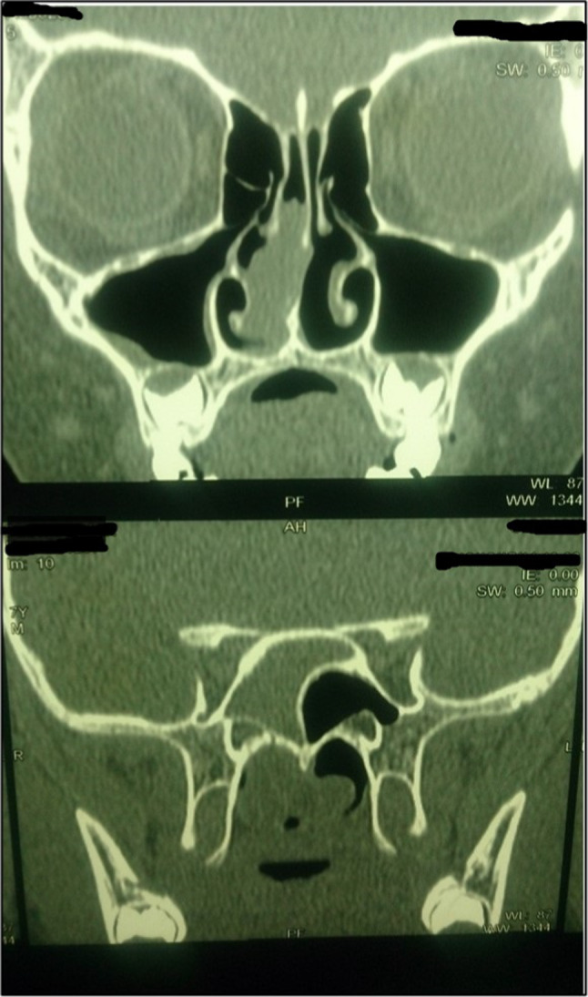
CT images showing soft tissue density involving right sphenoid sinus and choana, without bony erosion
Fig. 3.

Completely excised specimen with nasal, choanal and sphenoidal attachments
Post-operative follow-up at 6 months showed a widened sphenoid ostium with normal sinus mucosa, without any recurrence.
Discussion
Sphenochoanal polyp is a rare entity and is mostly seen in the young adults [2]. It arises from an inflamed sinus mucosa and is benign in nature. Most of the reports in literature are of adults with only a few cases reported in the paediatric age group [3–5].
The youngest reported case is that of an incidentally detected sphenochoanal polyp in a 3 year 8 month old child during adenotonsillectomy, which was later found to regress naturally [6]. Others have reported symptomatic sphenochoanal polyps in slightly older children [7–9].
Thorough ENT examination is mandatory, which helps in establishing the site of origin. In some cases, endoscopy alone may not give adequate information – especially in case of a large polyp or in children who may not allow endoscopy in the clinic. In such cases, imaging plays an important role to find the site of origin, anatomical variations and to rule out differentials [9]. The most common differentials include antrochoanal polyp, hypophyseal tumor, granulomatous lesion in adults and meningoencephalocele in a child. CT scan of the sinuses can provide adequate information on the sinus involved, erosion of sinus walls or skull base which can be quite helpful in ruling out differentials.
Surgery is the mainstay of treatment and should aim at complete removal of polyp including the nasal, choanal and sinus components especially the mucosal attachment and widening the sinus ostium—to avoid recurrence. One must also keep in mind the important structures in the vicinity of the sinus [10] to avoid catastrophic effects.
Conclusion
Sphenochoanal polyp, although a rare entity, must be kept in mind as inaccurate diagnosis can lead to incomplete excision of the polyp, persistence of symptoms and disease recurrence.
Funding
No funding was needed in preparation of this Case report.
Compliance with Ethical Standards
Conflict of interest
No conflict of Interest to be declared.
Ethical Approval
Consent taken from the Institute for publishing photographs and writing the case report.
Footnotes
Publisher's Note
Springer Nature remains neutral with regard to jurisdictional claims in published maps and institutional affiliations.
References
- 1.Frosini P, Picarella G, De Campora E. Antrochoanal polyp: analysis of 200 cases. Acta Otorhinolaryngol Ital. 2009;29(1):21–26. [PMC free article] [PubMed] [Google Scholar]
- 2.Al-Qudah MA. Sphenochoanal polyp: current diagnosis and management. Ear Nose Throat J. 2010;89:311–317. [PubMed] [Google Scholar]
- 3.Jadia S, Goyal R, Biswas R. Nasal mass mimicking antrochoanal polyp. BMJ Case Rep. 2010 doi: 10.1136/bcr.12.2009.2578. [DOI] [PMC free article] [PubMed] [Google Scholar]
- 4.Bist SS, Kumar R, Varshney S, Bisht M. Isolated sphenochoanal polyp: a rare clinical entity. Indian J Otolaryngol Head Neck Surg. 2007;59(1):56–57. doi: 10.1007/s12070-007-0016-3. [DOI] [PMC free article] [PubMed] [Google Scholar]
- 5.Dabholkar JP, Nair DR, Sharma A. Sphenochoanal polyp: a rare diagnosis in nasal obstruction. Indian J Otolaryngol Head Neck Surg. 2008;60(3):271–273. doi: 10.1007/s12070-008-0091-0. [DOI] [PMC free article] [PubMed] [Google Scholar]
- 6.Lim WK, Sdralis T. Regression of a sphenochoanal polyp in a child. Laryngoscope. 2004;114(5):903–905. doi: 10.1097/00005537-200405000-00022. [DOI] [PubMed] [Google Scholar]
- 7.Çeçen A, Kemal O, Atmaca S, Kavaz E. Isolated sphenochoanal polyp: report of three cases. Hippokratia. 2017;21(3):150–153. [PMC free article] [PubMed] [Google Scholar]
- 8.Tosun F, Yetiser S, Akcam T, Ozkaptan Y. Sphenochoanal polyp: endoscopic surgery. Int J Pediatr Otorhinolaryngol. 2001;58(1):87–90. doi: 10.1016/s0165-5876(00)00468-7. [DOI] [PubMed] [Google Scholar]
- 9.Lessa MM, Voegels RL, Pádua F, Wiikmann C, Romano FR, Butugan O. Sphenochoanal polyp: diagnose and treatment. Rhinology. 2002;40(4):215–216. [PubMed] [Google Scholar]
- 10.Kumral TL, Yildirim G, Uyar Y. Sphenochoanal polyps and the optic nerve. Clin Pract. 2012;2(1):e10. doi: 10.4081/cp.2012.e10. [DOI] [PMC free article] [PubMed] [Google Scholar]