

Abstract
Tuberculosis is the most common infectious disease in the world and the lingual location represents less than 1% of extra-pulmonary forms. We report a case of primary lingual tuberculosis which was presented to us in the form of a cold abscess in a 46-year-old alcohol and tobacco drinker weaned for 01 years. Confirmation was histological after biopsy of the lesion. Treatment with anti-tuberculosis drugs for 06 months resulted in a cure. Primary tuberculosis of the tongue is rare and can take several macroscopic forms, including cold abscess which is exceptional. A histological examination after biopsy will make the diagnosis.
Keywords: Tuberculosis, Cold abscess, Tongue, Oral cavity
Introduction
Tuberculosis is the most common infectious disease in the world, with an incidence rate that keeps soaring. This is presumably due to its connection with the AIDS pandemic and the development of a strain of multi-resistant Mycobacterium tuberculosis [1].
Tuberculosis of the oral cavity represents less than 1% of extra-pulmonary locations [2]. Despite the fact that Oral Tuberculosis may be primary, affecting any organ, it is most frequently secondary to a primary lung focus, through contaminated sputum or haematogenous spread. Although it is normally considered rare, the lingual location is the one that is most frequently found [3].
Generally, it appears as a unilateral lingual ulceration; its presentation in a cold abscess form is unusual. Biopsy followed by histological examination allows a diagnosis to be made.
We report an exceptional case of cold tubercular abscess of the tongue.
Observation
A 46-year-old man that weaned himself off alcohol and cigarettes 1 year ago was admitted to our department for glossodynia, localized swelling of the tongue, upper cervical swelling and unstated weight loss. The onset of the symptoms went back to 2.5 months, 2 weeks after an accidental bite of the tongue. It began with a painful lingual swelling associated with an upper cervical swelling increasing progressively in volume. We specify that the patient has been convicted throughout 14 months.
We did not record any notion of tuberculosis contagion or evening–night fever. No antituberculosis-vaccine was carried out.
Through examination, we found swelling of the mobile tongue (Fig. 1) at the level of its anterior third covered with a healthy mucosa, about 1.5 cm long, well limited and slightly sensitive as well as a submental lymphadenopathy of about 2.5 cm, which is soft, mobile and inflammatory.
Fig. 1.

Swelling of the mobile tongue at the level of its anterior third covered with a healthy mucosa, about 1.50 cm long, well limited
We did not find any peculiarities or abnormalities afterwards.
The MRI (Fig. 2) made it possible to objectify a small abscess collected of 11 × 13 mm located on the anteromedial part of the movable tongue as well as a submental lymphadenopathy of 2.2 cm long axis.
Fig. 2.
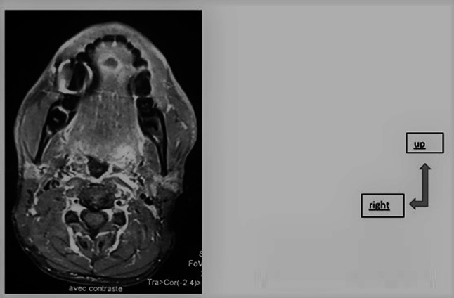
Axial MRI showing the lingual abcess
The result of fine-needle aspiration of the lymphadenopathy and lingual mass showed an aspect in favor of a necrotizing granulomatous lesion suggestive of tuberculosis.
Given the strong suspicion of tuberculosis, a biopsy was performed on the tongue, resulting in lingual tuberculosis.
A tuberculosis assessment was subsequently carried out including a chest X-ray that reported normal, a retroviral serology resulting negative, and an intradermal reaction to tuberculin returned positive at 15 mm.
The diagnosis of primary lingual cold tuberculous abscess was retained and anti-tuberculosis treatment was started according to the following scheme: 02 months of RHEZ (rifampicin, isoniazid, ethambutol, and pyrazinamide) then 04 months of RH (rifampicin and isoniazid). The evolution was favorable. Five months into the treatment, we noticed the disappearance of adenopathy and lingual swelling. After six months of follow-up, there was no sign of relapse.
Discussion
Lingual tuberculosis is secondary to infection with Mycobacterium tuberculosis and much more rarely Mycobacterium bovis [4].
In the literature, oral tuberculosis sites represent 0.05–05%. They are most often secondary and affect the elderly. The primitive forms are unusual and affect the young subject [5].
When it is primary, as in our case, transmission is direct by respiratory droplets or after ingestion of contaminated unpasteurized milk [1, 2].
The lining of the oral cavity constitutes a natural barrier to the invasion of the Mycobacterium [5]. Indeed, the cleaning action of saliva, the presence of salivary enzymes and tissue antibodies, the local saprophytic flora and the thickness of the epithelial wall are several factors opposing contamination [5, 6]. Nevertheless, poor oral health, inflammation, tooth extraction or trauma (as in the case of our patient), may constitute a gateway to the disease [5]. Low socio-economic status, smoking, poor hygiene and overcrowding are also other risk factors of the disease [2, 4, 7].
The average age found in the literature is 45, with a clear predominance of men [7].
The reasons for consultation are diverse and nonspecific; they may be glossodynia, odynophagia or even dysphagia [3, 7].
Alteration of general condition is quite common due to anorexia and weight loss caused by lesions in the mouth [1].
On examination, the tuberculous lesion can take several forms including ulcerations, tuberculoma or fissure [1, 4]. In our patient the disease presented itself as a cold abscess which is an exceptional and historical form, often secondary to the rupture of a tuberculoma [4, 8].
Submental or submandibular lymphadenopathy can be found in primary forms, as happened to our patient.
In our patient’s case, in addition to benign tumors of the tongue, the differential diagnosis unveiled a neoplastic submucosal lesion, proving once again that lingual squamous carcinoma cell is one of the most frequent cancers of the upper aero-digestive tract [3].
Despite fine needle aspiration is a relevant examination, coming back positive in 40% of cases [7], it does not diminish the interest of a biopsy, which in our opinion is the definitive test to confirm tuberculosis.
Biopsy allows to highlight a gigantocellular epithelioid granuloma with central caseous necrosis on histological examination and tuberculosis bacilli on the longer-lasting bacteriological examination [4].
In our case, the histology made it possible to make a quick diagnosis and start the treatment.
The healing of our patient shows the effectiveness of the anti-tuberculosis treatment over a period of 06 months and the good prognosis of this disease [5, 7]. However, when the treatment is not successful, it would be advisable to consider associating it with carcinoma [7].
Conclusion
Lingual tuberculosis is rare and it commonly appears as unilateral lingual ulceration. The clinical presentation in the form of a cold abscess is rarely reported in the literature. The biopsy followed by an anatomopathological examination allows the diagnosis to be made. The prognosis is generally good with anti-tuberculosis treatment.
Author contributions
HY writer, AD writer, CN writer, AK Writer.
Compliance with ethical standards
Conflict of interest
There is no conflict of interest between the authors.
Patient consent
The patient was consenting to the publication of the article.
Footnotes
Publisher's Note
Springer Nature remains neutral with regard to jurisdictional claims in published maps and institutional affiliations.
Contributor Information
Hussein Younes, Email: toyounes1989@hotmail.com.
Cire Ndiaye, Email: ndiayecire@hotmail.com.
Abdoulaye Dieye, Email: adieye90@gmail.com.
Abdoulaye Keita, Email: akeita66@gmail.com.
References
- 1.d’Elbée J-M, Bernard N, Vandenhende M-A, et al. Ulcération linguale avec dysphagie à l’origine du diagnostic d’une tuberculose pulmonaire. Médecine Buccale Chir Buccale. 2012;18:39–43. doi: 10.1051/mbcb/2011146. [DOI] [Google Scholar]
- 2.Hasan S, Khan MA (2011) Tuberculosis—a common disease with uncommon oral features Report of two cases with a detailed review of literature. Proc World Med Conf 156–166
- 3.Estomba CMC, da Costa ASA, Schmitz TR, Lago PV. Base of tongue tuberculosis: a case report. Iran J Otorhinolaryngol. 2015;27:239. [PMC free article] [PubMed] [Google Scholar]
- 4.Girszyn N, Belmekki A, Duterque M, et al. Tuberculose linguale au cours d’une tuberculose disséminée. Ann Dermatol Vénéréologie. 2005;132:368–369. doi: 10.1016/S0151-9638(05)79285-1. [DOI] [PubMed] [Google Scholar]
- 5.Ito F, Andrade C, Vargas P, et al. Primary tuberculosis of the oral cavity. Oral Dis. 2005;11:50–53. doi: 10.1111/j.1601-0825.2004.01055.x. [DOI] [PubMed] [Google Scholar]
- 6.Bhandarkar PD, Kasbekar VG, Shah RP, Hakim PP. Primary tuberculous ulcer of the tongue. Trop Doct. 1993;23:41–42. doi: 10.1177/004947559302300125. [DOI] [PubMed] [Google Scholar]
- 7.Kakisi OK, Kechagia AS, Kakisis IK, et al. Tuberculosis of the oral cavity: a systematic review. Eur J Oral Sci. 2010;118:103–109. doi: 10.1111/j.1600-0722.2010.00725.x. [DOI] [PubMed] [Google Scholar]
- 8.Yadav SPS, Agrawal A, Gulia JS, et al. Tuberculoma of the tongue presenting as Hemimacroglossia. Case Rep Med. 2012;2012:1–3. doi: 10.1155/2012/548350. [DOI] [PMC free article] [PubMed] [Google Scholar]