

A 38–year–old male refugee camp tenant presented with symptoms of upper respiratory infection. On clinical examination, Vesicular breathing was asymmetric and lower over the base of the left lung. Breath sounds were symmetrical and no adventitious sounds were heard. Vital signs were normal. His white blood cell count was 7.8 × 10⁹ per L. Biochemical analysis yielded no findings. The SARS-CoV-2 rapid antigen test was negative as was the PCR test.
Chest radiography showed a left-sided paracardiac mass, which was not obliterating the cardiac silhouette (Fig. 1A). To characterize the mass, the patient underwent a chest CT scan which revealed a left-sided, thoracic ectopic kidney. There was incomplete external rotation of the pelvicalyceal system and the ureter, and anomalous intrathoracic origin of the left renal vessels (Fig. 1B, C). Additional findings included atelectasis of adjacent lung parenchyma, owing to compression exerted by the ectopic kidney. Part of the spleen was also situated in the chest cavity.
Fig. 1.
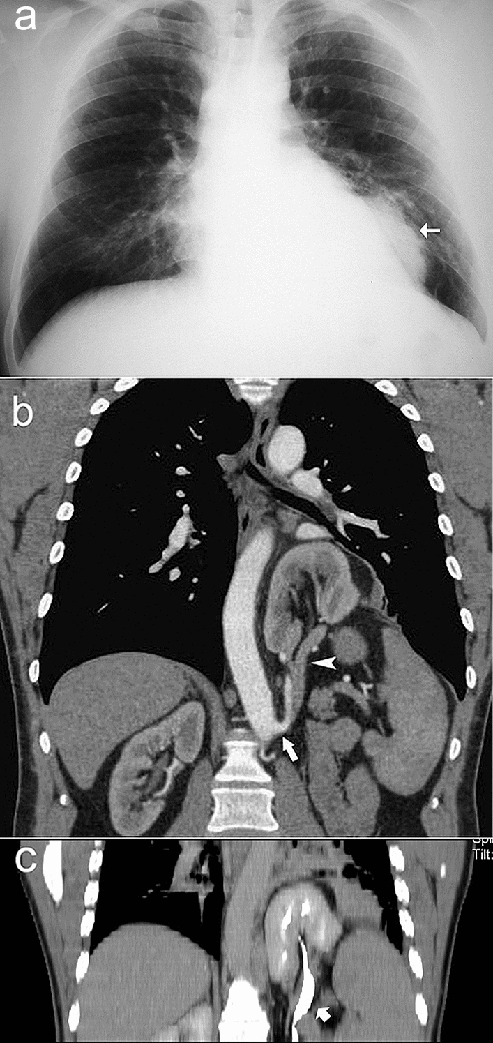
Intrathoracic kidney. A Chest radiograph shows a radiodense mass (arrow), which is not obliterating the cardiac silhouette, suggesting posterior location. B Chest CT scan with intravenous contrast shows left thoracic kidney with anomalous renal vasculature. Note the ascending renal artery branch (arrow) arising from the aorta, and renal vein (arrowhead). C 3D-reconstructed coronal CT image shows left intrathoracic kidney and proximal portion of ureter (thick arrow)
In embryonic life, kidney development occurs in three main steps, the pronephros, the mesonephros, and the metanephros. The metanephros normally ascends from its lower lumbar position to its final position. Renal ectopia most frequently occurs in the pelvis owing to failure of the metanephros to migrate during fetal development. Conversely, thoracic renal ectopia is exceedingly rare, accounting for about 0.0001% of ectopic kidneys [1]. This congenital anomaly is characterized by partial or complete migration of the kidney into the posterior mediastinum, with or without diaphragmatic hernia. Ectopic kidney is seen mostly on the left side, and males are affected more often than women [2]. Because thoracic kidneys are usually asymptomatic, the anomaly is often incidentally discovered on imaging studies and requires no particular treatment. Occasionally, thoracic kidneys may manifest with respiratory, gastrointestinal, or genitourinary symptoms, necessitating surgical correction (with closure of the diaphragmatic defect). Kidney function is usually preserved, as in our patient, who was treated only for his respiratory infection.
On imaging, thoracic kidney may present in association with gross anatomic abnormalities including variable rotation anomaly, elongation of the ureter, high derivation of the renal vessels, and medial deviation of the lower pole of the kidney.
Author contributions
The authors contributed equally to conceptualization, analysis and writing of this manuscript.
Funding
No funding was received to support this work.
Data Availability
The patient agreed to publication of his data and images in an anonymous form.
Declarations
Conflict of interest
Authors declare no conflict of interest.
Ethical statement
All procedures performed in studies involving human participants were in accordance with the ethical standards of the institutional and/or national research committee and with the 1964 Helsinki declaration and its later amendments or comparable ethical standards.
Informed consent
Informed consent was obtained from the patient included in the case report.
Footnotes
Publisher's Note
Springer Nature remains neutral with regard to jurisdictional claims in published maps and institutional affiliations.
References
- 1.Carrasco A, Castro R. Right diaphragmatic eventration with an intrathoracic kidney: case report and review of the literature. Case Rep Surg. 2018 doi: 10.1155/2018/2631391. [DOI] [PMC free article] [PubMed] [Google Scholar]
- 2.Peñafiel-Freire DM, Hernández-Martín S, Iceta-Lizarraga A, et al. Intrathoracic ectopic kidney. Ann Pediatr (Engl Ed) 2020;92(2):113–114. doi: 10.1016/j.anpedi.2019.01.005. [DOI] [PubMed] [Google Scholar]
Associated Data
This section collects any data citations, data availability statements, or supplementary materials included in this article.
Data Availability Statement
The patient agreed to publication of his data and images in an anonymous form.