

Abstract
Background and Aim:
The brain is one of the most complex and crucial organs of our body. Its health is a matter of concern for all individuals as the number of aged people is increasing gradually in the world. Carica papaya is a ubiquitous plant, and its different parts possess neuroprotective effects against various neurodegenerative diseases. However, its brain anti-aging effects have remained uninvestigated. Therefore, this study has examined the brain anti-aging strength of C. papaya pulp and seeds extracts in D-galactose-induced aging rats.
Methods:
The rats were intraperitoneally injected with 150 mg/kg of D-galactose for 8 consecutive weeks to induce brain aging. In parallel, the rats of papaya pulp and papaya seed treated groups were injected with 150 mg/kg papaya pulp extract and 150 mg/kg papaya seed extract, respectively. The negative control group was only injected with 0.9% saline, whereas in the rats of the positive control group along with D-galactose 100 mg/kg VC was injected. After the treatment period, different neurobehavioral, neurochemical, and antioxidant analyses were performed to unmask the anti-aging strength of C. papaya pulp and seeds extracts.
Results:
C. papaya pulp and seed extracts significantly improved cognitive learning skills, memory, and muscular strength in aging rats while reducing stress and anxiety levels. Moreover, they enhanced neurotransmitters concentration and reduced oxidative stress. However, the anti-aging effects of C. papaya pulp were more significant than seeds.
Conclusion:
These results suggest that both C. papaya pulp and seed extracts possess neuroprotective effects against brain aging or age-related brain deteriorations but the age-protecting capability of C. papaya pulp is higher than C. papaya seeds. Therefore, it could be utilized as a component to design a novel brain anti-aging drug.
Relevance for Patients:
Brain aging is a natural process that every individual experiences in his life. The regular consumption of C. papaya can improve the quality of life by protecting neurons from age-related deteriorations.
Keywords: anti-aging, brain, Carica papaya pulp, Carica papaya seeds, d-galactos
1. Introduction
The brain is recognized as one of the largest organs of our body that regulates all functions of life [1]. During the whole lifetime, it helps an individual to make sense of the whole world and understand the complex processes of nature. Its health is a matter of concern for all individuals as a healthy brain is the basic need of a healthy life. Brain aging is considered one of the most important issues in the modern era as the proportion of aged people is increasing gradually in the world. Pursuant to the United Nations World Population survey (conducted in 2015), it is predicted that the ratio of aged people will almost get double from 12% to 22% in 2050 [2]. Hence, there is an imminent need to unmask the anti-aging properties of natural phytochemicals so that the quality of life can be made better.
Medicinal plants are the inexplicable gift of nature. Historically, plants have been utilized to improve various health conditions. Especially, dietetic vegetables and fruits are viewed to perform a crucial function in the prevention and reduction of numerous ailments as they contain a wide set of phytochemicals, vitamins, and minerals [3]. For instance, a number of plant-based products are found to exert remarkable neuroprotective effects against different neurodegenerative diseases, that is, Parkinson’s disease, Alzheimer’s disease, epilepsy, and Huntington’s disease [4,5]. Similarly, their regular consumption lowers the risk of many health ailments, including heart diseases and cancers, which are ranked as the two major causes of the world’s mortality [6].
Carica papaya is one of such miraculous gifts of nature that remain available all year round. It has been widely found in both tropical and subtropical countries of the world [7]. Its broad distribution is mainly attributed to the longevity and profusion of its seeds. Conventionally, it has been used as an important constituent in folk medicine and with the passage of time more of its health benefits have been unveiled. In light of recent literature, it is evident that C. papaya possesses neuroprotective potential, as it can protect the brain from non-alcoholic steatohepatitis, stress-induced gastric mucosal lesion, Alzheimer’s disease, and traumatic epilepsy [8]. Banala and companions reported that papaya leaf extract could diminish fluoride-induced neurodegeneration in rat’s brain by inhibiting reactive oxygen species generation and microglial activation [9]. In the same fashion, fermented papaya preparation (FPP) was found to protect the brain from oxidative stress by activating Nrf2 to produce antioxidants and detoxification molecules from astrocytes [8]. It also caused reduction of epileptogenic monoamine secretion from neurons to inhibit epilepsy in in vivo model. Similarly, in the ischemia-reperfusion model, FPP maintained oxidative balance in the brain [10]. Lycopene, an important constituent of C. papaya, has been found to exert prophylactic role in the central nervous system, as it can protect the brain from different ailments such as Huntington’s disease, ischemia, epilepsy, Parkinson’s disease, depression, and Alzheimer’s disease [4]. It is also reported to improve the memory and cognitive ability of rats in aging, diabetes mellitus, fat-rich diet, and colchicine subjection [4]. Despite having tremendous neuroprotective effects, the brain anti-aging effects of its pulp and seeds have remained uninvestigated.
Keeping in view all these factors, in this study, the brain anti-aging potential of C. papaya pulp and seeds has been unveiled by studying different neurobehavioral and neurochemical alterations, and antioxidant enzymes level. Moreover, the anti-aging strength of C. papaya pulp and seeds has also been compared with an aim to help researchers in designing a potential anti-aging drug.
2. Materials and Methods
2.1. Chemicals
All chemical reagents exploited in the current experimental study were of analytical grade, and all solutions were freshly prepared before starting the experiments. D-galactose and Vitamin C (VC) (L-ascorbic acid) were ordered from the Sigma-Aldrich (USA). The ELISA kits used for the detection of acetylcholine (ACh) and acetylcholinesterase (AChE) were bought from Novus Biologicals (Centennial, USA), gamma-aminobutyric acid (GABA) ELISA kit was manufactured by MyBioScience (San Diego, CA, USA). Superoxide dismutase (SOD), glutathione peroxidase (GPX), and catalase (CAT) colorimetric assay kits were purchased from BioVision (US).
2.2. Papaya pulp extract (PPE) and papaya seed extract (PSE) preparation
Completely matured ripened papaya fruits were purchased from the local market. After washing, it was peeled off, and the pulp was divided into small pieces. It was then homogenized in distilled water followed by drying at 37°C water bath for 8 h. Afterward, centrifugation was carried out at 27°C for 20 min at 200 g. Then, the supernatant was carefully removed and finally lyophilized [11]. Similarly, for the preparation of PSE, the seeds were removed from papaya, followed by washing, drying, and crushing. These were then kept in distilled water for extraction at 70°C for 3 days. After filtration, it was finally lyophilized, which produced a bright brown color powder [12].
2.3. Animals and treatment
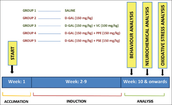
Thirty albino Wistar rats (age: 3 – 4 months) weighing 140 – 180 g were utilized for this experimental study. All of the rats were placed in separate cages to reduce the effects of social connection in a balanced and maintained room temperature (22 ± 2°C) along with a 12:12 h dark and light cycle. All rodents were handled properly according to guidelines of the National Institute of Health Guide for Care and Use of Laboratory Animals [13], and the experimental protocols were approved by the Institutional Ethics Review Board (JUW/IERB/SCI-ARA-011/2022). After 7 days of the acclimation period, the rats were randomly distributed into five groups (n = 6 for each group): (1) Negative control (normal saline treated) group, (2) D-galactose-treated group, (3) positive control (D-galactose and VC treated) group, (4) D-galactose- and PPE-treated group, and (5) D-galactose- and PSE-treated group. The rats of Group 1 were intraperitoneally injected with 0.9% saline, while the rats of Groups 2, 3, 4, and 5 were intraperitoneally injected with 150 mg/kg D-galactose [14], 150 mg/kg D-galactose plus 100 mg/kg VC [15], 150 mg/kg D-galactose plus 150 mg/kg PPE, and 150 mg/kg D-galactose plus 150 mg/kg PSE for 8 consecutive weeks, respectively (Figure 1). The concentrations of PPE and PSE were set as the same as the concentration of D-galactose.
Figure 1. A brief summary of the experimental design. After the acclimation period, the rats were randomly divided into 5 groups. The rats of group 1 were used as negative control whereas group 3 was used as positive control.
2.4. Behavior analysis
2.4.1. Food intake and body weight
The body weight and food consumption of the rats were monitored every week. For measuring the food intake, rats were provided with a measured quantity of a balanced diet, and the leftover food was measured at subsequent servings.
2.4.2. Morris water maze (MWM) test
MWM test was conducted to assess spatial memory and learning skills of aging rats. The apparatus was circular in shape, having 37 cm in height and 45 cm in diameter, while water (23 ± 2°C) was poured at an altitude of 12 cm. The tank was divided into four quadrants, that is, north, east, south, and west. The escape platform contained a flat metallic top with a diameter of 8 cm, which was kept 2 cm underneath the water’s surface in the north-west quadrant. Moreover, powdered milk was added to the water to make the platform invisible. This experiment was comprised three sessions: Training, learning, and testing. The cutoff time for the initial two sessions was 120 s and for the testing session (probe trial) was 60 s. In this test, the spatial memory parameters (latency time to reach the platform location, number of crossings made the over target quadrant, and time spent in the target quadrant) were all recorded. After testing each rat, the equipment was cleaned with 70% ethanol to remove urine or fecal boli.
2.4.3. Forced swim test (FST)
This test was performed to assess the physical power, fatigue, and endurance of rats [16]. The assessment involved placing rats separately in a transparent glass container (width 20 cm, height 56 cm), which was filled with clean tap water to a level of around 22 cm (25 ± 2°C), for about 6 min [17]. The water altitude was selected to prevent the rat from reaching the base of the container, while simultaneously avoiding escape from the equipment. The rat was regarded as immobile when it stopped struggling to swim or escape. In the last 4 min of the test, immobility time was recorded when the rat made no more efforts to escape or to keep its head above the surface of the water. After testing each rat, the equipment was cleaned with 70% ethanol to remove urine or fecal boli.
2.4.4. Elevated plus maze (EPM) test
EPM test was used to elucidate anxiety in rats, and the method followed was the same as affirmed previously [18]. The equipment was of plus sign shape that contained four arms of equivalent length and width (length 50 cm and width 10 cm), while the altitude of the arms from the ground was about 60 cm. In the equipment, two contradictory arms were without walls, that is, open, while the remaining two parallel were closed, that is, having walls attached to each other by the middle region of 5 cm2. The walls of the closed arm were 40 cm in length. The compound’s anxiolytic effect was observed by placing the rat in the middle region of the equipment in front of the open arm for around 5 min. In this experiment, the time spent by the rat in open and closed arms was calculated. After testing each rat, the equipment was cleaned with 70% ethanol to remove urine or fecal boli.
2.4.5. Kondziela’s inverted screen test (KIST)
It was used to measure muscle strength in rats [19]. For this, the rats were placed separately on the top of a square mashed wire screen which was rotated at 180°. The total time of the test was 2 min. Rats were positioned in the middle of the wire mesh, and then, it was inverted. The time elapsed when the rat falls off from the inverted screen was noted on the arbitrary scale of 1-5 (falling during 1–10 s = 1, 10–25 s = 2, 25–60 s = 3, 60–90 s = 4, and more than 90 s = 5) [20]. After testing each rat, the equipment was cleaned with 70% ethanol to remove urine or fecal boli.
2.4.6. Decapitation and organ index
After behavioral analysis, the rats were anesthetized by intraperitoneal injection of 0.1% sodium pentobarbital [21]. The rats were then decapitated using guillotine apparatus and the skull was opened within 1 min to take the brain out. It was then immediately immersed in ice-cold PBS. The brain was then microdissected to obtain the hippocampus and cortex for further analysis. These regions were stored at −2°C until processed. Moreover, other organs such as the liver, spleen, kidneys, and heart were also taken out for organ indexing. The organ coefficient was measured by dividing the organ weights by the body weight of the rats.
2.4.7. Neurochemical analysis
For neurochemical analysis, the ACh, AChE activity, and GABA concentration were evaluated in the hippocampus and cortex of the brain using a commercially available ELISA kit. Briefly, the tissues were homogenized and then centrifuged at 12,000 rpm for 30 min at 4°C, followed by the collection of the supernatant and determination of the ACh content, AChE activity, and GABA level in accordance with the manufacturer’s instructions.
2.4.8. Oxidative stress analysis
The activities of SOD, GPX, and CAT were determined using commercially available kits. In short, the activity of SOD was calculated on the basis of its capability to form a red-colored product by decreasing the oxidation of hydroxylamine. The GPX activity was determined by a yellowish-colored complex, produced by the reaction between the reduced glutathione and dinitrobenzoic acid. Similarly, the CAT activity was elucidated by measuring a yellow-colored product generated by the reaction between ammonium molybdate and residual hydrogen peroxide. The enzyme activities were represented as U/mg (unit/mg tissue protein) of brain tissue.
2.5. Statistical analysis
The data are presented as mean ± S.D. The results of neurobehavioral, neurochemical, and oxidative stress analysis were examined by one-way analysis of variance for every parameter following Tukey’s post hoc test to find comparison within groups. P < 0.05 or P < 0.01 was considered statistically significant results.
3. Results
3.1. PPE and PSE restored the body weight of rats decreased by D-galactose
The impact of D-galactose induction on the body weights of the rats is represented in Figure 2A. It is evident from the figure that after 6 weeks, rats induced with D-galactose experienced a considerable (P < 0.01) decrease in weights as compared to control. PPE and PSE remarkably reversed the effect of D-galactose by restoring weight loss. Furthermore, the weight-restoring potency of both PPE and PSE was almost similar (Figure 2A). Similarly, there was a notable difference in the amount of food intake between D-galactose-induced rats and control rats (Table 1) (P < 0.01), which may lead to weight loss. PPE and PSE recovered this behavior, and therefore, the weight of the rats was recovered.
Figure 2. Effect of PPE and PSE on the body weight (A) and organ coefficient (B) of aging rats. For all experiments, results are represented as mean ± SD (n = 6 for each group). Significant alterations are expressed as **P < 0.01 related to control group. #P<0.05 and ##P < 0.01 related to D-galactose group.

Table 1. Effect of D-galactose induction and C. papaya pulp and seed administration on the food intake of rats.
| Groups | 1 | 2 | 3 | 4 | 5 |
|---|---|---|---|---|---|
|
|
|
|
|
|
|
| (Control) | (D-galactose)** | (D-galactose+ VC)## | (D-galactose+ PPE)## | (D-galactose+ PSE)## | |
| Week 1 | 16.23±1.03 | 13.53±1.03 | 15.93±0.79 | 16.05±0.78 | 15.81±0.96 |
| Week 2 | 17.3±0.94 | 10.53±0.72 | 16.73±0.77 | 16.75±0.75 | 16.56±0.82 |
| Week 3 | 17.75±0.94 | 12±0.66 | 17.33±0.76 | 17.41±0.67 | 17.01±1.03 |
| Week 4 | 18.21±0.96 | 9.48±0.78 | 17.88±0.79 | 17.85±0.75 | 17.5±0.9 |
| Week 5 | 18.61±1.02 | 11.9±0.75 | 18.31±0.85 | 18.16±0.87 | 17.66±1.09 |
| Week 6 | 19.26±1.06 | 12.26±0.83 | 18.55±0.87 | 18.56±0.79 | 18.31±1.04 |
| Week 7 | 19.86±1.26 | 10.68±0.84 | 18.91±1.08 | 18.98±0.8 | 19.21±1.52 |
| Week 8 | 20.33±1.61 | 11.01±0.89 | 19.61±1.58 | 19.46±1.24 | 19.75±1.5 |
n = 6 for each group.
P < 0.01 related to control group.
P < 0.01 related with D-galactose group. C. papaya: Carica papaya
3.2. PPE and PSE improved the organ coefficient of D-galactose-induced rats
After 8 weeks of D-galactose-induced aging, a slight decrease in the size of the brain, spleen, and liver was observed as compared to the negative control. Whereas, the sizes of both kidneys and heart remained unaffected. It is evident from Figure 2B that PPE and PSE attenuated the effect of D-galactose. However, in this experiment, the efficacy of PPE was higher than PSE.
3.3. PPE and PSE ameliorated spatial memory of D-galactose-induced rats in MWM test
In this study, the MWM test was conducted to elucidate the cognitive ability and spatial memory of the rats. Analysis of escape latency data revealed that in all groups, the mean latency to locate the platform was gradually reduced with the passage of trails (Figure 3A). Nevertheless, D-galactose-treated rats took more time to find the platform in comparison to the control (P < 0.01). It indicates that D-galactose causes impairment of spatial memory in rats, and thus, rats took more time to reach the target quadrant. Similarly, the number of crossings over the targeted quadrant and the duration of time spent in the target quadrant was also significantly reduced (P < 0.01) on D-galactose treatment related to the other groups (Figure 3B and C). On the contrary, concurrent administration with PPE and PSE, this damaging effect was remarkably ameliorated as in Groups 4 and 5, no. of crossing over platform position and time spent in the platform quadrant were remarkably increased (Figure 3B and C). On comparing PPE with PSE, it was found that PPE was more efficient (P < 0.01) in recovering D-galactose-induced age-related memory deficits than PSE.
Figure 3. Effects of PPE and PSE on age-associated behavior alterations. PPE and PSE improved spatial memory and learning in MWM test by decreasing the escape latency (A), increasing the duration of time spent in the target quadrant (B), and the number of crossing the target quadrant (C). PPE and PSE reduced age-related stress in FST (D), anxiety in EPM test (E), and fatigue in KIST (F). For all experiments, results are represented as mean ± SD (n = 6 for each group). Significant alterations are expressed as **P < 0.01 related to control group. P < 0.05 and ##P < 0.01 related to D-galactose group. ++P<0.01 related to PPE group.

3.4. PPE and PSE protected D-galactose-induced rats from stress in FST
FST was performed to determine stress-like behavior in D-galactose induced rats. In rats, stress-like behavior is described as the cessation of continuously escape-directed attitude, when placed in an inescapable water-filled container. As shown in Figure 3D, D-galactose-treated rats spent more time at rest as compared to control (P < 0.01), which indicates the presence of stress in this group of rats. Treatment with PPE and PSE reduced this resting period of aged rats by enhancing their efforts to escape out of the water, but PPE remained more significant than PSE (P < 0.01). Moreover, its efficacy was almost similar to positive control. Hence, PPE can significantly improve aging-associated depression-like symptoms in rats.
3.5. PPE and PSE decreased the anxiety-like behavior of D-galactose-induced rats in EPM test
Anxiety disorders are closely linked with brain aging. Aged people experience more anxiety and thus avoid exploring novel and anxious environment compared to healthy individuals. Similar results were obtained after treating rats with D-galactose in the EPM test. The results of the EPM test are represented in Figure 3E, which clearly shows that D-galactose-induced rats avoided the open arm as they spent more time in the closed arm than rats of the control group (P < 0.01). This open-arm avoidance was suppressed by PPE, and PSE treatment as rats of these two groups spent their time exploring a new environment with almost similar fear as control. Furthermore, in this test, the efficacy of PSE was significantly (P < 0.01) lower than PPE and positive control. Furthermore, the effect of PPE was similar to positive control. Hence, PPE is more effective to cure age-related anxiety than PSE.
3.6. PPE and PSE enhanced muscular strength of D-galactose-induced rats in KIST
Physical power decline and tiredness are common symptoms of aging. KIST was used to determine and compare PPE and PSE capability to increase muscular strength and decrease fatigue-like symptoms in D-galactose-induced rats. Usually, healthy rats score maximum in this test. Following treatment with D-galactose, the muscular strength of the rats was significantly reduced as they fell down earlier than the control (P < 0.01) (Figure 3F). When rats were administered with PPE and PSE, the age-related physical decline was significantly (P < 0.01) improved as the rats of these groups spent a long time to fall down than the D-galactose-induced group, whereas no significant difference was observed between the activities of the PPE-treated group and the PSE group (Figure 3F). Therefore, it can be stated that both PPE and PSE can improve physical endurance and physical activities in aged people.
3.7. PPE and PSE protected the cholinergic system of D-galactose-induced rats
According to Figure 4A-D, it is evident that D-galactose damaged the cholinergic system of the brain by upregulating the activity of AChE while downregulating ACh expression in the cortex and hippocampus regions of the brain. Compared to control, there was a significant (P < 0.01) difference between the expressions of these two important components of the cholinergic system in the D-galactose-treated group. PPE and PSE weakened these damaging effects of D-galactose by decreasing AChE (P < 0.01) and increasing ACh concentration (P < 0.01). Moreover, the effect of PPE was more significant (P < 0.01 for both AChE activity and ACh level) than PSE and positive control (Figure 4A-D) in improving ACh-mediated cognitive impairment.
Figure 4. Effects of PPE and PSE on the neurotransmitters level of aging rats. PPE and PSE improved cholinergic system of aging rats by increasing ACh level and decreasing AChE activity in hippocampus (A and B) and cortex (C and D) of the brain. Likewise, PPE and PSE increased GABA concentration in the hippocampus (E) and cortex (F) of the D-galactose-treated rats. For all experiments, results are represented as mean ± SD (n = 6 for each group). Significant alterations are expressed as **P < 0.01 related to control group. #P < 0.05 and ##P < 0.01 related to D-galactose group. ++P < 0.01 related to PPE group.

3.8. PPE and PSE increased GABA level of D-galactose-induced rats
The level of GABA was measured to detect anxiety-like behavior of rats as it helps to suppress the feelings of anxiety. There was a remarkable reduction in the level of GABA in the hippocampus and cortex regions of D-galactose-treated rats as compared to control (P < 0.01) (Figure 4E and F). However, coadministration of PPE and PSE markedly reversed this aging effect of D-galactose by increasing GABA content in the brain (P < 0.01 for PPE and P < 0.05 for PSE). Moreover, the brain anti-aging effects of PPE were more beneficial than PSE as the level of GABA content increased by PPE was significantly higher than PSE (P < 0.01) and almost similar to positive control.
3.9. PPE and PSE increased the level of antioxidant enzymes in the brains of D-galactose-induced rats
In this study, SOD, GPX, and CAT were used as markers to detect oxidative stress in D-galactose-treated rats. A significant (P < 0.01) reduction was observed in the activity of these enzymes in the hippocampus of D-galactose-treated rats as compared to control rats (Figure 5A-C). These results clearly indicate that the brain aging model was established successfully. Following treatment with PPE and PSE, this damaging effect was relieved, as rats of these groups showed a remarkable increase in the activities of SOD (P < 0.01), GPX (P < 0.01), and CAT (P < 0.01). Moreover, the efficacy of PPE was found to be more significant (P < 0.05 for SOD and P < 0.01 for GPX and CAT) than PSE in protecting the brain from age-induced oxidative destruction.
Figure 5. Effects of PPE and PSE on the brain oxidative stress. PPE and PSE enhanced SOD (A), GPX (B), and CAT (C) activities in aging rats to protect oxidative stress. For all experiments, results are represented as mean ± SD (n = 6 for each group). Significant alterations are expressed as **P < 0.01 related to control group. ##P < 0.01 related to D-galactose group. +P < 0.05 related to PPE and ++P < 0.01 related to PPE group.

4. Discussion
Aging is a definite natural phenomenon that every individual experiences in his life. This is an irreversible process that ultimately leads to the deterioration of tissue or organ. Moreover, it is known as a crucial factor in cognitive decline and brain impairment [22]. Therefore, the identification of anti-aging compounds has become a matter of concern in the medical community [23]. D-galactose is a monosaccharide that is easy to obtain and simple to use. The chronic induction of rodents with D-galactose produces a brain aging model that highly resembles humans’ natural aging process, such as cognitive deficits, reduced anti-oxidase activity, and neurodegeneration. Moreover, it causes accelerated aging, with fewer chances of cancer and a high survival rate for animals [15]. In this study, D-galactose was used successfully to induce brain aging in rats as during the whole experimental period no death among the rats was observed.
VC or ascorbic acid is an essential key micronutrient that the human body cannot synthesize. It is a potent first-line antioxidant that helps to prevent the human body from oxidative stress by removing free radicals. Accumulating evidence suggests that it plays a fundamental role in extending life span by delaying the process of aging or by inhibiting the hallmarks of aging [24]. Furthermore, it is evident that it protects the brain from cognitive decline and neurodegenerative diseases [24,25]. Due to its higher ratio of oxidative metabolism, the brain needs a higher concentration of VC compared with other organs of the body [26]. Its deficiency in the brain leads to vascular dysfunction, memory impairment, and decreased angiogenesis [25]. Considering these factors, in this study, VC was used as a positive control to elucidate and contrast the brain anti-aging potential of PPE and PSE.
Body weight determination and organ indexing are two simple, commonly used parameters to elucidate D-galactose-induced animal models. After 8 weeks of induction with 150 mg/kg of D-galactose, a significant reduction was observed in the weights and organ sizes of the rats. These findings are in line with the previous findings that D-galactose consecutive administration negatively influences the weight of the rats [15,27]. The brain, heart, kidneys, and spleen are crucial organs of the body. The increased concentration of D-galactose results in glucose metabolism disorder that leads to the improper functioning of these important organs [28]. In this study, similar results have been obtained. However, on treatment with PPE and PSE, these hallmarks of aging were recovered. Hence, papaya pulp and seeds can be used to improve age-associated weight and organ function loss.
Aging is highly associated with behavioral alterations such as increased stress, depression, anxiety, impaired learning, and loss of memory. Therefore, in the current experimental study, various neurobehavioral experiments were conducted to determine the anti-aging potential of PPE and PSE. In the MWM test, spatial memory and cognitive functions were evaluated. The results indicated that coadministration of PPE or PSE with D-galactose decreased the escape latency, increased the duration of time spent in the target quadrant, and the number of crossings over the target quadrant. Thus, it is evident that PPE and PSE can improve age-associated cognitive deficits and the learning ability of rats. Moreover, FST was carried out to unveil the anti-stress effects of PPE and PSE. The results demonstrated that both of these possess the potential to overcome age-related stress in rats. In normal conditions, none or little anxiety could be produced when animals investigate the new environment. However, more anxiety could be produced when the animals are aging during the same exploration process [29]. In the EPM test, D-galactose-treated rats showed enhanced anxiety with less willingness to explore the opened arm path, whereas, in the same experiment, PPE and PSE administrated rats spent more time in open arms, suggesting their potential to reduce age-related anxiety. Physical activity decline and accelerated levels of fatigue are common symptoms of aging. Similar results have been obtained in KIST as D-galactose-treated rats showed less muscular strength and fell down earlier. The present study represents that PPE and PSE effectively attenuated the effect of D-galactose by increasing the falling latency of rats. Overall, in all behavioral assessments, PPE, PSE, and VC proved to be effective to ameliorate brain aging effects of D-galactose, but the efficacy of PPE was remarkably higher than PSE and VC.
The cholinergic system has a significant impact on modulating learning skills and memory. ACh and AChE are two important components of the cholinergic system [27]. Mounting pieces of evidence suggest that the D-galactose induction in rats impairs this system by reducing the concentration of ACh while increasing AChE activity [27,30]. The increased level of AChE leads to the degradation of ACh, which, in turn, leads to impaired memory and cognitive functions [30]. PPE and PSE markedly improved the cholinergic system by reducing AChE activity while increasing ACh concentration. These results are in line with the results of the MWM test that PPE and PSE led to improved cognitive skills and memory.
GABA is an amino acid neurotransmitter that possesses an inhibitory role in the brain [31]. When it binds with its receptor, a calming effect is produced that helps to reduce the feelings of stress and anxiety. Growing consensus indicates that the low level of GABA is linked with major depressive disorder [32]. The findings of this study highlight that chronic induction with D-galactose led to the reduced level of GABA in the hippocampus and cortex of the rats’ brain, which is in accordance with previous investigations [31,33]. However, PPE and PSE significantly increased its concentration, suggesting their potential impact on amino acid neurotransmitters of the brain. These results are consistent with the results of the FST and EPM test that PPE and PSE can overcome D-galactose-mediated stress and anxiety.
Brain aging is highly associated with oxidative stress. Being the most active organ of the body, it highly depends on oxygen to produce energy. Among various theories to describe the brain aging mechanism, the free radical theory describes this process best [34]. SOD, GPX, and CAT are regarded as the most critical antioxidant enzymes to protect our bodies from oxidative damage [31]. Therefore, in the present study, these were used as a marker to detect brain aging. The results of this study suggested that on induction with D-galactose, there was enhanced oxidative stress, as indicated by the decreased activities of SOD, GPX, and CAT. These results are in accordance with the previous research that D-galactose treatment leads to oxidative stress in the brain [22,31]. In PPE- and PSE-treated groups, enhanced activities of these enzymes were found, which indicate that PPE and PSE may protect brain aging by inhibiting oxidative stress.
Along with unveiling brain anti-aging effects of PPE and PSE, the present research also compared the anti-aging potential of PPE and PSE. Although both of them possessed the strength to overcome age-associated cognitive deficits and physical power deterioration in the D-galactose-induced model, the efficacy of PPE was significantly greater than PSE and positive control. In a few experiments, such as body weight determination, organ indexing, and KIST, both PPE and PSE harbored almost similar effects, but in most remaining of the experiments, that is, MWM test, FST, EPM test, neurochemical analysis, and oxidative stress determination, the effectiveness of PPE was remarkably higher. Thus, it can be stated that PPE can be potentially used to reduce or delay brain aging effects by modulating neurotransmitters level and increasing the level of antioxidant enzymes. Furthermore, C. papaya pulp can be utilized to develop a promising brain anti-aging medication.
5. Conclusion
The results of this study suggest neuroprotective effects of both C. papaya pulp and seed extracts against brain aging or age-related brain deteriorations. Coadministration of C. papaya pulp or seed extracts along with D-galactose substantially improved age-deteriorated behaviors in albino rats, that is, enhanced learning and cognitive skills, improved memory, reduced depression and anxiety, and increased muscular strength. Along with ameliorating age-impaired behaviors, C. papaya pulp and seeds extract also modulated neurotransmitters level by improving the functioning of the cholinergic system and increasing GABA level in the cortex and hippocampus of the brain. Similarly, C. papaya pulp and seeds extracts attenuated neuron degradation by protecting them from oxidative damage. Although both pulp and seed extracts were proved to be significant in protecting brain aging, the age-protecting capability of C. papaya pulp was much higher than C. papaya seeds in most of the experiments.
Acknowledgments
The authors are thankful to the Jinnah University for Women, Karachi, Pakistan to provide the laboratory facility to conduct this study.
Funding
This work was supported by the Fundamental Research Funds for the Central Universities (buctrc201910), Young Elite Scientists Sponsorship Program by Xinjiang Association for Science and Technology (2021), Beijing-Tianjin-Hebei Basic Research Cooperation Special Project (19JCZDJC65800(Z)), and the Scientific and Technological Research Project of Xinjiang Production and Construction Corps (2022AB022).
Conflicts of Interest
All authors declare no conflicts of interest.
References
- [1].Derbyshire E. Brain Health across the Lifespan:A Systematic Review on the Role of Omega-3 Fatty Acid Supplements. Nutrients. 2018;10:1094. doi: 10.3390/nu10081094. [DOI] [PMC free article] [PubMed] [Google Scholar]
- [2].United Nations. In:World Population Prospects:The 2015 Revision. United States: United Nations; 2015. Department of Economic and Social Affairs, Population Division. [Google Scholar]
- [3].Burlando B, Palmero S, Cornara L. Nutritional and Medicinal Properties of Underexploited Legume Trees from West Africa. Crit Rev Food Sci Nutr. 2019;59:178–88. doi: 10.1080/10408398.2018.1551776. [DOI] [PubMed] [Google Scholar]
- [4].Chen D, Huang C, Chen Z. A Review for the Pharmacological Effect of Lycopene in Central Nervous System Disorders. Biomed Pharmacother. 2019;111:791–801. doi: 10.1016/j.biopha.2018.12.151. [DOI] [PubMed] [Google Scholar]
- [5].Ratheesh G, Tian L, Venugopal JR, Ezhilarasu H, Sadiq A, Fan TP, et al. Role of Medicinal Plants in Neurodegenerative Diseases. Biomanuf Rev. 2017;2:2. [Google Scholar]
- [6].Heron M, Anderson RN. Changes in the Leading Cause of Death:Recent Patterns in Heart Disease and Cancer Mortality. NCHS Data Brief. 2016;254:1–8. [PubMed] [Google Scholar]
- [7].Khadam S, Afzal U, Gul H, Hira S, Satti M, Yaqub A, et al. Phytochemical Screening and Bioactivity Assessment of Leaves and Fruits Extract of Carica Papaya . Pak J Pharm Sci. 2019;32:1941–8. [PubMed] [Google Scholar]
- [8].Murakami S, Miyazaki I, Asanuma M. Neuroprotective Effect of Fermented Papaya Preparation by Activation of nrf2 Pathway in Astrocytes. Nutr Neurosci. 2018;21:176–84. doi: 10.1080/1028415X.2016.1253171. [DOI] [PubMed] [Google Scholar]
- [9].Banala RR, Nagapuri KK, Mohd KP, Reddy MM, Karnati PR. Carica Papaya Leaf Extract as a Neuroprotective Agent against Behavioral and Neurotransmitter Changes in Brain of the Rat Treated with Sodium Fluoride in Pre-and Post-Natal Periods. Pharmacogn Mag. 2018;14:123–31. [Google Scholar]
- [10].Logozzi M, Di Raimo R, Mizzoni D, Fais S. Anti-Aging and Anti-Tumor Effect of FPP®Supplementation. Eur J Transl Myol. 2020;30:8905. doi: 10.4081/ejtm.2019.8905. [DOI] [PMC free article] [PubMed] [Google Scholar]
- [11].Anuar NS, Zahari SS, Taib IA, Rahman MT. Effect of Green and Ripe Carica Papaya Epicarp Extracts on Wound Healing and During Pregnancy. Food Chem Toxicol. 2008;46:2384–9. doi: 10.1016/j.fct.2008.03.025. [DOI] [PubMed] [Google Scholar]
- [12].Ghaffarilaleh V, Fisher D, Henkel R. Carica Papaya Seed Extract Slows Human Sperm. J Ethnopharmacol. 2019;241:111972. doi: 10.1016/j.jep.2019.111972. [DOI] [PubMed] [Google Scholar]
- [13].National Research Councill. Guide for the Care and Use of Laboratory Animals. 8th ed. Washington, DC: The National Academies Press; 2011. [Google Scholar]
- [14].Banji OJ, Banji D, Ch K. Curcumin and Hesperidin Improve Cognition by Suppressing Mitochondrial Dysfunction and Apoptosis Induced by D-Galactose in Rat Brain. Food Chem Toxicol. 2014;74:51–9. doi: 10.1016/j.fct.2014.08.020. [DOI] [PubMed] [Google Scholar]
- [15].Yuan S, Yang Y, Li J, Tan X, Cao Y, Li S, et al. Ganoderma Lucidum Rhodiola Compound Preparation Prevent D-Galactose-Induced Immune Impairment and Oxidative Stress in Aging Rat Model. Sci Rep. 2020;10:19244. doi: 10.1038/s41598-020-76249-1. [DOI] [PMC free article] [PubMed] [Google Scholar]
- [16].Fatemi I, Khaluoi A, Kaeidi A, Shamsizadeh A, Heydari S, Allahtavakoli MA. Protective Effect of Metformin on D-Galactose-Induced Aging Model in Mice. Iran J Basic Med Sci. 2018;21:19–25. doi: 10.22038/IJBMS.2017.24331.6071. [DOI] [PMC free article] [PubMed] [Google Scholar]
- [17].Liu R, Fu Z, Zhang F, Mao Q, Luan C, Han X, et al. Effect of Yellow Rice Wine on Anti-Aging Ability in Aged Mice Induced by D-Galactose. Food Sci Hum Wellness. 2020;9:184–91. [Google Scholar]
- [18].Naqvi F, Haider S, Batool Z, Perveen T, Haleem DJ. Sub-Chronic Exposure to Noise Affects Locomotor Activity and Produces Anxiogenic and Depressive Like Behavior in Rats. Pharmacol Rep. 2012;64:64–9. doi: 10.1016/s1734-1140(12)70731-4. [DOI] [PubMed] [Google Scholar]
- [19].Kondziella W. A New Method for the Measurement of Muscle Relaxation in White Mice. Arch Int Pharmacodyn Ther. 1964;152:277–84. [PubMed] [Google Scholar]
- [20].Tabassum S, Haider S, Ahmad S, Madiha S, Parveen T. Chronic Choline Supplementation Improves Cognitive and Motor Performance via Modulating Oxidative and Neurochemical Status in Rats. Pharmacol Biochem Behav. 2017;159:90–9. doi: 10.1016/j.pbb.2017.05.011. [DOI] [PubMed] [Google Scholar]
- [21].Lu Z, Yang T, Wang L, Qiu Q, Zhao Y, Wu A, et al. Comparison of Different Protocols of Morris Water Maze in Cognitive Impairment with Heart Failure. Brain Behav. 2020;10:e01519. doi: 10.1002/brb3.1519. [DOI] [PMC free article] [PubMed] [Google Scholar]
- [22].Ma J, Wang H, Liu B, Shan Y, Zhou H, Qi X, et al. Combination of Chick Embryo and Nutrient Mixture Prevent D-Galactose-Induced Cognitive Deficits, Immune Impairment and Oxidative Stress in Aging Rat Model. Sci Rep. 2019;9:4092. doi: 10.1038/s41598-019-40953-4. [DOI] [PMC free article] [PubMed] [Google Scholar]
- [23].Sun K, Yang P, Zhao R, Bai Y, Guo Z. Matrine Attenuates D-Galactose-Induced Aging-Related Behavior in Mice via Inhibition of Cellular Senescence and Oxidative Stress. Oxid Med Cell Longev. 2018;2018:7108604. doi: 10.1155/2018/7108604. [DOI] [PMC free article] [PubMed] [Google Scholar]
- [24].Monacelli F, Acquarone E, Giannotti C, Borghi R, Nencioni A. Vitamin C, Aging and Alzheimer's Disease. Nutrients. 2017;9:670. doi: 10.3390/nu9070670. [DOI] [PMC free article] [PubMed] [Google Scholar]
- [25].Travica N, Ried K, Sali A, Scholey A, Hudson I, Pipingas A. Vitamin C Status and Cognitive Function:A Systematic Review. Nutrients. 2017;9:960. doi: 10.3390/nu9090960. [DOI] [PMC free article] [PubMed] [Google Scholar]
- [26].Dixit S, Bernardo A, Walker JM, Kennard JA, Kim GY, Kessler ES, et al. Vitamin C Deficiency in the Brain Impairs Cognition, Increases Amyloid Accumulation and Deposition, and Oxidative Stress in APP/PSEN1 and Normally Aging Mice. ACS Chem Neurosci. 2015;6:570–81. doi: 10.1021/cn500308h. [DOI] [PMC free article] [PubMed] [Google Scholar]
- [27].Zhou Y, Zhao F, Gao L, Du G, Zhang X, Qin X. Licorice Extract Attenuates Brain Aging of D-Galactose Induced Rats through Inhibition of Oxidative Stress and Attenuation of Neuronal Apoptosis. RSC Adv. 2017;7:47758–66. [Google Scholar]
- [28].Tang T, He B. Treatment of D-Galactose Induced Mouse Aging with Lycium Barbarum Polysaccharides and its Mechanism Study. Afr J Tradit Complement Altern Med. 2013;10:12–7. doi: 10.4314/ajtcam.v10i4.3. [DOI] [PMC free article] [PubMed] [Google Scholar]
- [29].Perna G, Iannone G, Alciati A, Caldirola D. Are Anxiety Disorders Associated with Accelerated Aging?A Focus on Neuroprogression. Neural Plastic. 2016;2016:8457612. doi: 10.1155/2016/8457612. [DOI] [PMC free article] [PubMed] [Google Scholar]
- [30].Liu J, Chen D, Wang Z, Chen C, Ning D, Zhao S. Protective Effect of Walnut on D-Galactose-Induced Aging Mouse Model. Food Sci Nutr. 2019;7:969–76. doi: 10.1002/fsn3.907. [DOI] [PMC free article] [PubMed] [Google Scholar]
- [31].Deng S, Lu H, Chi H, Wang Y, Li X, Ye H. Neuroprotective Effects of OMO within the Hippocampus and Cortex in a D-Galactose and Aβ25-35 Induced Rat Model of Alzheimer's Disease. Evid Based Complement Alternat Med. 2020;2020:1067541. doi: 10.1155/2020/1067541. [DOI] [PMC free article] [PubMed] [Google Scholar]
- [32].Fogaça MV, Duman RS. Cortical GABAergic Dysfunction in Stress and Depression:New Insights for Therapeutic Interventions. Front Cell Neurosci. 2019;13:87. doi: 10.3389/fncel.2019.00087. [DOI] [PMC free article] [PubMed] [Google Scholar]
- [33].Gu X, Zhou Y, Hu X, Gu Q, Wu X, Cao M, et al. Reduced Numbers of Cortical GABA-Immunoreactive Neurons in the Chronic D-Galactose Treatment Model of Brain Aging. Neurosci Lett. 2013;549:82–6. doi: 10.1016/j.neulet.2013.06.021. [DOI] [PubMed] [Google Scholar]
- [34].Kandlur A, Satyamoorthy K, Gangadharan G. Oxidative Stress in Cognitive and Epigenetic Aging:A Retrospective Glance. Front Mol Neurosci. 2020;13:41. doi: 10.3389/fnmol.2020.00041. [DOI] [PMC free article] [PubMed] [Google Scholar]