Abstract
Background
Atrial fibrillation (AF) is associated with stroke and MRI features of cerebral tissue damage but its impact on levels of serum neurofilament light chain (sNFL), an established biochemical marker of neuroaxonal damage, is unknown.
Methods and Results
In this observational study, sNFL was analyzed in 280 patients with AF and 280 controls without AF matched for age, sex, and diabetes status within the STABILITY (Stabilization of Atherosclerotic Plaque by Initiation of Darapladib Therapy) trial. None of the patients had a history of previous stroke or transient ischemic attack. Patients with a diagnosis of AF were divided into two groups based on if they were in AF rhythm at the time of blood sampling (AF ECG+, n=74), or not (AF ECG−, n=206). Multiple linear regression analysis was performed to adjust for clinical risk factors. In patients with AF, the levels of sNFL were 15% (AF ECG+) and 10% (AF ECG−) higher than in the control group after adjustment for clinical risk factors, P=0.047 and 0.04, respectively. There was no association between anticoagulation treatment and sNFL levels.
Conclusions
sNFL was elevated in patients with AF compared with matched controls without AF. Ongoing AF rhythm was associated with even higher levels of sNFL than in patients with a diagnosis of AF but currently not in AF rhythm. Anticoagulation treatment did not affect sNFL levels.
Trial Registration
Keywords: atrial fibrillation, brain health, ECG, neurofilament light chain, pathophysiology
Subject Categories: Atrial Fibrillation, Biomarkers, Clinical Studies
Nonstandard Abbreviations and Acronyms
- AF
atrial fibrillation
- BMI
body mass index
- eGFR
estimated glomerular filtration rate
- NT‐proBNP
N‐terminal pro B‐type natriuretic peptide
- sNFL
serum neurofilament light chain
- STABILITY
Stabilization of Atherosclerotic Plaque by Initiation of Darapladib Therapy
- TIA
transient ischemic attack
Clinical Perspective.
What Is New?
Serum neurofilament light chain, an established biomarker of neuroaxonal damage, was elevated in patients with atrial fibrillation (AF) compared with matched controls without AF, independent of established clinical risk factors.
Highest serum neurofilament light chain levels were seen in patients currently in AF rhythm at the time of blood sampling.
Anticoagulation treatment did not affect serum neurofilament light chain levels.
What Are the Clinical Implications?
These findings support a link between the AF rhythm per se and brain tissue damage other than stroke, and highlight the need of preventative treatment strategies beyond anticoagulation for patients with AF.
Atrial fibrillation (AF) is the most common sustained cardiac arrhythmia globally, affecting approximately 4% of the adult population. 1 It is associated with up to a 5‐fold increased risk for stroke, 2 and stroke prevention with oral anticoagulation constitutes an important part of treatment for AF. Beyond stroke, it is known that AF has a negative impact on brain health, and is associated with an increased prevalence of MRI lesions and cognitive impairment. 3 , 4 , 5 , 6 , 7 , 8 , 9 , 10 The link between AF and these conditions is less well defined than the well‐established association between AF and stroke. Hitherto, it has been difficult to determine the relative importance of the factors contributing to impaired brain health in patients with AF, and as a consequence we do not know if and how it can be prevented.
Neurofilaments are the predominant cytoskeletal component in large‐diameter myelinated axons but are scarcely expressed in the neural soma. 11 Pathological processes damaging axons release neurofilaments into the cerebrospinal fluid and subsequently blood. Neurofilaments are heteropolymers that are composed of four subunits. One of them, neurofilament light chain (NFL), has been extensively characterized and used as a biochemical marker of neuronal damage in cerebrospinal fluid in a variety of neurological diseases, including amyotrophic lateral sclerosis, multiple sclerosis (MS), stroke, and active cerebral small vessel disease. 12 , 13 Highly sensitive methods now allow for measurement of NFL in peripheral blood. 14 , 15 , 16 Levels of serum NFL (sNFL) remain elevated for several months after injury to the central nervous system and correlate well to imaging estimates of cerebral tissue damage. 17 , 18 , 19 , 20 , 21 , 22 Therefore, it could be a suitable biomarker for cerebral tissue damage also in patients with AF.
To explore the connection between AF and cerebral tissue damage, we measured sNFL in a cohort of patients without prior stroke or transient ischemic attack (TIA) included in the STABILITY (Stabilization of Atherosclerotic Plaque by Initiation of Darapladib Therapy) trial (ClinicalTrials.gov ID NCT00799903) 23 and compared patients with AF with matched controls without AF.
Methods
Study Cohort
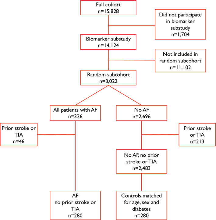
STABILITY was a randomized controlled trial comparing darapladib 160 mg versus placebo in 15 828 patients with stable coronary heart disease. Serum and plasma aliquots from blood samples obtained at randomization were available from 14 124 patients. In a previous nested case control study of 4127 patients that included all patients with events and a random cohort of patients with and without events within the STABILITY trial, a large array of established and novel biochemical markers of cardiovascular disease and inflammation were investigated. 24 From the random cohort used in that study (n=3022), all patients with a diagnosis of AF and without history of stroke and/or TIA were included in the present analysis (n=280). From the same random cohort, a control group without a diagnosis of AF and without history of stroke and/or TIA (n=280) was matched for age, sex, and diabetes status in a 1:1 ratio and included in the present analysis (Figure 1). Blood samples and 12‐lead ECGs were collected at the same time (at randomization visit). AF was defined as a history of chronic or paroxysmal AF, atrial flutter, or an ECG showing AF at randomization in STABILITY. To investigate the association between the current heart rhythm and sNFL levels, the patients with a diagnosis of AF were subdivided into 2 separate groups based on if they had ECG‐confirmed AF rhythm at randomization (AF ECG+, n=74), or not (AF ECG−, n=206).
Figure 1. Flowchart of patient selection.

AF indicates atrial fibrillation; and TIA, transient ischemic attack.
Ethical Approval
The STABILITY trial and its sub studies were performed in accordance with the Declaration of Helsinki, and was approved by the appropriate ethics committees at all sites and all patients provided written informed consent. Institutional review board number 2008/319, Uppsala, Sweden.
Biochemical Analyses
Venous blood samples were obtained at randomization in the morning after 9 hours of fasting, and were stored at −80 °C until biochemical analyses. The serum concentration of NFL was determined with Single Molecule Array (Simoa®) on the SR‐X™ Biomarker Detection System (Quanterix). The analyses were performed at the department of Clinical Chemistry, Uppsala University Hospital, by an experienced biomedical analyst that was blinded to the patient data. The lower limit of detection was 0.0552 pg/mL and the lower limit of quantification was 0.316 pg/mL. Estimated glomerular filtration rate (eGFR) and NT‐proBNP (N‐terminal pro B‐type natriuretic peptide) were determined through established assays as previously described. 25 , 26
Statistical Analysis
A sample size calculation was done prior to sNFL analysis, using a 2‐sided t test with significance level of 0.05, power 80% and standard deviation of 23 pg/mL. 27 Using all available cases and controls (ie, 280 patients with AF, and 280 without AF, both without prior stroke or TIA) we were able to show a difference in means of 5.46 pg/mL between patients with and without AF, with a power of 80%. This was considered an acceptable difference to proceed with the analyses. Propensity score matching by the variables age, sex, and diabetes status was performed prior to sNFL analysis in order to increase the power and lower the risk of confounding compared with selecting a random sample of controls without AF. 28 Propensity score matching was performed using the closest neighbor method with the R package MatchIt.
To investigate the associations between sNFL and patient characteristics, multiple linear regression analysis was performed. Adjustments were made in two steps: Model 1 included AF (defined as a history of chronic or paroxysmal AF, atrial flutter, or an ECG showing AF at randomization), age, sex, diabetes status, body mass index (BMI), eGFR, congestive heart failure, smoking (defined as current smoker of 5 or more cigarettes per day or previous smoker last 3 months), polyvascular disease (defined as coexistent arterial disease in at least 2 arterial territories; cerebral, carotid, peripheral), peripheral artery disease and treatment with oral anticoagulation before randomization. Model 2 included the variables in Model 1 and, in addition, the cardiac biomarker NT‐proBNP. Even though the patients were matched for age, sex and diabetes status, these variables were included in the models to increase the power of the analysis and the precision of the estimates. Patients with incomplete data on risk factors were omitted from the multiple linear regression analysis. Due to skewed distributions, the biomarkers sNFL and NT‐proBNP were log‐transformed using the natural logarithm before analysis. To aid in interpretation of the linear regression results the beta estimates were retransformed, herein called expBeta=exp(beta), which for a given predictor represents the proportion change in geometric mean of sNFL on its original scale. Baseline characteristics are presented as median (interquartile range) for continuous variables and percent (number) for discrete variables. P<0.05 from 2‐sided tests indicated statistical significance. All statistical analyses were performed using R version 4.0.2 (R Foundation for Statistical Computing, Vienna, Austria). All authors had full access to all the data in the study and takes responsibility for its integrity and the data analysis.
Data Availability
The data underlying this article will be shared on reasonable request to the corresponding author. Study documents are available at www.clinicalstudydatarequest.com.
Results
Baseline Characteristics
Baseline characteristics for patients with and without a diagnosis of AF matched for age, sex, and diabetes status are presented in Table 1. In both groups, the median age was 69 years (IQR 63, 75), 86% were men and 73% had hypertension. Congestive heart failure was more common in patients with AF compared with controls (31% versus 8.2%). Polyvascular disease, peripheral artery disease and prior myocardial infarction were also more common in patients with AF compared with controls (12% versus 10%; 7.5% versus 4.6%; and 54% versus 50%). A total of 74 (26%) of the patients with a diagnosis of AF had ECG‐verified AF (AF ECG+) at randomization. The majority of the patients were on treatment with angiotensin‐converting enzyme inhibitor (ACEi)/angiotensin II receptor blocker, statins and beta blockers. The levels of NT‐proBNP were higher in the group of patients with AF compared with the control group without AF.
Table 1.
Baseline Patient Characteristics
| Characteristic | Control group | Atrial fibrillation |
|---|---|---|
| Atrial fibrillation |
No (n=280) |
Yes (n=280) |
| Age, median (IQR), y | 69 (63–75) | 69 (63–75) |
| Male sex, % (n) | 86% (242) | 86% (241) |
| BMI, median (IQR), kg/m2 | 28.2 (25.9–32.4) | 29.0 (26.0–32.0) |
| Weight, median (IQR), kg | 82.5 (72.8–96.8) | 85.0 (76.7–97.8) |
| Systolic blood pressure, median (IQR), mm Hg | 129 (119–141) | 133 (122–144) |
| Diastolic blood pressure, median (IQR), mm Hg | 76.0 (70.0–83.0) | 79.0 (71.8–85.0) |
| ECG showing AF, % (n) | 0% (0) | 26% (74) |
| Diabetes, % (n) | 39% (109) | 40% (112) |
| eGFR, median (IQR), mL/min per 1.73 m2 | 72 (60–78) | 72 (60–78) |
| Congestive heart failure, % (n) | 8.2% (23) | 31% (86) |
| Smoking, % (n)* | 14% (38) | 12% (33) |
| Hypertension, % (n) | 73% (205) | 73% (205) |
| Polyvascular disease, % (n)† | 10% (28) | 12% (34) |
| Peripheral artery disease, % (n) | 4.6% (13) | 7.5% (21) |
| Prior myocardial infarct, % (n) | 50% (139) | 54% (150) |
| Prior PCI or CABG, % (n) | 79% (222) | 79% (222) |
| Treatment with | ||
| ACEi or ARB, % (n) | 76% (213) | 79% (222) |
| Statin, % (n) | 96% (269) | 98% (274) |
| Beta blockers, % (n) | 76% (214) | 81% (228) |
| Aspirin, % (n) | 96% (269) | 81% (228) |
| Oral anticoagulants before randomization, % (n) | 3.6% (10) | 34% (94) |
| Blood biomarkers | ||
| NFL, median (IQR), pg/mL | 18.6 (13.7–26.8) | 21.9 (15.3–30.7) |
| NT‐proBNP, median (IQR), ng/L | 170 (96.8–307) | 465 (200–980) |
Continuous variables are presented as median (interquartile range) and discrete and categorical variables are presented as percent (number). ACEi indicates angiotensin‐converting enzyme inhibitor; ARB, angiotensin II receptor blocker; BMI, body mass index; CABG, coronary artery bypass graft; eGFR, estimated glomerular filtration rate; IQR, interquartile range; NFL, neurofilament light chain; NT‐proBNP, N‐terminal pro B‐type natriuretic peptide; and PCI, percutaneous coronary intervention.
Smoking was defined as current smokers of 5 or more cigarettes per day or previous smoker last 3 months.
Polyvascular disease was defined as coexistent arterial disease in at least 2 arterial territories; cerebral, carotid, peripheral.
Levels of sNFL in Relation to AF
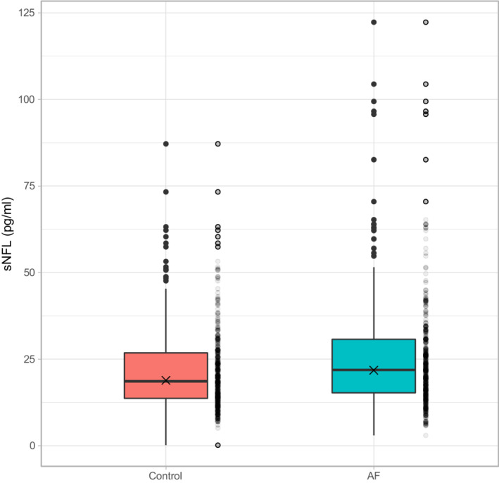
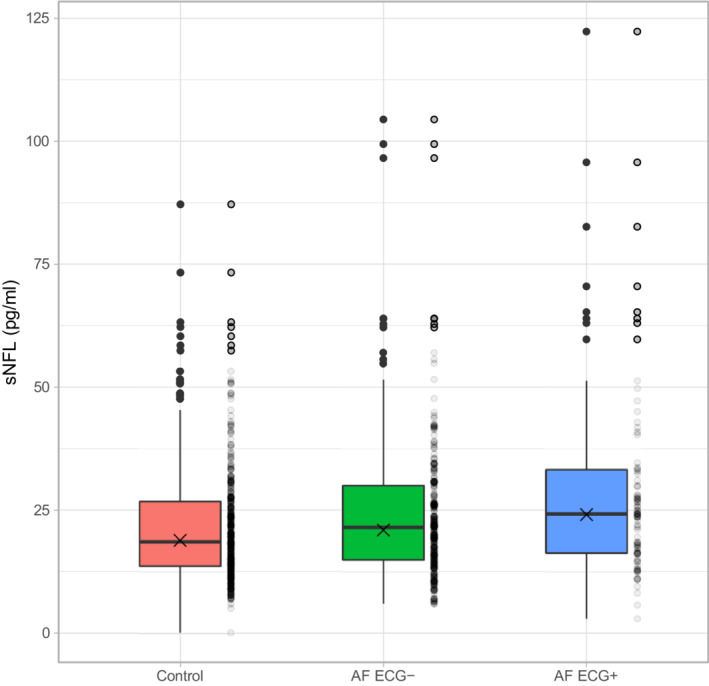
The levels of sNFL were higher in the AF group compared with the control group, geometric mean 21.8 versus 18.9 pg/mL, P=0.003 (Figure 2). Within the AF group, the levels of sNFL were 24.1 pg/mL in the AF ECG+ group, and 21.0 pg/mL in the AF ECG− group. Both AF ECG+ and AF ECG−, had higher sNFL levels than the control group, AF ECG+ P=0.001 and AF ECG− P=0.04 (Figure 3).
Figure 2. Levels of sNFL in patients without AF (control) and with AF.

Box represent interquartile range (IQR), whiskers min‐max within 1.5xIQR, thick line represents median and X marks the geometric mean. AF indicates atrial fibrillation; and sNFL, serum neurofilament light chain.
Figure 3. Levels of sNFL in patients without AF (control), with AF but without current AF‐rhythm on ECG (AF ECG−), and AF with current AF‐rhythm on ECG (AF ECG+).

Box represent interquartile range (IQR), whiskers min‐max within 1.5xIQR, thick line represents median and X marks the geometric mean. AF indicates atrial fibrillation; and sNFL, serum neurofilament light chain.
Multiple Linear Regression Analysis
The result of the multiple linear regression analysis is presented in Table 2. Seven patients had missing data on BMI, GFR and smoking and were excluded from the analysis. In Model 1, in which established clinical risk factors were included, AF, higher age, diabetes status, lower BMI, and lower eGFR were associated with higher levels of sNFL. In AF ECG+ patients the geometric mean of sNFL was 15% higher compared with the control group (expBeta 1.15, P=0.047). In AF ECG− patients, the geometric mean of sNFL was 10% higher compared with the control group (expBeta 1.10, P=0.04). Sex, congestive heart failure, smoking, polyvascular disease, peripheral artery disease, and treatment with oral anticoagulation before randomization were not associated with sNFL levels. In Model 2, where adjustment for NT‐proBNP was also made, the effect of AF on sNFL levels were attenuated and became non‐significant.
Table 2.
Result of Multiple Linear Regression Analysis
| Variable |
Model 1 R2=0.358 |
Model 2 R2=0.385 |
||
|---|---|---|---|---|
| expBeta (CI) | P value | expBeta (CI) | P value | |
| Atrial fibrillation | ||||
| ECG− | 1.10 (1.01–1.21) | 0.04 | 1.05 (0.958–1.15) | 0.3 |
| ECG+ | 1.15 (1.00–1.32) | 0.047 | 0.993 (0.855–1.15) | 0.9 |
| Age, by increase of 10 y | 1.28 (1.21–1.35) | <0.001 | 1.25 (1.19–1.32) | <0.001 |
| Male sex (reference female) | 0.897 (0.800–1.01) | 0.06 | 0.919 (0.820–1.03) | 0.1 |
| Diabetes | 1.18 (1.08–1.28) | <0.001 | 1.18 (1.08–1.28) | <0.001 |
| eGFR, by increase of 10 (mL/min per 1.73 m2) | 0.900 (0.878–0.924) | <0.001 | 0.912 (0.889–0.936) | <0.001 |
| BMI, by increase of 5, kg/m2 | 0.927 (0.892–0.963) | <0.001 | 0.935 (0.901–0.971) | <0.001 |
| Congestive heart failure | 1.02 (0.919–1.13) | 0.7 | 0.995 (0.899–1.10) | 0.9 |
| Smoking* | 1.03 (0.906–1.16) | 0.7 | 1.02 (0.901–1.15) | 0.8 |
| Polyvascular disease† | 1.10 (0.921–1.32) | 0.3 | 1.09 (0.915–1.30) | 0.3 |
| Peripheral artery disease | 0.939 (0.740–1.19) | 0.6 | 0.951 (0.754–1.20) | 0.7 |
| Treatment with oral anticoagulants before randomization | 1.04 (0.923–1.16) | 0.5 | 0.994 (0.886–1.12) | 0.9 |
| log(NT‐proBNP), by increase of 1 | 1.11 (1.06–1.15) | <0.001 | ||
Result of multiple linear regression analysis. Two models were fitted, Model 1 and Model 2 (Model 1 with the addition of cardiac biomarker NT‐proBNP). sNFL was log‐transformed prior to analysis and for easier interpretation the Beta‐values have been retransformed to expBeta (ie, eBeta). ExpBeta corresponds to the proportional increase or decrease in geometric mean of sNFL for each variable (eg, an increase in age with 10 years corresponds to a multiplicative increase of sNFL with 1.28 in Model 1). AF, higher age and diabetes were associated with increased levels of sNFL, whereas higher BMI and eGFR were associated with decreased levels of sNFL. Sex, congestive heart failure, smoking, polyvascular disease, peripheral artery disease, and treatment with oral anticoagulants were not associated with sNFL‐levels. The addition of NT‐proBNP to the model (Model 2) removed the association between AF and sNFL. AF indicates atrial fibrillation; BMI, body mass index; eGFR, estimated glomerular filtration rate; NT‐proBNP, N‐terminal pro B‐type natriuretic peptide; and sNFL, serum neurofilament light chain.
Smoking was defined as current smokers of 5 or more cigarettes per day or previous smoker last 3 months.
Polyvascular disease was defined as coexistent arterial disease in at least 2 arterial territories; cerebral, carotid, peripheral.
Discussion
The main finding of the present study was that in patients with chronic coronary artery disease, the levels of sNFL were higher in patients with AF compared with matched controls without AF, independent of clinical risk factors. The highest levels of sNFL were observed in patients with a diagnosis of AF and ECG‐verified AF at randomization. Beyond AF, the clinical risk factors with most influence on sNFL levels were age, diabetes, BMI, and eGFR. Sex, congestive heart failure, smoking, polyvascular disease, peripheral artery disease, and oral anticoagulation treatment did not affect the levels of sNFL significantly. However, when additionally adjusting for NT‐proBNP, which also is elevated in patients with AF, the association between AF and sNFL was attenuated and became non‐significant in this limited material.
NFL is an abundant cytoskeletal protein in the axonal compartment of neurons, and an established, yet unspecific, biochemical marker of neuroaxonal damage. 29 Elevated sNFL is present in many different neurological diseases and conditions that damage the nervous system regardless of the mechanism, 12 and predicts brain atrophy in eg, traumatic brain injury 22 and Alzheimer's disease. 30 In this study, sNFL concentrations were 15% higher in patients with ECG‐verified AF at randomization and 10% higher in patients with a history of AF but not in AF at randomization, compared with matched controls without AF and after adjustments for clinical risk factors known to affect sNFL. This result confirmed our hypothesis that AF is associated with an increased serum concentration of NFL. An increase of 10 or 15% of sNFL levels corresponds to an increase in chronological age of 2.3 and 3.3 years respectively, using data from a previous study on a normal aging population. 31 As none of the patients in our study had a history of stroke and/or TIA, the increased levels of sNFL in patients with AF are most likely due to other processes compromising the integrity of the brain tissue. Proposed mechanisms include microembolism originating from the heart and cerebral hypoperfusion due to the decreased and varying cardiac output in patients with AF. 32 In fact, AF has in previous studies been associated with MRI features of cerebral tissue damage other than stroke, eg, white matter hyperintensities, cortical microinfarcts, and atrophy, factors which in turn have been associated with higher sNFL. 5 , 6 , 7 , 8 , 9 , 33 The present findings support a direct connection between AF and cerebral tissue damage other than stroke, but it is still possible that our findings simply reflect the overall burden of a generalized cardiovascular disease affecting both the heart and the brain, and not AF per se. The observed increase of sNFL levels in patients with AF in the present study is modest compared with acute ischemic stroke where peak levels of 100–200 pg/mL appear in the first week, 19 , 21 and it is possible that the few outliers with values near or above 100 pg/mL represent recent silent cerebral infarcts.
About one‐third of the patients with AF were on oral anticoagulation treatment. If silent cerebral infarcts and microembolism were important factors contributing to the increased sNFL levels, an inverse association between oral anticoagulation treatment and sNFL would be expected. On the contrary, we could not observe any difference between untreated patients and patients on oral anticoagulation. This finding may indicate other mechanisms than thromboembolism, even though this should be interpreted with caution given the limited number of subjects and the lack of longitudinal sNFL measurements before and after initiation of oral anticoagulation treatment. The STABILITY study was performed between the years 2008 to2010, a time when the prevalence of oral anticoagulation treatment in patients with AF was generally lower compared with today. 34 This is reflected in the present study where in the AF group, only 34% were on oral anticoagulation treatment. Also, 3.6% of the patients in the control group were on anticoagulation treatment which might have attenuated any associations between oral anticoagulation treatment and sNFL.
sNFL was highest in patients currently in AF rhythm, which may reflect an increased prevalence of chronic AF, as patients with chronic AF are more likely to present with an ongoing AF rhythm than patients with paroxysmal AF. It has previously been reported that patients with persistent AF have a higher burden of MRI lesions, 6 , 7 and possibly are at higher risk of developing cognitive impairment, compared with patients with paroxysmal AF. 35 However, we could not explore this hypothesis due to the small sample size.
To the best of our knowledge, this is the first study to show that levels of sNFL are increased in patients with AF. In a previous study of patients with AF, sNFL levels were correlated to the CHA2DS2‐VASc score, vascular changes on MRI, and to some extent cognitive measurements. 27 However, no control group was available and conclusions of whether the levels were elevated due to AF could therefore not be made. In line with previous findings, the clinical risk factors associated with the sNFL levels in the present study were age, diabetes mellitus, BMI and renal function (eGFR). 27 , 31 , 36 , 37 , 38 Age was the strongest contributor to sNFL levels, where an increase in age of 10 years translated to an increase in sNFL of 25% in the fully adjusted model. In the current study, there was no association between sex and sNFL levels after adjustment for established clinical risk factors, which is consistent with a previous study of sNFL in a normal aging population. 31 Nonetheless, the low proportion of females (14%) in our study may have attenuated possible associations. We found no associations between levels of sNFL and congestive heart failure, polyvascular disease, peripheral artery disease, and smoking, where previous studies have reported conflicting results. 27 , 36 However, the low number of patients with polyvascular disease, peripheral artery disease, and smoking in both the AF group and the control group may have masked possible associations.
The natriuretic peptide NT‐proBNP is a biomarker for AF. NT‐proBNP reflects the effect of the AF rhythm on the heart and is prognostic for cardiovascular events and death in patients with AF 39 but also in patients with coronary heart disease. 40 Patients with AF have elevated levels of natriuretic peptides in comparison with controls 41 but the levels drop rapidly after restoration of AF to sinus rhythm. 42 , 43 In a recent study of 100 patients with AF undergoing electrical cardioversion, significant reductions in NT‐proBNP levels were associated with sinus rhythm at 30 days follow up. 44 The levels of NT‐proBNP were higher in the AF group compared with the control group in the present study. Similar to sNFL, the highest levels of NT‐proBNP were noticed in patients with ongoing AF rhythm (data not shown). To further investigate whether the sNFL levels were associated with the AF rhythm (ie, reflecting the effect of the AF rhythm on the brain), we extended the multiple linear regression analysis by adding NT‐proBNP. The addition of NT‐proBNP to the model attenuated the association between AF and sNFL, suggesting that sNFL (along with NT‐proBNP) may be linked to the AF rhythm, and the associated myocardial stretch. There was no association between a history of congestive heart failure (another source of NT‐proBNP release) and sNFL in the present study. However, no objective measures of heart function were available, limiting the interpretability of these findings.
Serum NFL is an emerging biomarker for neurologic diseases and is approaching clinical routine in the follow up of patients with MS. 45 Knowledge about confounding factors is important to be able to correctly interpret circulating levels of this biomarker when assessing a disease of interest, especially since neurofilaments are present in both the peripheral and central nervous system. How blood–brain permeability, liver function, renal clearance, and other concurrent diseases may affect the metabolism of sNFL is currently not fully understood. This study adds to the understanding of factors contributing to sNFL levels and indicates that there might be a reason to further study sNFL in patients with AF in relation to the risk of stroke and TIA.
This study included analyses of a large, prospective, closely monitored multinational cohort of patients with stable coronary heart disease and minimizing the risk of confounding by adjusting the analyses for a wide range of established prognostic variables including the strongly prognostic cardiac biomarker NT‐proBNP. However, this study has some limitations that should be mentioned. Brain imaging was not made in the STABILITY trial which limits the interpretation of the association between AF, sNFL and cerebral tissue damage. Furthermore, the observational setting does not permit firm mechanistic conclusions and the clinical importance of our findings remains to be elucidated. There is also a possibility that some patients with subclinical undiagnosed paroxysmal AF were included in the control population. However, if this was the case, it would have attenuated the differences between the AF and control group. Finally, the results may not be directly translated to the general AF population, since the study was performed with a clinical trial cohort of patients with stable coronary heart disease.
In patients with stable coronary heart disease, patients with concomitant AF had higher levels of sNFL compared with matched controls without AF. Highest sNFL levels were observed in patients with ongoing AF rhythm at the time of blood sampling. Oral anticoagulation treatment was not associated with sNFL levels. These findings provide biochemical support to the association between AF and cerebral tissue damage other than stroke, and add to the theory that the AF rhythm per se may give rise to further strain on cerebral tissue other than thromboembolism.
Sources of Funding
This work was supported by grants from the Regional Research Council Mid Sweden; Bissen Brainwalk Foundation; Selander Foundation; and Swedish State Support for Research (ALF‐agreement).
Disclosures
L.W. reports institutional research grants, consultancy fees, lecture fees, and travel support from AstraZeneca, Boehringer Ingelheim, Bristol‐Myers Squibb/Pfizer, and GlaxoSmithKline; institutional research grants from Merck&Co and Roche Diagnostics.
N.E. reports institutional research grants from Pfizer.
C.H. reports institutional research grants from GlaxoSmithKline; honoraria and research grants from Pfizer; consultant and advisory board fees from AstraZeneca, Bayer, Boehringer Ingelheim and Coala Life.
J.O. reports fees to his institution for consultant/advisory boards (including study steering committees and data safety monitoring boards) and lectures, from AstraZeneca, Bayer, Boehringer Ingelheim, Bristol‐Myers Squibb, Daichii Sankyo, Novartis, Pfizer, Portola, Roche Diagnostics, and Sanofi, outside the submitted work.
K.S., J.A., K.K., and J.B. reports no conflict of interest.
Acknowledgments
The authors thank biomedical analyst Asma Al‐Grety (sNFL‐analysis).
For Sources of Funding and Disclosures, see page 9.
References
- 1. Krijthe BP, Kunst A, Benjamin EJ, Lip GYH, Franco OH, Hofman A, Witteman JCM, Stricker BH, Heeringa J. Projections on the number of individuals with atrial fibrillation in the European Union, from 2000 to 2060. Eur Heart J. 2013;34:2746–2751. doi: 10.1093/eurheartj/eht280 [DOI] [PMC free article] [PubMed] [Google Scholar]
- 2. Wolf PA, Abbott RD, Kannel WB. Atrial fibrillation as an independent risk factor for stroke: the Framingham study. Stroke. 1991;22:983–988. doi: 10.1161/01.STR.22.8.983 [DOI] [PubMed] [Google Scholar]
- 3. Ott A, Breteler MMB, de Bruyne MC, van Harskamp F, Grobbee DE, Hofman A. Atrial fibrillation and dementia in a population‐based study. Stroke. 1997;28:316–321. doi: 10.1161/01.STR.28.2.316 [DOI] [PubMed] [Google Scholar]
- 4. Marzona I, O'Donnell M, Teo K, Gao P, Anderson C, Bosch J, Yusuf S. Increased risk of cognitive and functional decline in patients with atrial fibrillation: results of the ONTARGET and TRANSCEND studies. Can Med Assoc J. 2012;184:E329–E336. doi: 10.1503/cmaj.111173 [DOI] [PMC free article] [PubMed] [Google Scholar]
- 5. Kobayashi A, Iguchi M, Shimizu S, Uchiyama S. Silent cerebral infarcts and cerebral White matter lesions in patients with nonvalvular atrial fibrillation. J Stroke Cerebrovasc Dis. 2012;21:310–317. doi: 10.1016/j.jstrokecerebrovasdis.2010.09.004 [DOI] [PubMed] [Google Scholar]
- 6. Gaita F, Corsinovi L, Anselmino M, Raimondo C, Pianelli M, Toso E, Bergamasco L, Boffano C, Valentini MC, Cesarani F, et al. Prevalence of silent cerebral ischemia in paroxysmal and persistent atrial fibrillation and correlation with cognitive function. J Am Coll Cardiol. 2013;62:1990–1997. doi: 10.1016/j.jacc.2013.05.074 [DOI] [PubMed] [Google Scholar]
- 7. Stefansdottir H, Arnar DO, Aspelund T, Sigurdsson S, Jonsdottir MK, Hjaltason H, Launer LJ, Gudnason V. Atrial fibrillation is associated with reduced brain volume and cognitive function independent of cerebral infarcts. Stroke. 2013;44:1020–1025. doi: 10.1161/STROKEAHA.12.679381 [DOI] [PMC free article] [PubMed] [Google Scholar]
- 8. Wang Z, van Veluw SJ, Wong A, Liu W, Shi L, Yang J, Xiong Y, Lau A, Biessels GJ, Mok VCT. Risk factors and cognitive relevance of cortical cerebral microinfarcts in patients with ischemic stroke or transient ischemic attack. Stroke. 2016;47:2450–2455. doi: 10.1161/STROKEAHA.115.012278 [DOI] [PubMed] [Google Scholar]
- 9. Mayasi Y, Helenius J, McManus DD, Goddeau RP, Jun‐O'Connell AH, Moonis M, Henninger N. Atrial fibrillation is associated with anterior predominant white matter lesions in patients presenting with embolic stroke. J Neurol Neurosurg Psychiatry. 2018;89:6–13. doi: 10.1136/jnnp-2016-315457 [DOI] [PMC free article] [PubMed] [Google Scholar]
- 10. Conen D, Rodondi N, Müller A, Beer JH, Ammann P, Moschovitis G, Auricchio A, Hayoz D, Kobza R, Shah D, et al. Relationships of overt and silent brain lesions with cognitive function in patients with atrial fibrillation. J Am Coll Cardiol. 2019;73:989–999. doi: 10.1016/j.jacc.2018.12.039 [DOI] [PubMed] [Google Scholar]
- 11. Lee MK, Cleveland DW. Neuronal intermediate filaments. Annu Rev Neurosci. 1996;19:187–217. doi: 10.1146/annurev.ne.19.030196.001155 [DOI] [PubMed] [Google Scholar]
- 12. Khalil M, Teunissen CE, Otto M, Piehl F, Sormani MP, Gattringer T, Barro C, Kappos L, Comabella M, Fazekas F, et al. Neurofilaments as biomarkers in neurological disorders. Nat Rev Neurol. 2018;14:577–589. doi: 10.1038/s41582-018-0058-z [DOI] [PubMed] [Google Scholar]
- 13. Barro C, Chitnis T, Weiner HL. Blood neurofilament light: a critical review of its application to neurologic disease. Ann Clin Transl Neurol. 2020;7:2508–2523. doi: 10.1002/acn3.51234 [DOI] [PMC free article] [PubMed] [Google Scholar]
- 14. Rissin DM, Kan CW, Campbell TG, Howes SC, Fournier DR, Song L, Piech T, Patel PP, Chang L, Rivnak AJ, et al. Single‐molecule enzyme‐linked immunosorbent assay detects serum proteins at subfemtomolar concentrations. Nat Biotechnol. 2010;28:595–599. doi: 10.1038/nbt.1641 [DOI] [PMC free article] [PubMed] [Google Scholar]
- 15. Gisslén M, Price RW, Andreasson U, Norgren N, Nilsson S, Hagberg L, Fuchs D, Spudich S, Blennow K, Zetterberg H. Plasma concentration of the neurofilament light protein (NFL) is a biomarker of CNS injury in HIV infection: a cross‐sectional study. EBioMedicine. 2016;3:135–140. doi: 10.1016/j.ebiom.2015.11.036 [DOI] [PMC free article] [PubMed] [Google Scholar]
- 16. Kuhle J, Barro C, Andreasson U, Derfuss T, Lindberg R, Sandelius Å, Liman V, Norgren N, Blennow K, Zetterberg H. Comparison of three analytical platforms for quantification of the neurofilament light chain in blood samples: ELISA, electrochemiluminescence immunoassay and Simoa. Clin Chem Lab Med. 2016;54. doi: 10.1515/cclm-2015-1195 [DOI] [PubMed] [Google Scholar]
- 17. Disanto G, Barro C, Benkert P, Naegelin Y, Schädelin S, Giardiello A, Zecca C, Blennow K, Zetterberg H, Leppert D, et al. Serum neurofilament light: a biomarker of neuronal damage in multiple sclerosis. Ann. Neurol. 2017;81:857–870. doi: 10.1002/ana.24954 [DOI] [PMC free article] [PubMed] [Google Scholar]
- 18. Siller N, Kuhle J, Muthuraman M, Barro C, Uphaus T, Groppa S, Kappos L, Zipp F, Bittner S. Serum neurofilament light chain is a biomarker of acute and chronic neuronal damage in early multiple sclerosis. Mult Scler J. 2019;25:678–686. doi: 10.1177/1352458518765666 [DOI] [PubMed] [Google Scholar]
- 19. Tiedt S, Duering M, Barro C, Kaya AG, Boeck J, Bode FJ, Klein M, Dorn F, Gesierich B, Kellert L, et al. Serum neurofilament light: A biomarker of neuroaxonal injury after ischemic stroke. Neurology. 2018;91:e1338–e1347. doi: 10.1212/WNL.0000000000006282 [DOI] [PubMed] [Google Scholar]
- 20. Onatsu J, Vanninen R, Jäkälä P, Mustonen P, Pulkki K, Korhonen M, Hedman M, Zetterberg H, Blennow K, Höglund K, et al. Serum neurofilament light chain concentration correlates with infarct volume but not prognosis in acute ischemic stroke. J Stroke Cerebrovasc Dis. 2019;28:2242–2249. doi: 10.1016/j.jstrokecerebrovasdis.2019.05.008 [DOI] [PubMed] [Google Scholar]
- 21. Pujol‐Calderón F, Zetterberg H, Portelius E, Löwhagen Hendén P, Rentzos A, Karlsson J‐E, Höglund K, Blennow K, Rosengren LE. Prediction of outcome after endovascular embolectomy in anterior circulation stroke using biomarkers. Transl Stroke Res. 2022;13:65–76. doi: 10.1007/s12975-021-00905-5 [DOI] [PMC free article] [PubMed] [Google Scholar]
- 22. Graham NSN, Zimmerman KA, Moro F, Heslegrave A, Maillard SA, Bernini A, Miroz J‐P, Donat CK, Lopez MY, Bourke N, et al. Axonal marker neurofilament light predicts long‐term outcomes and progressive neurodegeneration after traumatic brain injury. Sci Transl Med. 2021;13:eabg9922. doi: 10.1126/scitranslmed.abg9922 [DOI] [PubMed] [Google Scholar]
- 23. White HD, Held C, Stewart R, Tarka E, Brown R, Davies RY, Budaj A, Harrington RA, Steg PG, Ardissino D, et al. Darapladib for preventing ischemic events in stable coronary heart disease. N Engl J Med. 2014;370:1702–1711. doi: 10.1056/NEJMoa1315878 [DOI] [PubMed] [Google Scholar]
- 24. Wallentin L, Eriksson N, Olszowka M, Grammer TB, Hagström E, Held C, Kleber ME, Koenig W, März W, Stewart RAH, et al. Plasma proteins associated with cardiovascular death in patients with chronic coronary heart disease: a retrospective study. PLoS Med. 2021;18:e1003513. doi: 10.1371/journal.pmed.1003513 [DOI] [PMC free article] [PubMed] [Google Scholar]
- 25. Held C, White HD, Stewart RAH, Budaj A, Cannon CP, Hochman JS, Koenig W, Siegbahn A, Steg PG, Soffer J, et al. Inflammatory biomarkers interleukin‐6 and c‐reactive protein and outcomes in stable coronary heart disease: experiences from the STABILITY (stabilization of atherosclerotic plaque by initiation of darapladib therapy) trial. J Am Heart Assoc. 2017;6. doi: 10.1161/JAHA.116.005077 [DOI] [PMC free article] [PubMed] [Google Scholar]
- 26. Hagström E, Held C, Stewart RAH, Aylward PE, Budaj A, Cannon CP, Koenig W, Krug‐Gourley S, Mohler ER, Steg PG, et al. Growth differentiation factor 15 predicts all‐cause morbidity and mortality in stable coronary heart disease. Clin Chem. 2017;63:325–333. doi: 10.1373/clinchem.2016.260570 [DOI] [PubMed] [Google Scholar]
- 27. Polymeris AA, Coslovksy M, Aeschbacher S, Sinnecker T, Benkert P, Kobza R, Beer J, Rodondi N, Fischer U, Moschovitis G, et al. Serum neurofilament light in atrial fibrillation: clinical, neuroimaging and cognitive correlates. Brain Commun. 2020;2:1–13. doi: 10.1093/braincomms/fcaa166 [DOI] [PMC free article] [PubMed] [Google Scholar]
- 28. Rosenbaum PR, Rubin DB. The central role of the propensity score in observational studies for causal effects. Biometrika. 1983;70:41–55. doi: 10.1093/biomet/70.1.41 [DOI] [Google Scholar]
- 29. Yuan A, Rao MV, Veeranna NRA. Neurofilaments and neurofilament proteins in health and disease. Cold Spring Harb Perspect Biol. 2017;9:a018309. doi: 10.1101/cshperspect.a018309 [DOI] [PMC free article] [PubMed] [Google Scholar]
- 30. Preische O, Schultz SA, Apel A, Kuhle J, Kaeser SA, Barro C, Gräber S, Kuder‐Buletta E, LaFougere C, Laske C, et al. Serum neurofilament dynamics predicts neurodegeneration and clinical progression in presymptomatic Alzheimer's disease. Nat Med. 2019;25:277–283. doi: 10.1038/s41591-018-0304-3 [DOI] [PMC free article] [PubMed] [Google Scholar]
- 31. Khalil M, Pirpamer L, Hofer E, Voortman MM, Barro C, Leppert D, Benkert P, Ropele S, Enzinger C, Fazekas F, et al. Serum neurofilament light levels in normal aging and their association with morphologic brain changes. Nat Commun. 2020;11:812. doi: 10.1038/s41467-020-14612-6 [DOI] [PMC free article] [PubMed] [Google Scholar]
- 32. Madhavan M, Graff‐Radford J, Piccini JP, Gersh BJ. Cognitive dysfunction in atrial fibrillation. Nat Rev Cardiol. 2018;15:744–756. doi: 10.1038/s41569-018-0075-z [DOI] [PubMed] [Google Scholar]
- 33. Duering M, Konieczny MJ, Tiedt S, Baykara E, Tuladhar AM, Leijsen E van, Lyrer P, Engelter ST, Gesierich B, Achmüller M, et al. Serum neurofilament light chain levels are related to small vessel disease burden. J Stroke 2018;20:228–238. doi: 10.5853/jos.2017.02565. [DOI] [PMC free article] [PubMed] [Google Scholar]
- 34. Hsu JC, Maddox TM, Kennedy KF, Katz DF, Marzec LN, Lubitz SA, Gehi AK, Turakhia MP, Marcus GM. Oral anticoagulant therapy prescription in patients with atrial fibrillation across the spectrum of stroke risk. JAMA Cardiol. 2016;1:55–62. doi: 10.1001/jamacardio.2015.0374 [DOI] [PubMed] [Google Scholar]
- 35. Chen LY, Agarwal SK, Norby FL, Gottesman RF, Loehr LR, Soliman EZ, Mosley TH, Folsom AR, Coresh J, Alonso A. Persistent but not paroxysmal atrial fibrillation is independently associated with lower cognitive function. J Am Coll Cardiol. 2016;67:1379–1380. doi: 10.1016/j.jacc.2015.11.064 [DOI] [PMC free article] [PubMed] [Google Scholar]
- 36. Korley FK, Goldstick J, Mastali M, Van Eyk JE, Barsan W, Meurer WJ, Sussman J, Falk H, Levine D. Serum NfL (neurofilament light chain) levels and incident stroke in adults with diabetes mellitus. Stroke. 2019;50:1669–1675. doi: 10.1161/STROKEAHA.119.024941 [DOI] [PMC free article] [PubMed] [Google Scholar]
- 37. Akamine S, Marutani N, Kanayama D, Gotoh S, Maruyama R, Yanagida K, Sakagami Y, Mori K, Adachi H, Kozawa J, et al. Renal function is associated with blood neurofilament light chain level in older adults. Sci Rep. 2020;10:20350. doi: 10.1038/s41598-020-76990-7 [DOI] [PMC free article] [PubMed] [Google Scholar]
- 38. Manouchehrinia A, Piehl F, Hillert J, Kuhle J, Alfredsson L, Olsson T, Kockum I. Confounding effect of blood volume and body mass index on blood neurofilament light chain levels. Ann Clin Transl Neurol. 2020;7:139–143. doi: 10.1002/acn3.50972 [DOI] [PMC free article] [PubMed] [Google Scholar]
- 39. Hijazi Z, Wallentin L, Siegbahn A, Andersson U, Christersson C, Ezekowitz J, Gersh BJ, Hanna M, Hohnloser S, Horowitz J, et al. N‐terminal pro–B‐type natriuretic peptide for risk assessment in patients with atrial fibrillation. J Am Coll Cardiol. 2013;61:2274–2284. doi: 10.1016/j.jacc.2012.11.082 [DOI] [PubMed] [Google Scholar]
- 40. Lindholm D, Lindbäck J, Armstrong PW, Budaj A, Cannon CP, Granger CB, Hagström E, Held C, Koenig W, Östlund O, et al. Biomarker‐based risk model to predict cardiovascular mortality in patients with stable coronary disease. J Am Coll Cardiol. 2017;70:813–826. doi: 10.1016/j.jacc.2017.06.030 [DOI] [PubMed] [Google Scholar]
- 41. Ellinor PT, Low AF, Patton KK, Shea MA, MacRae CA. Discordant atrial natriuretic peptide and brain natriuretic peptide levels in lone atrial fibrillation. J Am Coll Cardiol. 2005;45:82–86. doi: 10.1016/j.jacc.2004.09.045 [DOI] [PubMed] [Google Scholar]
- 42. Jourdain P, Bellorini M, Funck F, Fulla Y, Guillard N, Loiret J, Thebault B, Sadeg N, Desnos M. Short‐term effects of sinus rhythm restoration in patients with lone atrial fibrillation: a hormonal study. Eur J Heart Fail. 2002;4:263–267. doi: 10.1016/S1388-9842(02)00004-1 [DOI] [PubMed] [Google Scholar]
- 43. Wożakowska‐Kapłon B. Effect of sinus rhythm restoration on plasma brain natriuretic peptide in patients with atrial fibrillation. Am J Cardiol. 2004;93:1555–1558. doi: 10.1016/j.amjcard.2004.03.013 [DOI] [PubMed] [Google Scholar]
- 44. Meyre PB, Aeschbacher S, Blum S, Voellmin G, Kastner PM, Hennings E, Kaufmann BA, Kühne M, Osswald S, Conen D. Biomarkers associated with rhythm status after cardioversion in patients with atrial fibrillation. Sci Rep. 2022;12:1680. doi: 10.1038/s41598-022-05769-9 [DOI] [PMC free article] [PubMed] [Google Scholar]
- 45. Bittner S, Oh J, Havrdová EK, Tintoré M, Zipp F. The potential of serum neurofilament as biomarker for multiple sclerosis. Brain. 2021;144:2954–2963. doi: 10.1093/brain/awab241 [DOI] [PMC free article] [PubMed] [Google Scholar]
Associated Data
This section collects any data citations, data availability statements, or supplementary materials included in this article.
Data Availability Statement
The data underlying this article will be shared on reasonable request to the corresponding author. Study documents are available at www.clinicalstudydatarequest.com.


