Abstract
A gene fusion system based on plasmid pBBR1MCS and the expression of green fluorescent protein was developed for Brucella suis, allowing isolation of constitutive and inducible genes. Bacteria containing promoter fusions of chromosomal DNA to gfp were visualized by fluorescence microscopy and examined by flow cytometry. Twelve clones containing gene fragments induced inside J774 murine macrophages were isolated and further characterized.
Brucella spp. are gram-negative bacteria responsible for human and animal brucellosis in a variety of mammalian hosts (12). Brucella melitensis, B. suis, and B. abortus are most frequently associated with the disease in humans and in animals of agricultural importance such as cows, sheep, pigs, and goats. A major characteristic of this intracellular pathogen is its ability to survive and replicate in the macrophages of the host (10), where it remains enclosed in phagocytic compartments. Little is known to date about the mechanisms and the factors that allow brucellae to survive within their host cells. In a murine model of infection, htrA, purE, and a recently described two-component system participate in multiplication within macrophages (3, 5, 17).
If the identification of specific virulence genes in brucellae is only at its beginning, this has been due partly to the lack of genetic tools and techniques until recently. A major step in the study of Brucella sp. genetics related to virulence was the introduction of a broad-host-range vector (8). Here we describe the development and application of a green fluorescent protein (GFP) reporter gene system that is becoming a powerful tool in the analysis of various bacterial pathogens (20).
The gene encoding kanamycin resistance was cloned as a blunted 1.2-kb PstI fragment from plasmid pUC4K into the SacI site on plasmid pFPV25 (18), in the orientation opposite to the promoterless gfp gene gfpmut3 (2). Both genes were excised as a 2.2-kb EcoRI-EcoRV fragment and cloned blunt ended into plasmid pBBR1MCS (8), which had been previously digested by BamHI-AatII and was devoid of its original chloramphenicol resistance gene. The resulting plasmid, with a size of 5.6 kb, was named pBBR1-KGFP (Table 1 describes the strains and plasmids used in this study). This construct had the advantage of being considerably smaller than the broad-host-range vector previously described (13), due to the use of a single reporter gene. For the construction of a B. suis promoter library, chromosomal DNA of B. suis was partially digested by Sau3A for various time intervals and size fractionated on agarose gel. Fragments ranging from 0.5 to 1.5 kb in size were purified by electroelution and inserted into pBBR1-KGFP linearized by the unique BamHI site immediately upstream of gfpmut3. Recombinant B. suis strains were obtained by electroporation (7, 9) and isolated as individual clones on TS agar containing kanamycin at 100 μg/ml after incubation at 37°C for 3 days. A total of 5,000 clones were analyzed for the expression of constitutive and inducible promoters.
TABLE 1.
Bacterial strains and plasmids used in this study
| Strain or plasmid | Characteristicsa | Source or reference |
|---|---|---|
| E. coli DH5αF′b | F′ endA1 hsdR17 supE44 thi-1 recA1 gyrA relA1 Φ80lacZΔM15 Δ(lacZYA-argF) U169 | Life Technologies |
| B. suisc | ||
| 1330 | Biotype 1; ATCC 23444 | ATCCd |
| 1330-BBR1-KGFP | 1330 strain carrying pBBR1-KGFP; no gfp expression | This study |
| Plasmids | ||
| pUC4K | Contains Kanr cassette flanked by polylinker | Pharmacia |
| pBBR1MCS | Broad-host-range cloning vector (Cmr) | 8 |
| pFPV25 | Promoter trap vector containing gfpmut3 in plasmid pED350 (Ampr) | 18 |
| pBBR1-KGFP | pBBR1MCS derivative; Kanr; promoterless gfpmut3 from pFPV25 | This study |
Ampr, ampicillin resistance; Cmr, chloramphenicol resistance; Kanr, kanamycin resistance.
E. coli strains were grown in Luria-Bertani broth supplemented with the appropriate antibiotics at 37°C.
B. suis strains were grown at 37°C in TS supplemented with antibiotics when appropriate.
ATCC, American Type Culture Collection.
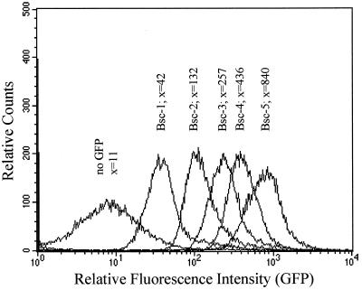
Constitutively expressed promoters of B. suis led to the formation of yellow-greenish colonies on agar plates after incubation for 3 days at 37°C, which were easily distinguished from the white colonies not expressing gfp. GFP-positive clones among the 5,000 clones analyzed were therefore visible to the naked eye on solid medium, and constitutive fluorescence of these clones was verified individually by fluorescence microscopy on glass slides. Seven percent of the library was found to be constitutively fluorescent, and as expected, the fluorescence intensities of the clones varied depending on the strength of the respective promoters. Five constitutively fluorescent clones were further analyzed with a FACScalibur flow cytometer (Becton Dickinson), confirming variable gfp expression depending on promoter strength (Fig. 1). Clone Bsc-5, with the strongest gfp expression measured, contained a fragment of 800 bp showing no significant homology to sequences in the GenBank database. Other clones contained Brucella promoters where the degree of expression varied with growth phase, as measured by fluorescence-activated cell sorter analysis during the log and stationary phases (data not shown). All clones maintained their fluorescence when passaged on nonselective solid medium, which is in agreement with the high stability of pBBR1MCS in brucellae (4).
FIG. 1.
Flow cytometry analysis of gfp expression by native B. suis promoters. Cell Quest software (Becton Dickinson) was used for the quantitation of fluorescence. Constitutive promoters of different strengths resulted in variable levels of fluorescence intensity. x, mean relative fluorescence intensity of B. suis clones Bsc-1, Bsc-2, Bsc-3, Bsc-4, and Bsc-5.
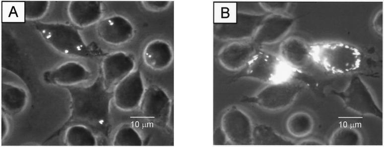
One major application of our approach is the study of live or killed brucellae within their host cells and of the cellular processes involved in intracellular multiplication of the pathogen. Murine macrophages and the macrophage cell line J774A.1 have become a well-established model system in the interaction with Brucella spp. (3, 6, 11, 15). J774 cells were seeded onto glass coverslips, infected with clone Bsc-5 for 30 min at a multiplicity of infection of 20, and washed three times in phosphate-buffered saline prior to incubation in RPMI medium (supplemented with 10% fetal calf serum) for up to 48 h. Cells were then fixed with 3.7% formaldehyde. Intracellular multiplication of B. suis was visualized by fluorescence microscopy (Leica DM IRB) at 24 and 48 h postinfection (Fig. 2A and B). Flow cytometry analysis and cell sorting of macrophages containing fluorescent B. suis by differential fluorescence induction (19) have been unsuccessful in our studies, as brucellae are very small, with a volume of 0.6 μm3 or less, and rates of infection are low. GFP fluorescence of intracellular bacteria did not exceed cellular background fluorescence and was too low for detection with a flow cytometer.
FIG. 2.
Intracellular B. suis Bsc-5 constitutively expressing gfp during infection of J774.A1 macrophages. Infected cells and multiplying bacteria were visualized at 24 h (A) and 48 h (B) postinfection by phase-contrast and epifluorescence microscopy using a fluorescein isothiocyanate filter.
Little is known about Brucella sp. gene expression in the intracellular environment, and studies have always been performed with previously defined and characterized genes (7, 11). To identify B. suis promoters that were strongly induced intracellularly compared to expression in RPMI cell culture medium or tryptic soy broth (TS), pools of 10 clones each of the promoter library were used to infect J774 macrophages in 96-well microtiter plates in the presence of kanamycin. At 24 and 48 h, the wells were screened under fluorescence microscopy for those containing intracellularly fluorescent bacteria. In a second round of infection, clones from positive wells were added individually to J774 cells. Fluorescence of positive clones was compared to that obtained with free bacteria in medium, and only the clones fluorescing exclusively intracellularly were retained for further characterization. In about 3% of the clones containing promoters inducing gfp expression, activation of these promoter sequences was visible exclusively intracellularly. We have isolated 12 B. suis clones of this type (Table 2). GFP-mediated fluorescence of these clones was detectable neither prior to infection nor after macrophage lysis, serial plating of released bacteria onto TS agar in the presence of kanamycin, and incubation at 37°C until colony formation (data not shown). Sequence analysis of the transcriptional fusions to gfp and alignments of the putatively encoded proteins to the Swissprot database using the BLASTx algorithm (1) revealed that five of these fragments showed no significant homology to sequences in the databases; six others showed homology to nucleotide-binding proteins and proteins of various transport systems with amino acid similarities ranging from 59 to 83% (Table 2).
TABLE 2.
Identified promoter fusions of B. suis genes to gfp specifically induced in J774 murine macrophages
| B. suis clone | Homologous genea | P(N)b | % Similarity (aa overlap)c | GenBank accession no. |
|---|---|---|---|---|
| 19A10 | nikA periplasmic binding protein-dependent transport system (E. coli) | 8.0 × 10−78 | 76 (223) | X73143 |
| 21C3 | GTP-binding protein (Caulobacter crescentus) | 2.3 × 10⁻³¹ | 81 (90) | AF084242 |
| 27C3 | livH leucine transport system (E. coli) | 1.2 × 10−41 | 83 (105) | U00039 |
| 28B10 | artI arginine-binding protein (E. coli) | 6.9 × 10⁻⁵ | 72 (36) | D90724 |
| 29H5 | gguA sugar uptake (Agrobacterium tumefaciens) | 1.3 × 10−44 | 82 (106) | U91632 |
| 30B2 | ABC transporter (Haemophilus influenzae) | 1.7 × 10⁻²² | 59 (72) | U32748 |
| 9H1 | nrdH/nrdI glutaredoxin-like protein (M. tuberculosis) | 6.1 × 10⁻¹⁵ | 80 (89) | Z83866 |
Based on BLASTx alignment.
Smallest-sum probability.
aa, amino acids.
Further work has to be done on the isolation and characterization, including insertional inactivation, of genes specifically induced by intramacrophagic brucellae, especially in the early phase of infection. Despite the fact that screening of the genome of B. suis is still in progress, our results showed that an important portion of the genes isolated so far, and induced intracellularly over at least 24 h, were potentially involved in the import of metabolic components from the eukaryotic host cells into the bacteria, hence possibly contributing to the rapid intramacrophagic multiplication of the latter. It is interesting to speculate that intracellular bacteria may also activate thioredoxin- or glutaredoxin-like systems used to counter oxidative stress by maintaining a reducing environment in the cytoplasm, as described for Escherichia coli (16). Indeed, one of the gene fragments isolated was highly homologous to the nrdH/nrdI glutaredoxin-like system from Mycobacterium tuberculosis.
The stable expression of gfp in B. suis will allow study of the interactions of this pathogen with its host cell, the macrophage. A constitutive B. suis GFP may, for example, be useful in the further characterization of the acidified phagosomes (15) and of the intracellular trafficking of Brucella-containing vacuoles (14). In addition, we have identified differentially expressed genes of B. suis strongly induced within the macrophage. This approach should accelerate the characterization of B. suis virulence factors and contribute to a better understanding of how brucellae avoid the bactericidal mechanisms of the macrophage.
Acknowledgments
We are grateful to Véronique Maurin and Françoise Porte for helpful discussion and comments on the use of the flow cytometer and fluorescence microscopy. We acknowledge M. T. Alvarez-Martinez for communication of B. suis sequencing data. Plasmid pFPV25 was a kind gift of Raphael Valdivia.
This work was supported in part by grant PL 980089 from the European Union.
REFERENCES
- 1.Altschul S F, Gish W, Miller W, Myers E W, Lipman D J. Basic local alignment search tool. J Mol Biol. 1990;215:403–410. doi: 10.1016/S0022-2836(05)80360-2. [DOI] [PubMed] [Google Scholar]
- 2.Cormack B P, Valdivia R H, Falkow S. FACS-optimized mutants of the green fluorescent protein (GFP) Gene. 1996;173:33–38. doi: 10.1016/0378-1119(95)00685-0. [DOI] [PubMed] [Google Scholar]
- 3.Crawford R M, van de Verg L L, Yuan L, Hadfield T L, Warren R L, Drazek E S, Houng H-S H, Hammack C, Sasala K, Polsinelli T, Thompson J, Hoover D L. Deletion of purE attenuates Brucella melitensisinfection in mice. Infect Immun. 1996;64:2188–2192. doi: 10.1128/iai.64.6.2188-2192.1996. [DOI] [PMC free article] [PubMed] [Google Scholar]
- 4.Elzer P H, Kovach M E, Phillips R W, Robertson G T, Peterson K M, Roop R M., II In vivo and in vitro stability of the broad-host-range cloning vector pBBR1MCS in six Brucellaspecies. Plasmid. 1994;33:51–57. doi: 10.1006/plas.1995.1006. [DOI] [PubMed] [Google Scholar]
- 5.Elzer P H, Phillips R W, Kovach M E, Peterson K M, Roop R M., II Characterization of a Brucella abortus high-temperature-requirement A (htrA) deletion mutant. Infect Immun. 1994;62:4135–4139. doi: 10.1128/iai.62.10.4135-4139.1994. [DOI] [PMC free article] [PubMed] [Google Scholar]
- 6.Gross A, Spiesser S, Terraza A, Rouot B, Caron E, Dornand J. Expression and bactericidal activity of nitric oxide synthase in Brucella suis-infected murine macrophages. Infect Immun. 1998;66:1309–1316. doi: 10.1128/iai.66.4.1309-1316.1998. [DOI] [PMC free article] [PubMed] [Google Scholar]
- 7.Köhler S, Teyssier J, Cloeckaert A, Rouot B, Liautard J P. Participation of the molecular chaperone DnaK in intracellular growth of Brucella suiswithin U937-derived phagocytes. Mol Microbiol. 1996;20:701–712. doi: 10.1111/j.1365-2958.1996.tb02510.x. [DOI] [PubMed] [Google Scholar]
- 8.Kovach M E, Phillips R W, Elzer P H, Roop II R M, Peterson K M. pBBR1MCS: a broad-host-range cloning vector. BioTechniques. 1994;16:800–802. [PubMed] [Google Scholar]
- 9.Lai F, Schurig G G, Boyle S M. Electroporation of a suicide plasmid bearing a transposon into Brucella abortus. Microb Pathog. 1990;9:363–368. doi: 10.1016/0882-4010(90)90070-7. [DOI] [PubMed] [Google Scholar]
- 10.Liautard J P, Gross A, Dornand J, Köhler S. Interactions between professional phagocytes and Brucellaspp. Microbiologia SEM. 1996;12:197–206. [PubMed] [Google Scholar]
- 11.Lin J, Ficht T A. Protein synthesis in Brucella abortusinduced during macrophage infection. Infect Immun. 1995;63:1409–1414. doi: 10.1128/iai.63.4.1409-1414.1995. [DOI] [PMC free article] [PubMed] [Google Scholar]
- 12.Morgan W J B, Corbel M J. Brucella infections in man and animal. In: Parker M T, Collier L H, editors. Topley and Wilson's principles of bacteriology, virology and immunity. 3. E. London, England: Arnold; 1990. pp. 547–570. [Google Scholar]
- 13.Ouahrani-Bettache S, Porte F, Teyssier J, Liautard J P, Köhler S. pBBR1-GFP: a broad-host-range vector for prokaryotic promoter studies. BioTechniques. 1999;26:620–622. doi: 10.2144/99264bm05. [DOI] [PubMed] [Google Scholar]
- 14.Pizarro-Cerda J, Méresse S, Parton R G, van der Goot G, Sola-Landa A, Lopez-Goni I, Moreno E, Gorvel J P. Brucella abortustransits through the autophagic pathway and replicates in the endoplasmic reticulum of nonprofessional phagocytes. Infect Immun. 1998;66:5711–5724. doi: 10.1128/iai.66.12.5711-5724.1998. [DOI] [PMC free article] [PubMed] [Google Scholar]
- 15.Porte F, Liautard J P, Köhler S. Early acidification of phagosomes containing Brucella suisis essential for intracellular survival in murine macrophages. Infect Immun. 1999;67:4041–4047. doi: 10.1128/iai.67.8.4041-4047.1999. [DOI] [PMC free article] [PubMed] [Google Scholar]
- 16.Ruddock L W, Klappa P. Oxidative stress: protein folding with a novel redox switch. Curr Biol. 1999;9:R400–R402. doi: 10.1016/s0960-9822(99)80253-x. [DOI] [PubMed] [Google Scholar]
- 17.Sola-Landa A, Pizarro-Cerda J, Grillo M J, Moreno E, Moriyon I, Blasco J M, Gorvel J P, Lopez-Goni I. A two-component regulatory system playing a critical role in plant pathogens and endosymbionts is present in Brucella abortusand controls cell invasion and virulence. Mol Microbiol. 1998;29:125–138. doi: 10.1046/j.1365-2958.1998.00913.x. [DOI] [PubMed] [Google Scholar]
- 18.Valdivia R, Falkow S. Bacterial genetics by flow cytometry: rapid isolation of Salmonella typhimuriumacid-inducible promoters by differential fluorescence induction. Mol Microbiol. 1996;22:367–378. doi: 10.1046/j.1365-2958.1996.00120.x. [DOI] [PubMed] [Google Scholar]
- 19.Valdivia R, Falkow S. Fluorescence-based isolation of bacterial genes expressed within host cells. Science. 1997;277:2007–2011. doi: 10.1126/science.277.5334.2007. [DOI] [PubMed] [Google Scholar]
- 20.Valdivia R H, Hromockyj A E, Monack D, Ramakrishnan L, Falkow S. Applications for green fluorescent protein (GFP) in the study of host-pathogen interactions. Gene. 1996;173:47–52. doi: 10.1016/0378-1119(95)00706-7. [DOI] [PubMed] [Google Scholar]


