Abstract
Aims
Our objective was describing an algorithm to identify and prevent vascular injury in patients with intrapelvic components.
Methods
Patients were defined as at risk to vascular injuries when components or cement migrated 5 mm or more beyond the ilioischial line in any of the pelvic incidences (anteroposterior and Judet view). In those patients, a serial investigation was initiated by a CT angiography, followed by a vascular surgeon evaluation. The investigation proceeded if necessary. The main goal was to assure a safe tissue plane between the hardware and the vessels.
Results
In ten at-risk patients undergoing revision hip arthroplasty and submitted to our algorithm, six were recognized as being high risk to vascular injury during surgery. In those six high-risk patients, a preventive preoperative stent was implanted before the orthopaedic procedure. Four patients needed a second reinforcing stent to protect and to maintain the vessel anatomy deformed by the intrapelvic implants.
Conclusion
The evaluation algorithm was useful to avoid blood vessels injury during revision total hip arthroplasty in high-risk patients.
Cite this article: Bone Jt Open 2022;3(11):859–866.
Keywords: Hip arthroplasty, Vascular System Injuries, Hip, Intrapelvic implants
Introduction
Vascular injuries during revision of total hip arthroplasty (revision THA) can be a catastrophic complication. The critical point generally occurs during the removal of intrapelvic components. It is difficult estimate the exact prevalence of vascular injury during revision THA. Shoenfeld et al 1 described an overall 7% mortality rate, and a 15% incidence of limb loss when an injury occurs. The main risk factors include revision procedures, left side procedures, and intrapelvic migration of the acetabular component of the hip prosthesis.
Because their proximity of the acetabulum, the external iliac, obturator, gluteal, and femoral vessels are at higher risk during revision THA. 2,3 According to Kawasaki et al, 4 the external iliac veins are located notably closer to the pelvis when compared to the arteries at all axial levels in CT scans. They are closest in the left hip, and in around 36% of the time, the iliac veins lay directly on the osseous surface of the pelvis.
To safely intrapelvic implants removal, it is essential to undertake thorough preoperative planning. Depending on the several factors, such as the time between the beginning of the symptoms, degree of osteolysis, or presence of infection, the component position can change dramatically from its original site. In other cases, a poorly performed THA with cement extravasation or screws mispositioning can impose high surgical difficulties.
Preoperative assessment is based on a complete radiological workup, and depending on the position of the implant, angio-CT should be ordered. In many cases, a retroperitoneal approach must be considered in presence of some residual bone shell, an intrapelvic foreign body, or a path deviation from normal in a vascular bundle or the ureter. 5
In patients with high risk of vascular injury during revision THA, one alternative to protect the vessels is perform a lower limb vascular access immediately before the surgery. In this situation, a guide wire can be inserted and “left in position”, to be used as necessary. In case of transoperative vascular injury, a balloon could be inflated or a vascular endoprosthesis (covered stent) could be promptly positioned. 6 Another alternative is insert the stent previously to the orthopaedic surgery, 7 also named by us as “preventive stenting”.
This article describes our step-by-step algorithm to evaluate the patient for revision THA with intrapelvic implants and their vascular injury risk. With this investigative approach, we were able to identify patients who are at high risk of injury to the iliac vessels and then decide who could benefit from a preventive stent insertion.
Methods
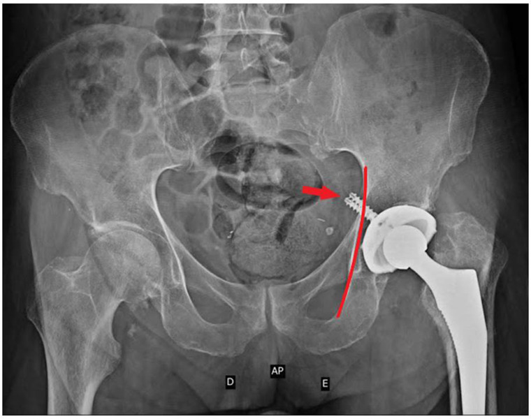
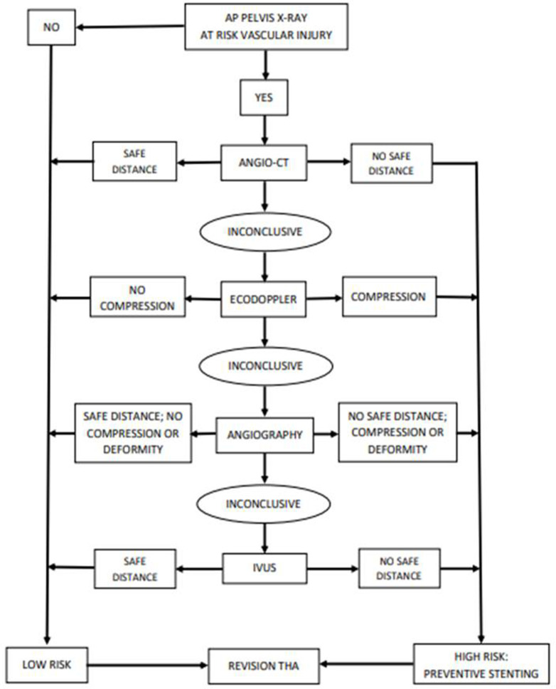
This study was approved by the Research Ethics Committee of Hospital de Clínicas de Porto Alegre, Brazil (no. 2020 to 0338). This study was conducted in accordance with guidelines by the National Institutes of Health. Between the patients submitted to revision THA in the period of 2015 to 2020, we identified ten with transoperative risk for vascular injury. We consider at risk to be when, in any radiological incidence (anteroposterior (AP), alar, or obturatriz), there were components or cement migration 5 mm beyond the ilioischial line (Figure 1). These ten patients were submitted to our investigative algorithm (vascular surgeon evaluation, angio-CT, ecodoppler, angiography, and intravascular ultrasound (IVUS)) and, after this reclassified as low or high risk (Figure 2); high risk means extremely close distance between orthopaedic device and blood vessel wall (< 5 mm and/or vessel deformity and/or vessel compression).
Fig. 1.
Pelvis anteroposterior radiograph demonstrating two screws (red arrow) overlapping the ilio ischiatic line (red line) > 5 mm. It is a red flag to vascular injury during revision total hip arthroplasty.
Fig. 2.
Flux diagram of progressive investigation of patients at vascular injury risk.
Those patients with a positive radiograph, i.e. a radiograph in which the ilioischial line is exceeded 5 mm or more by the orthopedic implant (screw, cage, cement, etc.), were submitted to abdomen and pelvis CT angiogram, followed by a vascular surgeon consultation. This evaluation was directed to a two-fold aim: to stablish the proximity between intrapelvic components to pelvic vessels in CT axial, coronal, and 3D reconstruction images, and to evaluate the inflammatory complex surrounding the pelvic components. If there was a clear tissue plane between those structures, the patient was cleared for surgery. On the other hand, if medial dislocation of vascular structures, a questionable plane or failure in obtaining clear images due to artefacts, the patient was submitted to a Duplex scan examination. Duple scan is ordered to evaluate images, flow velocities and configuration of arterial and venous waves. In case of technical difficulties (e.g. depth of the vessels, obesity, or overlying bowel gas) or a clear alteration in images or velocities, the patient was submitted for angiography. Most of the time, only venous angiography was performed since arterial lesions as compression or dislocation are rare and were easy to be ruled out in previous examinations.
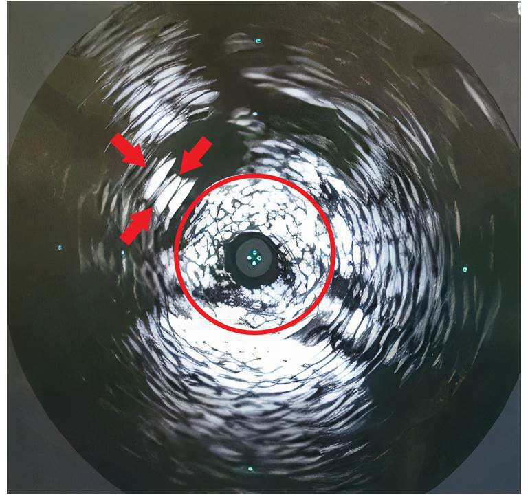
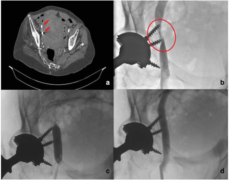
If it was not possible to observe a safe distance between vessels and orthopaedic implants, an IVUS was performed during the same procedure. IVUS allows the assessment of the vessel lumen, vessel wall, and their proximity to metallic components (Figure 3). In the absence of compression and identification of a tissue plane between vessel and component, the patient was cleared for surgery. When injury/dislocation of the iliac vein was depicted, a covered balloon-expandable nitinol stent was implanted. If the post-implantation IVUS examination showed a significant compression of the stent mesh, a second higher radial force venous dedicated stent was implanted inside the first stent (Figure 4).
Fig. 3.

Intravascular ultrasound image. Vein wall (red circle) is extremely close (< 5 mm) to the screw (red arrows).
Fig. 4.
a) CT pelvis showing intrapelvic screws (red arrows) on the right side.b) Venography demonstrating iliac external vein stenosis caused by screws. c) Venography showing balloon at the stenosis site.d) Venography after stent insertion. The image shows the full flow in iliac external vein. The patient was prescribed dual antiplatelet therapy with 100 mg of aspirin and 75 mg of clopidogrel daily for four weeks since the literature suggests that this a critical period o for early stent occlusion. Before surgery, antiplatelets were suspended one week before the planned surgery.
Results
After evaluation of the AP radiograph, alar oblique view, and obturator oblique view, 150 patients were evaluated and ten were identified as being at risk of vascular injury during transoperative time (Table I). All patients were submitted to our algorithm.
Table I.
High-risk patient profile.
| Sex | Age, yrs | Side | Initial risk | Final risk | Stent | Vessel at risk |
|---|---|---|---|---|---|---|
| Female | 78 | Right | High | Low | No | No vessel |
| Female | 59 | Right | High | High | Yes | Iliac extern vein |
| Female | 48 | Left | High | Low | No | No vessel |
| Female | 61 | Left | High | High | Yes | Iliac extern vein |
| Male | 72 | Right | High | Low | No | No vessel |
| Female | 66 | Right | High | Low | No | No vessel |
| Female | 56 | Left | High | High | Yes | Iliac extern vein |
| Female | 71 | Left | High | High | Yes | Iliac extern vein |
| Female | 67 | Right | High | High | Yes | Iliac extern vein |
| Female | 63 | Left | High | High | Yes | Iliac extern vein |
Six patients were recognized as being at high risk of vascular injury (distance ≤ 5 mm between vessels and implant), and a preventive preoperative stent was inserted before the revision THA. Four patients needed an inner reinforcing stent to recover vessel diameter.
In all patients submitted to stent insertion, the main vessel at risk was the extern iliac vein, particularly its transition to femoral common vein. There were no complications during stent insertion.
No patient submitted to stent insertion presented abnormal bleeding during surgery. There was no abnormal drop in red blood cell count and hematocrit in the postoperative period.
One patient presented deep venous thrombosis (DVT) in the ipsilateral leg seven days after the revision THA. It was treated with the institutional protocol for anticoagulation with success. There were no other complications.
Discussion
Despite being unusual, there are several reports of vascular injury during THA and revision THA. Abularrage et al 8 in 41,633 arthroplasties (knee and hip; primary and revision) identified lower limb arterial injuries in 0.20% of revision THAs. The risk factors identified were revision THA (odds ratio (OR) 2.7) and African American race. Shoenfeld et al 1 also considered that revision THA and intrapelvic implant migration were the main risk factors to vascular injury. It is estimated that 40% of vascular injuries in the hip occur during revision THA. 5,9
Most of the vascular injuries occured during revision THA intrapelvic implant removal, and were most commonly periprosthetic fibrosis, metallosis, and infection. Inflammatory tissue developing around the implants increases the vessel wall adherence and friability, resulting in higher vascular injury risk. Previous chronic vascular disease or irradiation could also contribute to worsening fragility of the vessel.
Revision THA planning begins with AP pelvis radiograph and lateral radiograph views. In a cadaveric study, Galat et al 10 found that the obturator and iliac oblique (Judet) views were most useful in defining screw position. The iliac oblique view clearly revealed screws that violated the quadrilateral surface and therefore were directed toward the obturator vessels and nerve. The obturator oblique view revealed screws that violated the anterior column and therefore were directed toward the external iliac vessels. The lateral view additionally clarified such screws by determining general anterior or posterior direction. However, we consider that the AP pelvis radiograph view can trigger the first red flag sign, mainly if any implant is breaking the ilioischial line.
It is important to highlight that some cement brands used in the past were not radiopaque. To avoid this pitfall, it is fundamental to observe in many radiological views if the characteristics of the femoral stem and the acetabular component are indicative of cemented implants. Review of the medical records, if available, for surgical description can also be helpful. The amount of extruded material is less important than the location of extrusion. This is particularly true in anteriorly extruded cement. This cement may not be as apparent on a standard AP view of the pelvis, but may place the femoral vessels at risk during extraction. 11
In some cases, the removal of intrapelvic implants, as reinforcement cages, cups, and metal back with important intrapelvic dislocation, is performed by abdominal approach. The surgical removal strategy depends on the implant shape, size, and location. The final result often is the increase in surgical time, complexity, and morbidity. 11
The initial goal in our algorithm was to identify radiological signs of risk for vascular injury during implant removal. Due to mortality and morbidity associated with acute vascular injuries or their late consequences, we considered high suspicion to be essential in all cases where any orthopaedic implant overtake the ilioischial line more than 5 mm.
We adopted 5 mm of distance in first x-rays view because this is the shortest measurement between the nearest blood vessel (extern iliac vein) and the bone landmarks, as observed by Kawasaki et al 4 in an angio-CT anatomical study. In these cases, after radiograph views, we always request an angio-CT. Periprosthetic join infection must be always investigated since there is an increased risk of vascular injury secondary to inflammatory local reaction. In addition, in cases in which a distance > 5 mm is not visualized between the closest blood vessel and the bone landmark, such as in superolateral migration with severe osteolysis without the presence of medially located metallic artifacts or cement, are of low risk for these lesions.
The angio-CT scan provides a better understanding of the relationship between major blood vessels and intrapelvic implants, as well as providing information of the relationship with other intrapelvic organs (bowel, bladder, ureter), and the extension of the inflammatory reaction caused by these components. 4,10 Whle it may have some limitations with metal or other materials that create artefacts, anatomical variant, or other local conditions (i.e. presence of gas in bowel), angio-CT still provides a lot of information without the necessity of invasive procedures.
In cases of inconclusive angio-CT, an arterial and venous duplex scan was ordered. However, in some patients, there are technical difficulties mainly because of deep vessels location, the presence of bowel gas, or an enlarged abdomen. When a good acoustic window was not possible to obtain, an indirect evaluation was performed. 12 Despite ultrasound limitations, we considered this method useful, especially because of its low cost and wide availability.
Another investigative tool is an invasive angiography and/or venography. Metallic artefacts may jeopardize a proper isualization of main vessels and, in this situation, an IVUS was performed. The IVUS allows us to identify the parietal vessel architecture and its relations with close structures (vessels, bone cement, or metal) up to 5 mm of distance. To our knowledge, this is the first study that uses IVUS to evaluate extrinsic vessels compression by metallic implants or cement.
If the safe distance between intrapelvic implant and vessel was 5 mm or less and/or there was a clear compression or lesion of the vessel, a percutaneous coated stent was inserted to protect the vessel and improve the blood flow.
Stenting can prevent acute (bleeding) and chronic (pseudoaneurysm and arteriovenous fistula) complications, as described by several authors. 9,13,14 On the other hand, preventive stenting was rarely reported in series or case reports. 7
In 2016, we began a pilot project with the main purpose of avoiding or diminishing the risk of vascular injury during revision THA. In our first two cases, immediately before the orthopaedic surgery, a sheath and guidewire were inserted in the vessel at risk. In the case of a vascular injury, a stent could be immediately placed. This procedure is very similar to that described by Asemota et al 6 in 2018. According to that author, the technique improved vascular visualization before revision THA, 2 allowed for precautionary placement of a vascular access sheath, 3 avoided arbitrary stent placement, and avoided the potential surgical complexity of repairing a stented vessel. 4
We have a different understanding related to Asemota et al. 6 First, the sheath and guidewire are located anterior in proximal thigh, and in cases of a posterolateral approach, it is necessary to change the patient position to implant the stent with precision. The time elapsed to place the patient in the proper position, ensure the presence of a vascular surgeon immediately (with all endovascular set of sheaths, guidewires and stents of several diameter and lengths), and the image setup may be very long; if an arterial lesion is present, this can be a life-threatening situation. Second, after stent placement, a dual antiplatelet therapy is necessary to avoid stent occlusion, therefore increasing the risk of bleeding specially when in combination with other anticoagulants. Third, in cases of arterial or venous laceration, it is difficult to cross the lesion with the stent and open surgery is sometimes necessary, increasing the risk. Fourth, an arbitrary stent placement could be avoided by an exhaustive preoperative investigation protocol.
The coated polyte-trafluoroethylene’s (PTFE) stent allows high security to revision THA. This stent decreases the risk of vessel laceration in transoperative time. In four patients, due to significative compression by the intrapelvic implant and gross secondary inflammatory reaction implantation of an inner reinforcing stent was necessary.
The arterial injury is the most common injury describing in the literature. In a review, Alshameeri et al 9 identified a total of 61 articles (51 individual case reports and ten case series). The vascular injuries have been reported in all the main vessels around the hip, and the most frequent vessels injured were the common femoral artery, extern iliac artery, profunda femoral artery, superior gluteal artery, and extern iliac vein. In their study, most injuries occurred in females and on the left side.
In our case series, the main vessel at risk was the vein, not the artery. More precisely, the external iliac vein transition to femoral common vein. Our findings match with the anatomical study conducted by Kawasaki et al. 4 These authors, in a well-conducted angio-CT study, evaluated the relation between the main vessel position and bone landmarks. According to their observations, the external iliac vessels were located closer to the pelvis as they exit the pelvic cavity and, in particular, the left side vessels were located closer to the pelvis than those of the right side at all levels. The external iliac veins were located notably closer to the pelvis than the arteries at all axial levels.
We agree with these observations, particularly because all our stents were inserted in external iliac veins. We believe that venous injuries have been under-reported in the literature. Venous bleeding can be stopped by the subsequent contained retroperitoneal haematoma that compress the vessel, and so be unnoticed during surgery. It seems possible that many postoperative lower limb oedemas and DVT could be result from a not diagnosed venous injury.
One patient developed DVT immediately after surgery. In this case, the interval between the stent placement and surgery was less than seven days, though we felt that the interval was overly short. After this case, we considered extremally necessary to wait at least 30 days between the vascular and the orthopaedic procedures. In this period, the patient must receive dual-platelet therapy.
In our orthopedics service, all the revisions performed used the posterolateral approach, and it is for this reason that we are careful to exclude risk factors for vascular injury. In the event of an arterial or venous injury, if the patient is in lateral decubitus, access to the iliac vessels is difficult, requiring a change of decubitus and prompt action by the vascular surgeon, aspects that increase time, bleeding, and risk to the patient.
Our study has many limitations. The low number of patients, absence of a control group, and a single surgical team study limits the use of recommendation widely. However, this investigation algorithm could be the first step in more robust multicentric studies to evaluate its efficiency in the vascular injury prevention.
In conclusion, the progressive investigation, beginning with a basic exam (radiograph), and the serial exams was useful in identify high risk patients to vascular injury during revision THA. The “preventive stent” could be an alternative to minimize vascular injury in revision THA.
Take home message
- In ten at-risk patients undergoing revision hip arthroplasty and submitted to our algorithm, six were recognized as being high risk to vascular injury during surgery.
- This article demonstrated it is useful to avoid blood vessels injury during revision total hip arthroplasty in high-risk patients.
Footnotes
Author contributions: C. V. Diesel: Conceptualization.
M. R. Guimarães: Software, Data curation.
S. M. Menegotto: Software, Data curation.
A.H Pereira: Software, Data curation.
A. A. Pereira: Software, Data curation.
L. H. Bertolucci: Software, Data curation, Writing – original draft.
E. C Freitas: Software, Data curation, Writing – original draft.
C. R. Galia: Supervision.
Funding statement: The authors received no financial or material support for the research, authorship, and/or publication of this article.
Open access funding: The authors report that the open access funding for this manuscript was self-funded.
Contributor Information
Cristiano V. Diesel, Email: cristianodiesel@gmail.com.
Marcelo R. Guimarães, Email: mrguima@yahoo.com.br.
Samuel M. Menegotto, Email: samuel_menegotto@yahoo.com.br.
Adamastor H. Pereira, Email: epereira@terra.com.br.
Alexandre A. Pereira, Email: ecfreitas@hcpa.edu.br.
Leonardo H. Bertolucci, Email: leonardohbertolucci@gmail.com.
Eduarda C. Freitas, Email: eduarda.freitas.hcpa@gmail.com.
Carlos R. Galia, Email: cgalia@hcpa.edu.br.
References
- 1. Shoenfeld NA, Stuchin SA, Pearl R, Haveson S. The management of vascular injuries associated with total hip arthroplasty. J Vasc Surg. 1990;11(4):549–555. 10.1016/0741-5214(90)90301-P [DOI] [PubMed] [Google Scholar]
- 2. Bach CM, Steingruber IE, Ogon M, Maurer H, Nogler M, Wimmer C. Intrapelvic complications after total hip arthroplasty failure. Am J Surg. 2002;183(1):75–79. 10.1016/s0002-9610(01)00845-5 [DOI] [PubMed] [Google Scholar]
- 3. Ovrum E, Dahl HK. Vessel and nerve injuries complicating total hip arthroplasty. Arch Orthop Trauma Surg (1978). 1979;95(4):267–269. 10.1007/BF00389697 [DOI] [PubMed] [Google Scholar]
- 4. Kawasaki Y, Egawa H, Hamada D, Takao S, Nakano S, Yasui N. Location of intrapelvic vessels around the acetabulum assessed by three-dimensional computed tomographic angiography: prevention of vascular-related complications in total hip arthroplasty. J Orthop Sci. 2012;17(4):397–406. 10.1007/s00776-012-0227-7 [DOI] [PubMed] [Google Scholar]
- 5. Girard J, Blairon A, Wavreille G, Migaud H, Senneville E. Total hip arthroplasty revision in case of intra-pelvic cup migration: designing a surgical strategy. Orthop Traumatol Surg Res. 2011;97(2):191–200. 10.1016/j.otsr.2010.10.003 [DOI] [PubMed] [Google Scholar]
- 6. Asemota D, Passano B, Feng JE, Novikov D, Anoushiravani AA, Schwarzkopf R. Preoperative optimization for vascular involvement complicating revision total hip arthroplasty. Arthroplast Today. 2018;4(4):411–416. 10.1016/j.artd.2018.02.003 [DOI] [PMC free article] [PubMed] [Google Scholar]
- 7. Tavares R de P, Arcenio Neto E, Taki W. Total hip revision arthroplasty of high-risk pelvic vascular injury associated with an endovascular approach: a case report. Rev Bras Ortop. 2018;53(5):626–631. 10.1016/j.rboe.2018.07.002 [DOI] [PMC free article] [PubMed] [Google Scholar]
- 8. Abularrage CJ, Weiswasser JM, Dezee KJ, Slidell MB, Henderson WG, Sidawy AN. Predictors of lower extremity arterial injury after total knee or total hip arthroplasty. J Vasc Surg. 2008;47(4):803–807. 10.1016/j.jvs.2007.11.067 [DOI] [PubMed] [Google Scholar]
- 9. Alshameeri Z, Bajekal R, Varty K, Khanduja V. Iatrogenic vascular injuries during arthroplasty of the hip. Bone Joint J. 2015;97-B(11):1447–1455. 10.1302/0301-620X.97B11.35241 [DOI] [PubMed] [Google Scholar]
- 10. Galat DD, Petrucci JA, Wasielewski RC. Radiographic evaluation of screw position in revision total hip arthroplasty. Clin Orthop Relat Res. 2004;419(419):124–129. 10.1097/00003086-200402000-00020 [DOI] [PubMed] [Google Scholar]
- 11. Fehring TK, Guilford WB, Baron J. Assessment of intrapelvic cement and screws in revision total hip arthroplasty. J Arthroplasty. 1992;7(4):509–518. 10.1016/s0883-5403(06)80072-0 [DOI] [PubMed] [Google Scholar]
- 12. Hui JZ, Goldman RE, Mabud TS, Arendt VA, Kuo WT, Hofmann LV. Diagnostic performance of lower extremity Doppler ultrasound in detecting iliocaval obstruction. J Vasc Surg Venous Lymphat Disord. 2020;8(5):821–830. 10.1016/j.jvsv.2019.12.074 [DOI] [PubMed] [Google Scholar]
- 13. Makar RR, Salem A, McGee H, Campbell D, Bateson P. Endovascular treatment of bleeding external iliac artery pseudo-aneurysm following control of haemorrhage with Sengstaken tube during revision total hip arthroplasty. Ann R Coll Surg Engl. 2007;89(5):W4–7. 10.1308/147870807X188452 [DOI] [PMC free article] [PubMed] [Google Scholar]
- 14. D’Angelo F, Piffaretti G, Carrafiello G, et al. Endovascular repair of a pseudo-aneurysm of the common femoral artery after revision total hip arthroplasty. Emerg Radiol. 2007;14(4):233–236. 10.1007/s10140-007-0605-1 [DOI] [PubMed] [Google Scholar]


