Abstract
Measurable residual disease (MRD) is the most powerful independent predictor of risk of relapse and long-term survival in adults and children with acute lymphoblastic leukemia (ALL). For almost all patients with ALL there is a reliable method to evaluate MRD, which can be done using multi-color flow cytometry, quantitative polymerase chain reaction to detect specific fusion transcripts or immunoglobulin/T-cell receptor gene rearrangements, and high-throughput next-generation sequencing. While next-generation sequencing-based MRD detection has been increasingly utilized in clinical practice due to its high sensitivity, the clinical significance of very low MRD levels (<10-4) is not fully characterized. Several new immunotherapy approaches including blinatumomab, inotuzumab ozogamicin, and chimeric antigen receptor T-cell therapies have demonstrated efficacy in eradicating MRD in patients with B-ALL. However, new approaches to target MRD in patients with T-ALL remain an unmet need. As our MRD detection assays become more sensitive and expanding novel therapeutics enter clinical development, the future of ALL therapy will increasingly utilize MRD as a criterion to either intensify or modify therapy to prevent relapse or de-escalate therapy to reduce treatment-related morbidity and mortality.
Introduction
Multi-agent chemotherapy and risk-adapted protocols revolutionized the management of pediatric acute lymphoblastic leukemia (ALL), and transformed this historically fatal disease into a curable entity in ~90% of children.1 Although outcomes continue to be inferior for adults with ALL relative to children, the successful adoption of pediatric-inspired intensive chemotherapy in the treatment of young adults (<40 years) with ALL has significantly improved overall survival with rates approaching 65-80%.2,3 Furthermore, novel immunotherapies such as the CD19-directed bi-specific T-cell engager blinatumomab, the anti-CD22 antibody-drug conjugate inotuzumab ozogamicin, and chimeric antigen receptor (CAR) T-cell therapies offer new treatment options for B-lineage ALL and are now being explored in the front-line setting.4-6 As our therapeutic armamentarium expands, accurate risk stratification at critical time points is essential to deliver optimal treatment to improve outcomes in adults with ALL. After standard multi-agent chemotherapy, the majority of adults will achieve complete morphological remission (CR).7, 8 However, response assessment is further refined by the evaluation of subclinical (not detected morphologically) measurable (also known as minimal) residual disease (MRD), which strongly and independently associates with outcomes across ALL studies and patient cohorts.9-12 Furthermore, MRD is increasingly incorporated into risk-adapted protocols in adult ALL populations, in which MRD may inform therapeutic decisions by identifying patients at high risk of relapse who may benefit from novel treatment approaches and/or allogeneic hematopoietic cell transplant (HCT), or patients with chemosensitive disease at lower risk of relapse who are likely to do well in the absence of HCT in first complete remission.13,14 The prognostic significance of MRD in adult ALL has been shown in both younger adults (<40 years) and older adults (>40 years).15,16
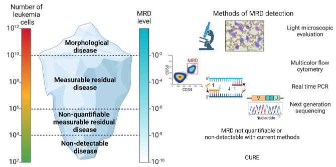
MRD is defined as measurable leukemia in a sample that is devoid of leukemia cells by morphological assessment with a light microscope. It is estimated that a patient presents with 1012 (a trillion) bone marrow leukemic blasts at diagnosis, and morphological assessment can detect approximately 1% blasts or more (Figure 1). Therefore, MRD theoretically represents anything less than 1010 cells (<1% blasts). In clinical practice, MRD is often used to describe any detectable leukemia below the traditional remission definition of 5% blasts by morphological assessment. However, as MRD detection assays have become more sensitive, it is generally recognized that an appropriate assay for the detection of ALL MRD in the clinic should be validated and reproducible at a sensitivity threshold of at least 10-4, or 0.01% leukemia cells in the bone marrow. The fundamental idea behind MRD interpretation is simple: the rate of decline in disease burden in response to systemic therapy is of prognostic value and a measure of risk for relapse, and intervening on lower levels of disease should result in improved outcomes. Herein, we review commonly used methods of MRD detection in ALL and provide clinical context and guidance to practising clinicians on how to interpret and intervene on MRD in adult ALL.
Figure 1.
Measurable residual disease assessment in acute lymphoblastic leukemia. Schematic representation of disease levels in acute lymphoblastic leukemia with corresponding measurable residual disease levels. This can resemble an “iceberg”, with different detection methods offering different levels of sensitivity and breadth of detecting residual disease. MRD: measurable residual disease; PCR: polymerase chain reaction.
Methods and technical aspects of measurable residual disease assessment in acute lymphoblastic leukemia
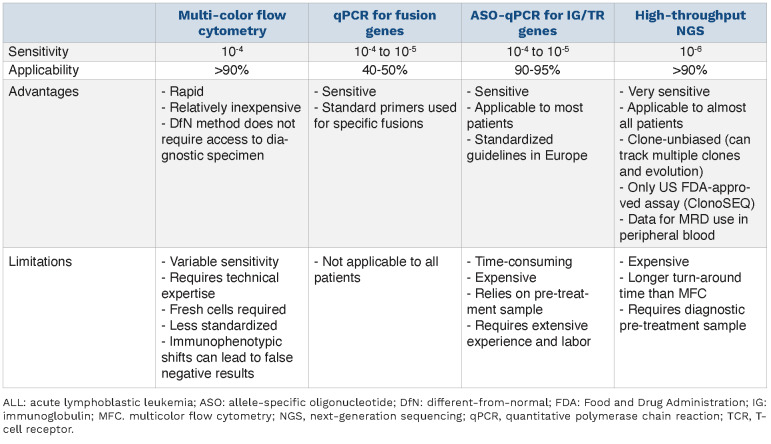
All MRD detection methods leverage features that are present exclusively in leukemic blasts to differentiate them from normal cells. Commonly used techniques include multicolor flow cytometry (MFC) to detect leukemic cells by immunophenotypic aberrancies, real-time quantitative polymerase chain reaction (qPCR) for detection of recurrent gene fusions (e.g., BCR-ABL1) or rearranged immunoglobulin (IG) and T-cell receptor (TCR) genes.17 A more recent technique which relies on high-throughput next-generation sequencing (NGS) may offer a more sensitive approach to detect IG and TCR rearrangements in ALL blasts.18 The main advantages and disadvantages of these MRD assessment methods are summarized in Table 1.
Multicolor flow cytometry
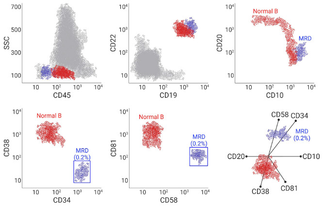
MFC is a fast and relatively inexpensive method that is broadly applicable to most ALL cases. It distinguishes leukemic cells based on their aberrant immunophenotype or leukemia-associated immunophenotype. A leukemia-associated immunophenotype can include antigen overor under-expression, asynchronous antigen expression, cross-lineage antigen expression, and ectopic pheno-types.16 It is necessary to obtain information about the immunophenotype at diagnosis in order to track it throughout the clinical course of an individual patient. However, these features may change under therapeutic pressure, and antigens may be lost or new antigens may be over-expressed as the leukemia evolves. To overcome this challenge, an alternative flow-based MRD approach named “different from normal” has been widely utilized.19 The “different from normal” approach involves a standardized panel of several markers that are used to distinguish leukemic cells from normal hematopoietic cells (Figure 2). The most common markers used to identify leukemic B lymphoblasts include CD10, CD19, CD45, CD34 and CD38. Leukemic cells often have high CD10 and low CD38 expression, which may distinguish them from hematogones. Aberrant myeloid marker expression (e.g., CD33, CD13, and CD15), or expression of CD9, CD73, and CD81 may also be helpful to define B-lymphoblasts.20 Clinical laboratories should be informed if the patient has received CD19-, CD20- or CD22-targeted therapies, as these markers may no longer be reliable in MRD detection for these patients. It is important to identify CD19 antigenic escape with flow MRD, since these patients would not benefit from further CD19-targeted therapies. In patients receiving anti-CD19 agents, other B-cell markers, such as CD20, CD22, and CD79a, can be used to identify the CD19-negative leukemia population, but can also lead to misidentification of normal B-cell precursors.21 Therefore, flow MRD should be used in conjunction with other methods of MRD evaluation (such as qPCR or NGS-based methods) for patients who have received anti-CD19 therapies. Clinicians and MRD laboratories should also be aware of the rare event of myeloid lineage switch after anti-CD19 therapies, which has been reported in both children and adults treated with blinatumomab or CAR T-cell therapies.22,23 This is more commonly observed in patients with KMT2A-rearranged ALL, but has also been seen in ALL with BCR-ABL1 trans-location.23 In these cases, flow cytometry may identify blasts expressing myeloid as opposed to lymphoid markers. Since these cases persistently harbor their cyto-genetic rearrangement at the time of myeloid relapse, complementing flow MRD with reverse transcriptase PCR (RT-PCR)-based MRD assessment can enable accurate diagnosis of this entity. Similarly, NGS-based MRD assessment may be helpful when flow-based MRD assessment has limitations in these cases. Finally, another recently described rare entity called switch ALL (swALL) may pose challenges for flow-based MRD detection.24 These precursor B-ALL arise from CD2+ lymphoblasts that do not harbor KMT2A rearrangements but have upregulated CEBPα activity. These cases are characterized by a switch between precursor B (CD19+ CD14–) and monocytoid (CD19– CD14+) immunophenotypes through a transdifferentiation mechanism involving alterations in the expression of CEBPα, PAX5, PU1 and GM-CSFR.25 This disease can be tracked by using IG gene rearrangements that are preserved throughout different switch states.
Table 1.
Comparison of techniques to measure residual disease in acute lymphoblastic leukemia.
CD34, TdT, CD7, cytoplasmic CD3, and CD1a are commonly utilized markers for flow-based MRD detection in T-ALL; however, MFC for residual T-lymphoblast detection is less developed compared to that for B-ALL.26
Although the sensitivity of MRD MFC from ALL bone marrow aspirate sampling approaches 10-4, results are dependent upon the quality of sample obtained, and the laboratory operator’s experience.27 The results of MFC-based MRD assessment may be optimized by treating samples with EDTA or heparin anti-coagulation and using 2-5 mL from the first pull of bone marrow aspirate. Since the degree of cellularity in the sample will affect the recorded number of events, up to 5 mL may be required for hypocellular remission samples. There is no evidence for unequal distribution of ALL cells in different parts of the bone marrow compartment as shown in studies of bilateral bone marrow sampling for MRD detection.28 Fresh samples sent for MFC should be processed within 24-48 hours of collection, and an advantage of MFC is the rapid reporting time for clinical results which may be returned to the clinician within 3 days of collection. Since MFC assays try to identify rare events, the number of events needed to be collected depends on the desired assay sensitivity and the optimal coefficient of variation as dictated by Poisson statistics.29 For ALL MRD, the recommended current threshold for clinical decisions is 0.01% (10-4) sensitivity. Thus, to obtain a coefficient of variation of 10%, 106 events must be acquired. Select reference laboratories can reach high sensitivity, as was reported in the PETHEMA study under the EuroFlow MRD Consortium.13,30 By using an optimized erythrocyte bulk-lysis protocol, bone marrow samples containing more than 107 cells can be lysed and resuspended in a small volume of buffer (~100 mL), with which one can achieve the 0.01-0.001% (10-4 to 10-5) sensitivity.19 This bulk-lysis protocol for sample preparation was combined with a two-tube next-generation flow approach by the EuroFlow Consortium, which also benefits from an optimized combination of fluoro-chromes and antibody reagents to increase specificity at very low MRD levels.31
Figure 2.
Multicolor flow cytometry as a method of measurable residual disease detection in acute lymphoblastic leukemia. An example showing multicolor flow cytometry of a bone marrow specimen, obtained after induction with chemotherapy plus rituximab (anti-CD20) in a patient with B-cell acute lymphoblastic leukemia (B-ALL). The measurable residual disease (MRD) population (0.2% of total events) is shown in blue, and distinguished from normal B cells and hematogones by its overexpression of CD10, CD58, and CD34, and underexpression of CD38 and CD81. CD20 expression is lost in leukemic blasts as a result of rituximab therapy. The radar plot visualization easily distinguishes the B-ALL MRD population.
Although MFC may be performed using peripheral blood, the ability to use blood as a source for MRD detection by MFC is limited by reduced sensitivity.
Real-time quantitative polymerase chain reaction
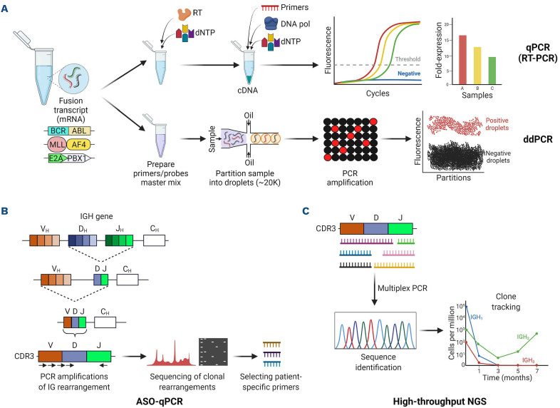
Chimeric gene fusions are major oncogenic drivers that can be found in ~40% of B-ALL, and some T-ALL cases. Since these rearrangements are oncogenic, they are often stable throughout the disease course, making them good targets for MRD assessment (Figure 3A). RT-PCR can be used to track BCR-ABL1, E2A-PBX1, KMT2A and CRLF2 rearrangements in B-ALL, as well as TAL1, TLX1, and TLX3 fusion transcripts in T-ALL.32 The advantage of fusion transcript detection is the availability of standardized universal primers specific for each fusion transcript, which simplifies the MRD detection process while offering a sensitivity of 10-4 to 10-5.33 Quantification of BCR-ABL1 transcripts is relatively straightforward, and p210 transcript quantification can be reported using an international standard (IS) because of the work done in chronic myeloid leukemia, while the IS score cannot be applied to the more common (in ALL) p190 transcript, so standard quantification of the BCR-ABL/ABL ratio is used. Fusion transcript detection with RT-PCR is an essential method of MRD assessment for patients with BCR-ABL and KMT2A rearrangements, since these patients may experience immunophenotypic shifts or myeloid lineage switch under the influence of CD19-directed antibody and CAR T-cell therapies.23
Figure 3.
Molecular methods of measurable residual disease detection in acute lymphoblastic leukemia. (A) Quantitative polymerase chain reaction (qPCR) and digital droplet PCR (ddPCR) can be used to detect fusion mRNA transcripts in acute lymphoblastic leukemia (ALL). (B) Allele-specific oligonucleotide qPCR (ASO-qPCR) leverages IG/TR gene rearrangements to detect measurable residual disease in ALL. It relies on identification of V(D)J sequences in a diagnostic sample, followed by the design of patient-specific primers for the sequences. (C) High-throughput next-generation sequencing (NGS) also targets specific V(D)J rearrangements, but it is fast and clone-unbiased as it uses multiplex PCR and does not require development of patient-specific primers.
Digital droplet PCR (ddPCR) is a third-generation PCR technology, which is based on separation of the sample into at least 20,000 water-oil emulsion droplets, followed by PCR amplification in each droplet. This technique is highly sensitive with improvement in the limit of detection and does not require a reference curve. Early studies suggest that ddPCR may have utility as a more sensitive MRD detection tool in Philadelphia chromosome-positive (Ph+) B-ALL.34,35
During early stages of lymphocyte development, B- and T-cell progenitors undergo somatic recombination of the variable (V), joining (J), and in some cases, diversity (D) gene segments of their IG and TCR genes, respectively.
Leukemic blasts arising from these precursor lymphoid cells contain clonal V(D)J rearrangements in more than 80% of cases.33 Given the specificity of the rearranged IG/TR DNA sequence for identification of leukemic cells, qPCR-based methods have been developed to track MRD. Allele-specific qPCR (ASO-qPCR) is a labor-intense procedure, which relies on the identification of “fingerprintlike” IG/TR V(D)J sequence(s) in a diagnostic leukemia sample, followed by development of patient-specific primers to assess for the presence of these specific sequence(s) in remission samples (Figure 3B).36 The initial characterization requires a panel of screening PCR using established primers for the complementarity-determining region 3 (CDR3), which is the hypervariable heavy chain region of IG/TR genes. This is then followed by Sanger sequencing of products to identify the clones and develop patient-specific primers. Depending on template availability, primer selection, and the amount of DNA in a given specimen, this patient-specific assay can have a sensitivity between 10-4 and 10-5.37 IG/TR-based methods are applicable to more than 90% of ALL cases, with reduced sensitivity in early T-precursor ALL, since the latter often arises from more immature progenitors that have not undergone TCR rearrangement. In addition to its laborious and time-consuming methodology, ASO-qPCR may be limited by the loss or emergence of new V(D)J sequences, leading to false negative MRD results.38
In a study in which both BCR-ABL1 level and IG/TR ASO-qPCR were monitored in the bone marrow of Ph+ B-ALL patients, overall concordance between the two methods was ~70%,39 but IG/TR was found to be more reliable at predicting outcomes. However, another study investigating the discrepancy between the two methods showed that some patients with persistent BCR-ABL1 transcript levels may in fact contain the fusion gene in myeloid cells, indicating a chronic myeloid leukemia-like stem cell disease, which may be missed by ASO/qPCR.40 In the GRAAPH-2014 study, 38% of adults with de novo Ph+ ALL had residual BCR-ABL1 positivity during treatment, related to BCR-ABL1 “clonal hematopoiesis”.41 The presence of BCR-ABL1 clonal hematopoiesis was not associated with poorer outcome, and patients with residual clonal hematopoiesis did not benefit from allogeneic HCT. Therefore, RT-PCR and IG/TR MRD techniques may complement each other and should be monitored simultaneously in Ph+ ALL patients in order to guide therapeutic decisions.
Several studies compared the sensitivity of MFC- and qPCR-based MRD assessment in peripheral blood versus bone marrow samples. In B-ALL, MRD can be up to three-logs lower in the peripheral blood than in bone marrow, indicating the importance of marrow assessments when these methods are used to track MRD.42 In contrast, MRD levels in T-ALL are roughly equivalent between the two sources, which may in part be explained by the thymic origin of this disease.43
High-throughput next-generation sequencing
Recent advances in NGS have led to a great interest in developing NGS-based MRD methods, which can potentially increase the sensitivity of detection. These assays include sequencing the IG/TR gene V(D)J rearrangements as in ASO-qPCR, but have the capability of simultaneously amplifying multiple combinations of rearranged IG/TR genes by multiplex PCR without the need for patient-specific primers (Figure 3C).44 Leveraging the high capacity of NGS, a larger picture of the IG/TR repertoire can be interrogated in one experiment, enabling the detection of clones that are at lower frequency at diagnosis but expand later in the disease trajectory.45 Unlike ASO-qPCR, NGS-based IG/TR MRD is not clone-biased, meaning that new clones may be identified as they emerge in remission and relapse samples if the necessary clonal evolution and new clone identification tools are included in the assessment.46 The sensitivity of NGS MRD approximates 0.0001% (10-6) in the bone mar-row;47 however, the significance of MRD below the traditional threshold of 10-4 has not been defined across different clinical scenarios. Similar to flow MRD, the sensitivity of NGS MRD depends on the amount of input, which is the amount of DNA in this method. The analysis of NGS MRD data also requires a bioinformatic pipeline. The U.S. Food and Drug Administration approved the use of ClonoSEQ NGS technology (Adaptive Biotechnologies, Seattle, WA, USA) as an MRD assessment method in ALL.48 Studies have demonstrated concordance of NGS-based ALL MRD assessment using bone marrow and peripheral blood; thus, NGS may potentially enable MRD quantification from peripheral blood without the need for frequent bone marrow sampling.49,50 In addition to RT-PCR and ASO-qPCR, NGS-based MRD may represent another platform to track the disease in the setting of immunophenotypic shifts or myeloid lineage switch. Since it is an IG/TR-based method, NGS MRD technology may have lower sensitivity in early T-precursor-ALL, as was the case for ASO-qPCR.
Emerging methods
Routine clinical MRD assessment for a given patient often includes at least two methods described above (e.g., flow MRD plus RT-PCR and/or IG/TR-based methods), which provide complementary information and are advantageous for overcoming the limitations of one method. EuroClonality-NGS DNA capture (EuroClonality-NDC) is an emerging assay that is designed as an integrated tool to characterize IG/TR rearrangements, chromosomal translocations, copy number alterations, and somatic mutations through a standardized pipeline.51 This assay is a robust tool providing a single workflow for detection of B- and T-cell clonality, as well as fusion transcripts and sequence variants.
As the technology advances, several innovative approaches are promising to improve the technical aspects of current MRD assessment methods, including new flow MRD methods (e.g., spectral flow cytometry which allows the simultaneous analysis of multiple surface markers), novel MFC panels, cell-free DNA-based methods, and novel NGS strategies (e.g., ALL-specific mutation detection). However, none of these techniques has robust clinical validation in ALL yet, therefore we will not discuss them within the scope of this review.
Clinical significance of measurable residual disease in adult acute lymphoblastic leukemia
Prognostication in ALL has historically relied on baseline leukemia characteristics including white blood cell count at diagnosis, immunophenotype, and cytogenetic abnormalities.52 Increasingly, studies have demonstrated that MRD response to front-line therapy outweighs traditional prognostic parameters and has emerged as the strongest independent predictor of ALL outcomes.30,53-55 In a meta-analysis of 39 publications, comprising 13,637 patients of all ages and ALL subtypes, achieving MRD negativity (by flow or qPCR) was associated with hazard ratios of 0.25 (0.24-0.33) for event-free survival and 0.28 (0.19-0.41) for overall survival.56 The authors also showed the significant prognostic utility of MRD at varying time points of assessment regardless of the MRD detection method used.
Monitoring measurable residual disease during frontline multi-agent chemotherapy
The addition of MRD to the response assessment following front-line multi-agent chemotherapy is standard of care in the management of ALL.57 Given that the traditional therapeutic course of ALL tends to include up to 12 months of intensive chemotherapy followed by 1.5-2.5 years of maintenance, understanding the optimal timing and frequency of MRD assessment is important to the delivery of high quality ALL care. Few studies have actually reported the utility of ongoing interval MRD monitoring in patients achieving MRD-negative response. The German Multicenter Study Group on Adult ALL (GMALL) evaluated a cohort of adults with ALL in first complete remission and found that the median time between emergence of MRD by ASO-qPCR and clinical relapse was approximately 4 months, suggesting that the interval between MRD assessment time-points should likely be 3 months or less, and that ongoing MRD monitoring may provide a potential window for clinical intervention prior to fulminant disease relapse.58 Given that the majority of ALL relapses occur within the first 3 years following diagnosis,59 MRD monitoring for early relapse detection is likely most critical during this time period.
Typically, MRD is first assessed at the completion of remission induction, which occurs 2-4 weeks following therapy initiation depending on the treatment regimen utilized. Patients who achieve MRD negativity (at least <10-4) at this early time point are considered to have chemosensitive disease, which is associated with excellent disease-free survival, and this information guides the initiation of consolidation therapy.2 For these patients, we generally recommend that MRD be assessed both following induction therapy and following consolidation therapy. For adults who receive a pediatric inspired regimen, current data suggest that persistence of MRD following consolidation therapy (usually 12-16 weeks into treatment) may be the time-point with greatest relevance for making a change in treatment. Alternative therapy for MRD persistence includes blinatumomab in patients with B-ALL, given the high rates of reported MRD clearance in the BLAST trial among patients with persistent MRD and good outcomes for responding patients, the majority of whom proceeded to allogeneic transplant.60 Currently, the U.S. Intergroup A041501 study (NCT03150693) is evaluating the incorporation of inotuzumab ozogamicin into a pediatric inspired regimen with the aim of early MRD eradication following induction therapy to improve event-free survival. Unfortunately, there is currently no proven MRD “eraser” for patients with T-ALL. For these patients, persistence of MRD following consolidation or within 3 months of therapy initiation is a signal to escalate therapy and to strongly consider allogeneic HCT in transplant-eligible adults. MRD should be assessed at shorter intervals, if possible, in patients with persistent, or newly detectable MRD, as impending clinical relapse is often likely.
NGS MRD offers a new and sensitive platform for MRD detection, yet we have limited data for the prognostic significance of low-level (between 10-4 and 10-6) MRD after multi-agent chemotherapy. The recent GMALL 07/2003 study looked at the outcomes of patients who had persistent NGS MRD below the 10-4 detection level after consolidation therapy.61 Patients with undetectable NGS-MRD had significantly better overall survival when compared to patients with positive NGS-MRD. Similarly, a recent retrospective analysis looking at 84 Ph-negative B-ALL patients in complete remission by flow MRD showed that 38% were MRD-positive by the ClonoSEQ NGS method.62 Patients who were MRD negative by the NGS method had significantly lower risk of relapse, which also translated into longer overall survival. Monitoring of MRD for Ph+ ALL may be performed by a variety of methods, but RT-PCR for BCR-ABL1 transcripts is convenient and commonly used in the USA. For adult patients with Ph+ ALL receiving intensive chemotherapy in combination with second- or third-generation tyrosine kinase inhibitors, MRD response at 3 months following therapy has been shown to be highly informative. In a retrospective study conducted by the group at the MD Anderson Cancer Center, among 85 adult Ph+ ALL patients who received hyper-CVAD plus a tyrosine kinase inhibitor, those who achieved complete molecular remission by RT-PCR at 3 months had an excellent overall survival even without HCT.63 A phase II study of hyper-CVAD plus ponatinib in Ph+ B-ALL demonstrated higher MRD-negative complete remission rates when compared to the hyper-CVAD plus dasatinib historic control group.64 The role of MRD response following chemotherapy-free/light regimens for Ph+ ALL remains unclear but MRD response will likely also be important in identifying patients in need of intensification and/or additional therapeutic interventions.
Measurable residual disease and allogeneic hematopoietic cell transplantation
Allogeneic HCT is currently indicated for adults with relapsed or refractory ALL or patients who demonstrate persistent MRD following frontline induction/consolidation therapy. Several studies have shown that achieving an MRD-negative state prior to allogeneic HCT improves relapse-free survival and overall survival. However, the depth of MRD response prior to traditional myeloablative allogeneic HCT may be important. For example, in an international study of 616 ALL patients who underwent allogeneic HCT, patients with low MRD had similar outcomes to those without MRD pre-trans-plant.65 MRD positivity after allogeneic HCT was more important than pre-HCT MRD positivity, thus close monitoring with sensitive techniques is warranted. In a smaller study of adult patients with ALL, the presence of pre-transplant MRD at >10-4 detected by an NGS-based method predicted a 7.7-times higher risk of relapse after transplantation.66 The prognostic impact of MRD has been demonstrated across transplant donor source and conditioning regimens.67,68 It is clear that further therapy should be considered to deepen response prior to allogeneic HCT if MRD is present at >10-4; an open question remains whether additional clearance of MRD present at <10-4 is necessary to achieve successful transplant outcomes.
Serial tracking of MRD following allogeneic HCT also predicts for impending disease relapse. In a study of 43 adult patients who underwent HCT for ALL, those with detectable qPCR-based MRD on day +100 had an 80% relapse risk compared to a 7% risk in patients with undetectable post-HCT MRD.69 In another small study of adult ALL HCT recipients, detection of MRD by NGS at any level following HCT heralded clinical relapse; the median time between MRD detection and relapse was 3 months.66 Finally, MRD tracking using peripheral blood following HCT in adult ALL has been shown to be feasible and convenient, to correlate closely with bone marrow NGS-based MRD assessment, and to predict for clinical relapse.49
Measurable residual disease in relapsed/refractory acute lymphoblastic leukemia and novel therapies
In addition to the robust evidence in newly diagnosed ALL, prognostic value of MRD for patients in second complete remission and beyond has also been demonstrated.70 MRD has also played an important role in establishing eligibility and monitoring treatment response in key studies of novel therapies such as blinatumomab, inotuzumab ozogamicin, and CAR T-cell therapy.4,5,71 Not only did MRD-positive ALL define the inclusion criteria for the BLAST trial, leading to an acceptance of MRD as an indication for blinatumomab in BALL, but MRD was also shown in secondary analyses to predict for relapse-free survival and overall survival in patients with relapsed/refractory ALL achieving complete remission following blinatumomab.72 In the landmark phase III INO-VATE trial comparing inotuzumab ozogamicin versus standard chemotherapy in relapsed/refractory B-ALL, the MRD-negative complete remission rate, assessed by MFC (10-4 sensitivity), was 46% in patients treated with inotuzumab.73 Patients who had MRD response had longer overall survival and progression-free survival than patients with persistent MRD. Among children receiving CAR T-cell therapy, real-world datasets have demonstrated improved survival and reduced toxicity following CAR T-cell therapy when administered to patients with lower level/MRD-positive disease, as opposed to patients in full clinical relapse. Although the MRD-negative response rate following CAR T-cell therapy in adult ALL is very high (approximated at 81% in a meta-analysis including 489 adults receiving CAR T-cell therapies across published trials),74 whether early MRD response to CAR T-cell therapy translates into long-term durable remissions in adult ALL patients remains unclear.
Re-emergence or persistence of MRD in patients with Ph+ BALL might be associated with the selection of clones harboring ABL1 kinase domain mutations (e.g., T315I).75 It is often difficult to identify the specific resistance mutation given the low level of the transcript in patients without morphological relapse. Blinatumomab has been shown to be effective in eradicating MRD in Ph+ ALL.76 A common strategy would be to combine blinatumomab with ponatinib, since the latter has activity against most of the common resistance mutations of ABL1.64
Conclusions
Incorporation of MRD assessment is a fundamental component of risk-directed therapy in ALL. For almost all patients with ALL there is a reliable method to assess residual disease, which remains the most powerful independent predictor of outcomes. Blinatumomab, inotuzumab ozogamicin and CAR T cells are highly effective immunotherapy approaches in B-ALL; each is currently being explored alone and in combinations as a mechanism for MRD eradication earlier in the course of disease. The development of novel, active agents for T-ALL patients with persistent MRD remains an unmet need. As MRD detection assays become increasingly more sensitive and additional novel therapeutics enter clinical development, the future of ALL management in adults will require nuanced and evidence-based, risk-adapted treatment approaches to guide intensification or de-escalation of therapy.
Acknowledgments
The figures in this review were prepared with BioRender. CS is supported by an American Society of Hematology Research Training Award for Fellows.
References
- 1.Pui CH, Evans WE. A 50-year journey to cure childhood acute lymphoblastic leukemia. Semin Hematol. 2013;50(3):185-196. [DOI] [PMC free article] [PubMed] [Google Scholar]
- 2.Stock W, Luger SM, Advani AS, et al. A pediatric regimen for older adolescents and young adults with acute lymphoblastic leukemia: results of CALGB 10403. Blood. 2019;133(14):1548-1559. [DOI] [PMC free article] [PubMed] [Google Scholar]
- 3.Wieduwilt MJ, Stock W, Advani A, et al. Superior survival with pediatric-style chemotherapy compared to myeloablative allogeneic hematopoietic cell transplantation in older adolescents and young adults with Ph-negative acute lymphoblastic leukemia in first complete remission: analysis from CALGB 10403 and the CIBMTR. Leukemia. 2021;35(7):2076-2085. [DOI] [PMC free article] [PubMed] [Google Scholar]
- 4.Kantarjian H, Stein A, Gokbuget N, et al. Blinatumomab versus chemotherapy for advanced acute lymphoblastic leukemia. N Engl J Med. 2017;376(9):836-847. [DOI] [PMC free article] [PubMed] [Google Scholar]
- 5.Kantarjian HM, DeAngelo DJ, Stelljes M, et al. Inotuzumab ozogamicin versus standard therapy for acute lymphoblastic leukemia. N Engl J Med. 2016;375(8):740-753. [DOI] [PMC free article] [PubMed] [Google Scholar]
- 6.Maude SL, Laetsch TW, Buechner J, et al. Tisagenlecleucel in children and young adults with B-cell lymphoblastic leukemia. N Engl J Med. 2018;378(5):439-448. [DOI] [PMC free article] [PubMed] [Google Scholar]
- 7.Kantarjian H, Thomas D, O'Brien S, et al. Long-term follow-up results of hyperfractionated cyclophosphamide, vincristine, doxorubicin, and dexamethasone (Hyper-CVAD), a doseintensive regimen, in adult acute lymphocytic leukemia. Cancer. 2004;101(12):2788-2801. [DOI] [PubMed] [Google Scholar]
- 8.Sive JI, Buck G, Fielding A, et al. Outcomes in older adults with acute lymphoblastic leukaemia (ALL): results from the international MRC UKALL XII/ECOG2993 trial. Br J Haematol. 2012;157(4):463-471. [DOI] [PMC free article] [PubMed] [Google Scholar]
- 9.Bartram J, Wade R, Vora A, et al. Excellent outcome of minimal residual disease-defined low-risk patients is sustained with more than 10 years follow-up: results of UK paediatric acute lymphoblastic leukaemia trials 1997-2003. Arch Dis Child. 2016;101(5):449-454. [DOI] [PubMed] [Google Scholar]
- 10.Borowitz MJ, Devidas M, Hunger SP, et al. Clinical significance of minimal residual disease in childhood acute lymphoblastic leukemia and its relationship to other prognostic factors: a Children's Oncology Group study. Blood. 2008;111(12):5477-5485. [DOI] [PMC free article] [PubMed] [Google Scholar]
- 11.Vora A, Goulden N, Wade R, et al. Treatment reduction for children and young adults with low-risk acute lymphoblastic leukaemia defined by minimal residual disease (UKALL 2003): a randomised controlled trial. Lancet Oncol. 2013;14(3):199-209. [DOI] [PubMed] [Google Scholar]
- 12.Bassan R, Bruggemann M, Radcliffe HS, Hartfield E, Kreuzbauer G, Wetten S. A systematic literature review and meta-analysis of minimal residual disease as a prognostic indicator in adult B-cell acute lymphoblastic leukemia. Haematologica. 2019;104(10):2028-2039. [DOI] [PMC free article] [PubMed] [Google Scholar]
- 13.Ribera JM, Morgades M, Ciudad J, et al. Chemotherapy or allogeneic transplantation in high-risk Philadelphia chromosome-negative adult lymphoblastic leukemia. Blood. 2021;137(14):1879-1894. [DOI] [PubMed] [Google Scholar]
- 14.Dhedin N, Huynh A, Maury S, et al. Role of allogeneic stem cell transplantation in adult patients with Ph-negative acute lymphoblastic leukemia. Blood. 2015;125(16):2486-2496; quiz 2586. [DOI] [PubMed] [Google Scholar]
- 15.Gokbuget N, Kneba M, Raff T, et al. Adult patients with acute lymphoblastic leukemia and molecular failure display a poor prognosis and are candidates for stem cell transplantation and targeted therapies. Blood. 2012;120(9):1868-1876. [DOI] [PubMed] [Google Scholar]
- 16.van Dongen JJ, van der Velden VH, Bruggemann M, Orfao A. Minimal residual disease diagnostics in acute lymphoblastic leukemia: need for sensitive, fast, and standardized technologies. Blood. 2015;125(26):3996-4009. [DOI] [PMC free article] [PubMed] [Google Scholar]
- 17.Chen X, Wood BL. How do we measure MRD in ALL and how should measurements affect decisions. Re: ireatment and prognosis? Best Pract Res Clin Haematol. 2017;30(3):237-248. [DOI] [PubMed] [Google Scholar]
- 18.Ladetto M, Bruggemann M, Monitillo L, et al. Next-generation sequencing and real-time quantitative PCR for minimal residual disease detection in B-cell disorders. Leukemia. 2014;28(6):1299-1307. [DOI] [PubMed] [Google Scholar]
- 19.Theunissen P, Mejstrikova E, Sedek L, et al. Standardized flow cytometry for highly sensitive MRD measurements in B-cell acute lymphoblastic leukemia. Blood. 2017;129(3):347-357. [DOI] [PMC free article] [PubMed] [Google Scholar]
- 20.Shaver AC, Seegmiller AC. B lymphoblastic leukemia minimal residual disease assessment by flow cytometric analysis. Clin Lab Med. 2017;37(4):771-785. [DOI] [PubMed] [Google Scholar]
- 21.Mikhailova E, Semchenkova A, Illarionova O, et al. Relative expansion of CD19-negative very-early normal B-cell precursors in children with acute lymphoblastic leukaemia after CD19 targeting by blinatumomab and CAR-T cell therapy: implications for flow cytometric detection of minimal residual disease. Br J Haematol. 2021;193(3):602-612. [DOI] [PubMed] [Google Scholar]
- 22.Haddox CL, Mangaonkar AA, Chen D, et al. Blinatumomab-induced lineage switch of B-ALL with t(4:11)(q21;q23) KMT2A/AFF1 into an aggressive AML: pre- and post-switch phenotypic, cytogenetic and molecular analysis. Blood Cancer J. 2017;7(9):e607. [DOI] [PMC free article] [PubMed] [Google Scholar]
- 23.Rossi JG, Bernasconi AR, Alonso CN, et al. Lineage switch in childhood acute leukemia: an unusual event with poor outcome. Am J Hematol. 2012;87(9):890-897. [DOI] [PubMed] [Google Scholar]
- 24.Slamova L, Starkova J, Fronkova E, et al. CD2-positive B-cell precursor acute lymphoblastic leukemia with an early switch to the monocytic lineage. Leukemia. 2014;28(3):609-620. [DOI] [PubMed] [Google Scholar]
- 25.Novakova M, Zaliova M, Fiser K, et al. DUX4r, ZNF384r and PAX5-P80R mutated B-cell precursor acute lymphoblastic leukemia frequently undergo monocytic switch. Haematologica. 2021;106(8):2066-2075. [DOI] [PMC free article] [PubMed] [Google Scholar]
- 26.van Dongen JJ, Lhermitte L, Bottcher S, et al. EuroFlow antibody panels for standardized n-dimensional flow cytometric immunophenotyping of normal, reactive and malignant leukocytes. Leukemia. 2012;26(9):1908-1975. [DOI] [PMC free article] [PubMed] [Google Scholar]
- 27.Kalina T, Flores-Montero J, van der Velden VH, et al. EuroFlow standardization of flow cytometer instrument settings and immunophenotyping protocols. Leukemia. 2012;26(9):1986-2010. [DOI] [PMC free article] [PubMed] [Google Scholar]
- 28.van der Velden VH, Hoogeveen PG, Pieters R, van Dongen JJ. Impact of two independent bone marrow samples on minimal residual disease monitoring in childhood acute lymphoblastic leukaemia. Br J Haematol. 2006;133(4):382-388. [DOI] [PubMed] [Google Scholar]
- 29.Hedley BD, Keeney M. Technical issues: flow cytometry and rare event analysis. Int J Lab Hematol. 2013;35(3):344-350. [DOI] [PubMed] [Google Scholar]
- 30.Ribera JM, Oriol A, Morgades M, et al. Treatment of high-risk Philadelphia chromosome-negative acute lymphoblastic leukemia in adolescents and adults according to early cytologic response and minimal residual disease after consolidation assessed by flow cytometry: final results of the PETHEMA ALL-AR-03 trial. J Clin Oncol. 2014;32(15):1595-1604. [DOI] [PubMed] [Google Scholar]
- 31.Pedreira CE, Costa ES, Lecrevisse Q, van Dongen JJ, Orfao A, EuroFlow Consortium. Overview of clinical flow cytometry data analysis: recent advances and future challenges. Trends Biotechnol. 2013;31(7):415-425. [DOI] [PubMed] [Google Scholar]
- 32.van Dongen JJ, Macintyre EA, Gabert JA, et al. Standardized RT-PCR analysis of fusion gene transcripts from chromosome aberrations in acute leukemia for detection of minimal residual disease. Report of the BIOMED-1 Concerted Action: investigation of minimal residual disease in acute leukemia. Leukemia. 1999;13(12):1901-1928. [DOI] [PubMed] [Google Scholar]
- 33.Bartram J, Patel B, Fielding AK. Monitoring MRD in ALL: methodologies, technical aspects and optimal time points for measurement. Semin Hematol. 2020;57(3):142-148. [DOI] [PubMed] [Google Scholar]
- 34.Ansuinelli M, Della Starza I, Lauretti A, et al. Applicability of droplet digital polymerase chain reaction for minimal residual disease monitoring in Philadelphia-positive acute lymphoblastic leukaemia. Hematol Oncol. 2021;39(5):680-686. [DOI] [PMC free article] [PubMed] [Google Scholar]
- 35.Coccaro N, Anelli L, Zagaria A, et al. Droplet digital PCR is a robust tool for monitoring minimal residual disease in adult Philadelphia-positive acute lymphoblastic leukemia. J Mol Diagn. 2018;20(4):474-482. [DOI] [PubMed] [Google Scholar]
- 36.van der Velden VH, van Dongen JJ. MRD detection in acute lymphoblastic leukemia patients using Ig/TCR gene rearrangements as targets for real-time quantitative PCR. Methods Mol Biol. 2009;538:115-150. [DOI] [PubMed] [Google Scholar]
- 37.van der Velden VH, Cazzaniga G, Schrauder A, et al. Analysis of minimal residual disease by Ig/TCR gene rearrangements: guidelines for interpretation of real-time quantitative PCR data. Leukemia. 2007;21(4):604-611. [DOI] [PubMed] [Google Scholar]
- 38.Correia RP, Bento LC, de Sousa FA, Barroso RS, Campregher PV, Bacal NS. How I investigate minimal residual disease in acute lymphoblastic leukemia. Int J Lab Hematol. 2021;43(3):354-363. [DOI] [PubMed] [Google Scholar]
- 39.Cazzaniga G, De Lorenzo P, Alten J, et al. Predictive value of minimal residual disease in Philadelphia-chromosome-positive acute lymphoblastic leukemia treated with imatinib in the European intergroup study of post-induction treatment of Philadelphia-chromosome-positive acute lymphoblastic leukemia, based on immunoglobulin/T-cell receptor and BCR/ABL1 methodologies. Haematologica. 2018;103(1):107-115. [DOI] [PMC free article] [PubMed] [Google Scholar]
- 40.Hovorkova L, Zaliova M, Venn NC, et al. Monitoring of childhood ALL using BCR-ABL1 genomic breakpoints identifies a subgroup with CML-like biology. Blood. 2017;129(20):2771-2781. [DOI] [PubMed] [Google Scholar]
- 41.Rathana KIM, Rousselot P, Cayuela JM, et al. Frequency and outcome of Philadelphia chromosome-positive acute lymphoblastic leukemia with BCR-ABL1 clonal hematopoiesis after blast clearance: results from the Graaph-2014 trial. Blood. 2021;138(Supplement 1):3478. [Google Scholar]
- 42.Coustan-Smith E, Sancho J, Hancock ML, et al. Use of peripheral blood instead of bone marrow to monitor residual disease in children with acute lymphoblastic leukemia. Blood. 2002;100(7):2399-2402. [DOI] [PubMed] [Google Scholar]
- 43.van der Velden VH, Jacobs DC, Wijkhuijs AJ, et al. Minimal residual disease levels in bone marrow and peripheral blood are comparable in children with T cell acute lymphoblastic leukemia (ALL), but not in precursor-B-ALL. Leukemia. 2002;16(8):1432-1436. [DOI] [PubMed] [Google Scholar]
- 44.Bartram J, Mountjoy E, Brooks T, et al. Accurate sample assignment in a multiplexed, ultrasensitive, high-throughput sequencing assay for minimal residual disease. J Mol Diagn. 2016;18(4):494-506. [DOI] [PubMed] [Google Scholar]
- 45.Bartram J, Goulden N, Wright G, et al. High throughput sequencing in acute lymphoblastic leukemia reveals clonal architecture of central nervous system and bone marrow compartments. Haematologica. 2018;103(3):e110-e114. [DOI] [PMC free article] [PubMed] [Google Scholar]
- 46.Wu J, Jia S, Wang C, et al. Minimal residual disease detection and evolved IGH clones analysis in acute B lymphoblastic leukemia using IGH deep sequencing. Front Immunol. 2016;7:403. [DOI] [PMC free article] [PubMed] [Google Scholar]
- 47.Faham M, Zheng J, Moorhead M, et al. Deep-sequencing approach for minimal residual disease detection in acute lymphoblastic leukemia. Blood. 2012;120(26):5173-5180. [DOI] [PMC free article] [PubMed] [Google Scholar]
- 48.Monter A, Nomdedeu JF. ClonoSEQ assay for the detection of lymphoid malignancies. Expert Rev Mol Diagn. 2019;19(7):571-578. [DOI] [PubMed] [Google Scholar]
- 49.Muffly L, Sundaram V, Chen C, et al. Concordance of peripheral blood and bone marrow measurable residual disease in adult acute lymphoblastic leukemia. Blood Adv. 2021;5(16):3147-3151. [DOI] [PMC free article] [PubMed] [Google Scholar]
- 50.Sala Torra O, Othus M, Williamson DW, et al. Next-generation sequencing in adult B cell acute lymphoblastic leukemia patients. Biol Blood Marrow Transplant. 2017;23(4):691-696. [DOI] [PMC free article] [PubMed] [Google Scholar]
- 51.Stewart JP, Gazdova J, Darzentas N, et al. Validation of the EuroClonality-NGS DNA capture panel as an integrated genomic tool for lymphoproliferative disorders. Blood Adv. 2021;5(16):3188-3198. [DOI] [PMC free article] [PubMed] [Google Scholar]
- 52.Wetzler M, Dodge RK, Mrozek K, et al. Prospective karyotype analysis in adult acute lymphoblastic leukemia: the Cancer and Leukemia Group B experience. Blood. 1999;93(11):3983-3993. [PubMed] [Google Scholar]
- 53.Bruggemann M, Raff T, Kneba M. Has MRD monitoring superseded other prognostic factors in adult ALL? Blood. 2012;120(23):4470-4481. [DOI] [PubMed] [Google Scholar]
- 54.Holowiecki J, Krawczyk-Kulis M, Giebel S, et al. Status of minimal residual disease after induction predicts outcome in both standard and high-risk Ph-negative adult acute lymphoblastic leukaemia. The Polish Adult Leukemia Group ALL 4-2002 MRD study. Br J Haematol. 2008;142(2):227-237. [DOI] [PubMed] [Google Scholar]
- 55.Bruggemann M, Raff T, Flohr T, et al. Clinical significance of minimal residual disease quantification in adult patients with standard-risk acute lymphoblastic leukemia. Blood. 2006;107(3):1116-1123. [DOI] [PubMed] [Google Scholar]
- 56.Berry DA, Zhou S, Higley H, et al. Association of minimal residual disease with clinical outcome in pediatric and adult acute lymphoblastic leukemia: a meta-analysis. JAMA Oncol. 2017;3(7):e170580. [DOI] [PMC free article] [PubMed] [Google Scholar]
- 57.Brown PA, Shah B, Advani A, et al. Acute lymphoblastic leukemia, Version 2.2021, NCCN clinical practice guidelines in oncology. J Natl Compr Canc Netw. 2021;19(9):1079-1109. [DOI] [PubMed] [Google Scholar]
- 58.Raff T, Gokbuget N, Luschen S, et al. Molecular relapse in adult standard-risk ALL patients detected by prospective MRD monitoring during and after maintenance treatment: data from the GMALL 06/99 and 07/03 trials. Blood. 2007;109(3):910-915. [DOI] [PubMed] [Google Scholar]
- 59.Stock W, Johnson JL, Stone RM, et al. Dose intensification of daunorubicin and cytarabine during treatment of adult acute lymphoblastic leukemia: results of Cancer and Leukemia Group B study 19802. Cancer. 2013;119(1):90-98. [DOI] [PMC free article] [PubMed] [Google Scholar]
- 60.Gokbuget N, Dombret H, Bonifacio M, et al. Blinatumomab for minimal residual disease in adults with B-cell precursor acute lymphoblastic leukemia. Blood. 2018;131(14):1522-1531. [DOI] [PMC free article] [PubMed] [Google Scholar]
- 61.Kotrova M, Koopmann J, Trautmann H, et al. Prognostic value of low-level MRD in adult acute lymphoblastic leukemia detected by low- and high-throughput methods. Blood Adv. 2022;6(10):3006-3010. [DOI] [PMC free article] [PubMed] [Google Scholar]
- 62.Short NJ, Kantarjian HM, Patel K, et al. Ultrasensitive next-generation sequencing-based measurable residual disease assessment in Philadelphia chromosome-negative acute lymphoblastic leukemia after frontline therapy: correlation with flow cytometry and impact on clinical outcomes. Blood. 2020;136(Suppl 1):26-28. [Google Scholar]
- 63.Short NJ, Jabbour E, Sasaki K, et al. Impact of complete molecular response on survival in patients with Philadelphia chromosome-positive acute lymphoblastic leukemia. Blood. 2016;128(4):504-507. [DOI] [PMC free article] [PubMed] [Google Scholar]
- 64.Jabbour E, Short NJ, Ravandi F, et al. Combination of hyper-CVAD with ponatinib as first-line therapy for patients with Philadelphia chromosome-positive acute lymphoblastic leukaemia: long-term follow-up of a single-centre, phase 2 study. Lancet Haematol. 2018;5(12):e618-e627. [DOI] [PMC free article] [PubMed] [Google Scholar]
- 65.Bader P, Salzmann-Manrique E, Balduzzi A, et al. More precisely defining risk peri-HCT in pediatric ALL: pre- vs post-MRD measures, serial positivity, and risk modeling. Blood Adv. 2019;3(21):3393-3405. [DOI] [PMC free article] [PubMed] [Google Scholar]
- 66.Logan AC, Vashi N, Faham M, et al. Immunoglobulin and T cell receptor gene high-throughput sequencing quantifies minimal residual disease in acute lymphoblastic leukemia and predicts post-transplantation relapse and survival. Biol Blood Marrow Transplant. 2014;20(9):1307-1313. [DOI] [PMC free article] [PubMed] [Google Scholar]
- 67.Baron F, Labopin M, Ruggeri A, et al. Impact of detectable measurable residual disease on umbilical cord blood transplantation. Am J Hematol. 2020;95(9):1057-1065. [DOI] [PubMed] [Google Scholar]
- 68.Yoon JH, Min GJ, Park SS, et al. Minimal residual disease-based long-term efficacy of reduced-intensity conditioning versus myeloablative conditioning for adult Philadelphiapositive acute lymphoblastic leukemia. Cancer. 2019;125(6):873-883. [DOI] [PubMed] [Google Scholar]
- 69.Spinelli O, Peruta B, Tosi M, et al. Clearance of minimal residual disease after allogeneic stem cell transplantation and the prediction of the clinical outcome of adult patients with high-risk acute lymphoblastic leukemia. Haematologica. 2007;92(5):612-618. [DOI] [PubMed] [Google Scholar]
- 70.Saygin C, Papadantonakis N, Cassaday RD, et al. Prognostic impact of incomplete hematologic count recovery and minimal residual disease on outcome in adult acute lymphoblastic leukemia at the time of second complete response. Leuk Lymphoma. 2018;59(2):363-371. [DOI] [PubMed] [Google Scholar]
- 71.Turtle CJ, Hanafi LA, Berger C, et al. CD19 CAR-T cells of defined CD4+:CD8+ composition in adult B cell ALL patients. J Clin Invest. 2016;126(6):2123-2138. [DOI] [PMC free article] [PubMed] [Google Scholar]
- 72.Zugmaier G, Gokbuget N, Klinger M, et al. Long-term survival and T-cell kinetics in relapsed/refractory ALL patients who achieved MRD response after blinatumomab treatment. Blood. 2015;126(24):2578-2584. [DOI] [PMC free article] [PubMed] [Google Scholar]
- 73.Jabbour E, Gokbuget N, Advani A, et al. Impact of minimal residual disease status in patients with relapsed/refractory acute lymphoblastic leukemia treated with inotuzumab ozogamicin in the phase III INO-VATE trial. Leuk Res. 2020;88:106283. [DOI] [PubMed] [Google Scholar]
- 74.Grover P, Veilleux O, Tian L, et al. Chimeric antigen receptor-T cell therapy in adults with B-cell acute lymphoblastic leukemia: a systematic review. Blood Adv. 2022;6(5):1608-1618. [DOI] [PMC free article] [PubMed] [Google Scholar]
- 75.Soverini S, De Benedittis C, Papayannidis C, et al. Drug resistance and BCR-ABL kinase domain mutations in Philadelphia chromosome-positive acute lymphoblastic leukemia from the imatinib to the second-generation tyrosine kinase inhibitor era: the main changes are in the type of mutations, but not in the frequency of mutation involvement. Cancer. 2014;120(7):1002-1009. [DOI] [PubMed] [Google Scholar]
- 76.Martinelli G, Boissel N, Chevallier P, et al. Complete hematologic and molecular response in adult patients with relapsed/refractory Philadelphia chromosome-positive B-precursor acute lymphoblastic leukemia following treatment with blinatumomab: results from a phase II, single-arm, multicenter study. J Clin Oncol. 2017;35(16):1795-1802. [DOI] [PubMed] [Google Scholar]


