Abstract
The presence of invasion-inhibitory activity that is regulated by the transcriptional activator ExsA of cytotoxic Pseudomonas aeruginosa has previously been proposed. The results of this study show that both ExoT and ExoS, known type III secreted effector proteins of P. aeruginosa that are regulated by ExsA, possess this activity. Invasion was reduced 94.4% by ExoT and 96.0% by ExoS. Invasion-inhibitory activity is not linked to ADP-ribosylation activity, at least for ExoS, since a noncatalytic mutant also inhibits uptake by an epithelial cell line (invasion was reduced 96.0% by ExoSE381A).
The earliest events in most Pseudomonas aeruginosa infections occur at the epithelium. Depending on their interaction with epithelial cells in vitro, clinical isolates of P. aeruginosa may be classed as cytotoxic or invasive, and these phenotypes correspond to distinct genotypes (10). Acute epithelial cell death induced by cytotoxic P. aeruginosa has been demonstrated to be due to the expression of ExoU, delivered via type III secretion, that is regulated by the transcriptional activator ExsA (7). Wild-type cytotoxic strains do not invade epithelial cells. An exoU mutant of P. aeruginosa (PA103exoU::Tn5Tc) is noncytotoxic but remains noninvasive (5, 12). A subsequent mutation in exsA results in an invasive phenotype (5). This result suggests that there is at least one ExsA-regulated gene encoding the inhibition of invasion in cytotoxic P. aeruginosa.
Inhibition of P. aeruginosa uptake by epithelial cells.
YopE and YopH, secreted via type III secretion by Yersinia spp., prevent uptake by eukaryotic cells (15). Furthermore, YopH is essential for antiphagocytic function (14). YopH is a protein tyrosine phosphatase (11) that has been demonstrated to prevent uptake of other microorganisms (26).
P. aeruginosa PA103 was used to demonstrate a similar capacity by cytotoxic P. aeruginosa to block uptake of other bacteria. To allow invasion levels to be quantified, a noncytotoxic mutant, PA103exoU (Table 1), was used for these experiments. Invasion of a rabbit corneal epithelial (RCE) cell line was determined as previously described (9). RCE cells were fed with modified SHEM (13), containing bovine pituitary extract (5 μg/ml) in place of cholera toxin. For most assays, cells were cultured on 24-well plates and used between 3 and 6 days after passage. Cells were washed with phosphate-buffered saline (PBS) and inoculated with 200 μl of bacterial suspension (in modified Eagle's medium with Earle's salts and l-glutamine, buffered with 1 M HEPES-NaOH [pH 7.6], 0.35 g of NaHCO3 per liter, and 6 g of bovine serum albumin per liter [Sigma, St. Louis, Mo.]). Infected cells were then incubated for 3 h at 37°C in 5% CO2. The bacterial suspension was then carefully aspirated, and the wells were washed with two sequential 0.5-ml aliquots of PBS (Sigma) to remove nonassociated bacteria. Adherent extracellular bacteria were killed by incubation for 1 h after the addition of 1 ml of gentamicin (200 μg/ml). The antibiotic was aspirated, and the excess was removed by one wash of 1 ml of PBS before the cells were lysed with 0.025% Triton X-100. The number of intracellular bacteria was determined by culturing serial dilutions of the lysate.
TABLE 1.
Strains of P. aeruginosa used in this study
| Strain | Relevant characteristic(s) | Reference |
|---|---|---|
| PA103exoU::Tc | PA103exoU; transposon mutant of cytotoxic strain; lacks ExoU | 7 |
| PA103exoU::Tc/exsA::Ω | PA103exoUexsA; lacks entire ExsA-regulated system | 5 |
| 6294 | Clinical isolate; invasive phenotype | 9 |
| 6487 | Clinical isolate; invasive phenotype | 9 |
| PA103exoUexoT::Tc pUCP | PA103exoUexoT; lacks ExoU and ExoT; contains control plasmid encoding Cbr | 17 |
| PA103exoUexoT::Tc | ExoT expressed in trans | 17 |
| pUCPexoT | ||
| PA103ΔexoUexoT::Tc | ExoS expressed in trans | 17 |
| pUCPexoS | ||
| PA103ΔexoUexoT::Tc | Noncatalytic mutant ExoS, expressed in trans | 17 |
| pUCPexoSE381A |
To demonstrate uptake inhibition by cytotoxic P. aeruginosa, PA103exoU was coincubated with invasive strains of P. aeruginosa. The uptake of invasive strains 6294 and 6487 and an invasive mutant of PA103 (PA103exoUexsA) (Table 1) was compared to the uptake of these strains in the presence of PA103exoU. An inoculum of 107 CFU was used for PA103 mutants, and the inoculum for strains 6294 and 6487 was reduced to 2 × 10⁶ CFU as these strains are significantly more invasive (10). As a control, the effect of PA103exoUexsA (which lacks invasion-inhibitory activity) on the invasion of 6294 was also examined. Different strains of internalized bacteria were distinguished in viable counts by plating duplicate aliquots on nonselective medium (MacConkey agar) and selective medium (tryptic soy agar [Difco, Detroit, Mich.] with 100 μg of tetracycline per ml or 10 μg of streptomycin per ml [Sigma]).
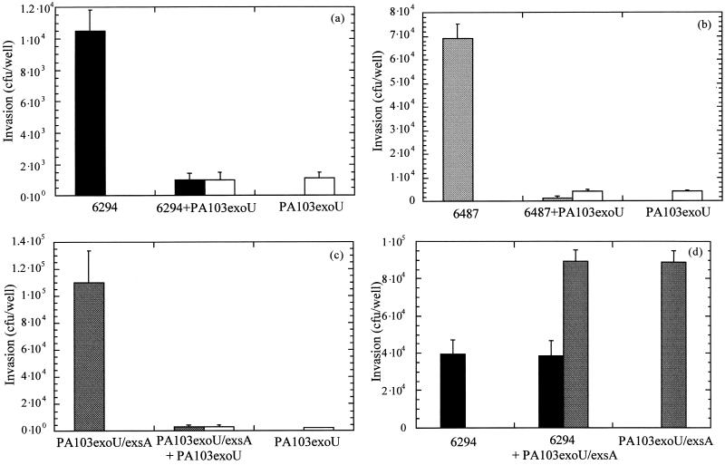
PA103exoU inhibited uptake of all three invasive P. aeruginosa strains tested (P < 0.001 for all strains) (Fig. 1a to c). In contrast, PA103exoUexsA was not able to inhibit uptake of 6294 (P = 0.062; Mann-Whitney U test) (Fig. 1d).
FIG. 1.
PA103exoU inhibits uptake of invasive strains
of P. aeruginosa. PA103exoU was coincubated with
invasive strains of P. aeruginosa during infection of RCE
cells, and uptake was quantified. PA103exoU (□) inhibited
the invasion of all strains assayed when coincubated with 6294 (■)
(a), 6487 (░⃞) (b), and PA103exoUexsA
( ) (c).
PA103exoUexsA (█) did not inhibit invasion when
coincubated with 6294 (■) (d).
Levels of bacterial association with RCE cells (i.e., the sum of adherent and invasive bacteria) were determined for both PA103exoU and PA103exoUexsA as previously described (9). Bacteria were incubated with RCE as described above, and then cells were washed vigorously three times with PBS before the cells were lysed with Triton X-100 and bacteria were enumerated. There was no significant difference between the abilities of PA103exoU and PA103exoUexsA to associate with cells (mean ± standard error, [1.40 ± 0.32] × 106 CFU/well compared to [1.19 ± 0.24] × 106 CFU/well, respectively; P = 0.1489 by the Mann-Whitney U test). This suggested that uptake inhibition by PA103exoU was not due to an inability to associate with the host cell membrane.
A likely candidate for an invasion inhibitor might be a Pseudomonas homolog to YopH, a type III effector that inhibits uptake of Yersinia. A TBLASTN 2.0.8 search of the published genome of P. aeruginosa failed to find a region homologous to YopH (1) or to the catalytic region that is invariantly conserved among protein tyrosine phosphatases such as YopH (11).
Of the type III secreted effector proteins that have been reported for P. aeruginosa (19, 20), only ExoU and ExoT are produced by the cytotoxic strain PA103. ExoU has been shown to be essential for acute cytotoxicity; ExoT is not required (7).
ExoT and ExoS possess invasion inhibitory activity.
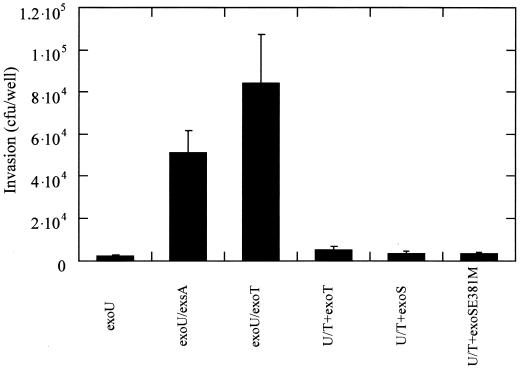
To investigate the role of ExoT in uptake inhibition, the ability of a noncytotoxic mutant of PA103 with a second mutation in exoT (PA103exoUexoT) (Table 1) to invade corneal epithelial cells was compared to that of PA103exoU. PA103exoUexoT invaded epithelial cells 40-fold more efficiently than PA103exoU (Fig. 2). When exoT was complemented back in trans (Table 1), inhibition of invasion was restored (Fig. 2).
FIG. 2.
Uptake of PA103exoU mutants by RCE cells. Bacterial strains were incubated for 3 h with RCE cells before the determination of intracellular bacteria. PA103exoU is noninvasive, and further mutation of exsA or exoT resulted in an invasive phenotype. Complementation with exoT, exoS, or exoSE381A restored invasion-inhibitory activity.
ExoT and ExoS belong to the ADP-ribosyltransferase family. Although they have 75% amino acid identity, ExoT possesses only 0.2% enzymatic activity compared to ExoS (18). Interestingly, both exoS and a noncatalytic version of exoS (Table 1) complemented the invasion-inhibitory activity that was lost by mutating exoT in PA103exoU (Fig. 2), suggesting that ADP-ribosylating activity is not required for the inhibition of uptake, at least by ExoS.
Actin microfilaments are involved in the ability of epithelial cells to take up P. aeruginosa (8). It was recently reported by Vallis and coworkers (17) that ExoT, ExoS, and the noncatalytic mutant ExoSE381A caused morphological changes to CHO cells without apparent membrane damage. In the present study, RCE cells were also found to display rounded morphology, but not trypan blue staining, after infection with bacteria expressing these proteins (data not shown). These changes to host cell morphology suggest that the cytoskeleton may be affected by these proteins, and this might explain the cells' loss of ability to take up bacteria.
ExoT inhibits uptake of an invasive strain of P. aeruginosa.
Unlike PA103exoU, PA103exoUexoT was not able to block uptake of the invasive strain 6294 by RCE cells (Table 2). Complementation with exoT in trans restored the ability of PA103exoUexoT to block RCE cell uptake of 6294. This result demonstrated that ExoT in a cytotoxic strain can function to block invasion of invasive P. aeruginosa.
TABLE 2.
Uptake of 6294 during coincubation with PA103 mutants
| Coincubation of 6294 with: | % Relative uptake of 6294 ± SDa |
|---|---|
| PA103exoU | 2.54 ± 1.20 |
| PA103exoU/exsA | 107.63 ± 12.63 |
| PA103exoU/exoT | 95.65 ± 23.01 |
| PA103exoU/exoT with exoT in trans | 1.09 ± 0.38 |
Uptake of 6294 when coincubated with PA103 mutant strains expressed as a percentage relative to uptake of 6294 incubated alone.
The results of this study demonstrated that both ExoT and ExoS can inhibit the uptake of a cytotoxic strain of P. aeruginosa by epithelial cells. Interestingly, invasive P. aeruginosa encodes and secretes both of these proteins, yet these strains invade efficiently. Furthermore, mutation of exsA in an invasive strain (PAO1) does not significantly affect its ability to invade (10). There are a number of possible explanations for this. P. aeruginosa produces exoenzyme S as a heterologous aggregate of ExoS and ExoT (17). Possibly, the interaction of these two effectors results in the loss of their invasion-inhibitory activity. Alternatively, invasive strains might also encode a suppressor of ExoT and ExoS or an effector that has a more dominant and positive effect on invasion than ExoT or ExoS.
A third possibility is that the ExsA-regulated type III secretion system is not activated by cell contact with corneal epithelial cells in invasive P. aeruginosa. Vallis and coworkers (16) have shown that there are differences in stimulation of the ExsA-regulated system between low-calcium conditions and the presence of serum or cell contact with CHO cells. Although ExoT and ExoS are secreted by invasive P. aeruginosa under conditions inducing growth (such as low calcium levels), they may not necessarily be secreted upon contact with corneal epithelial cells in culture.
What is the role of inhibition of invasion by ExoT in the pathogenesis of cytotoxic P. aeruginosa? There are considerable similarities between the type III secretion systems of P. aeruginosa and those of Yersinia. In this study, we have shown that ExoT inhibits uptake of P. aeruginosa, similar to the function of YopH of Yersinia spp. A strain of Yersinia pseudotuberculosis lacking YopH remains cytotoxic (14), and an exoT mutant of P. aeruginosa also remains cytotoxic (7). However, a yopH deletion mutant of Y. pseudotuberculosis is avirulent in a murine model of intraperitoneal infection, while an exoT mutant of P. aeruginosa remains virulent in an acute lung infection model (3, 7). The presence of ExoU, which is associated with acute cytotoxicity, may mask any effect of ExoT in this model. It is clear, however, that ExsA-regulated proteins other than ExoU are involved in virulence. An exsA mutant of invasive P. aeruginosa is avirulent or less virulent in acute infection models (4, 7). Invasive P. aeruginosa does not encode ExoU, indicating an important role for other effectors, at least in this class of P. aeruginosa strains at some stage of the infectious process.
ExoT is the only type III effector protein reported to date that is encoded by both invasive and cytotoxic P. aeruginosa strains. The conservation of this gene across strains would suggest an important role in survival; this may involve the control of phagocytosis by eukaryotic cells.
Acknowledgments
This work was supported by a Bausch & Lomb PostDoctoral Fellowship to B.A.C., grants AI-31665 and AI-01289 to D.W.F. from the National Institute of Allergy and Infectious Diseases, National Institutes of Health, and grant RO1-EY11221 to S.M.J.F. from the National Eye Institute, National Institutes of Health.
REFERENCES
- 1.Altschul S F, Madden T L, Schäffer A A, Zhang J, Zhang Z, Miller W, Lipman D J. Gapped BLAST and PSI-BLAST: a new generation of protein database search programs. Nucleic Acids Res. 1997;25:3389–3402. doi: 10.1093/nar/25.17.3389. [DOI] [PMC free article] [PubMed] [Google Scholar]
- 2.Andersson K, Carballeira N, Magnusson K-E, Persson C, Stendahl O, Wolf-Watz H, Fällman M. YopH of Yersinia pseudotuberculosisinterrupts early phosphotyrosine signaling associated with phagocytosis. Mol Microbiol. 1996;20:1057–1069. doi: 10.1111/j.1365-2958.1996.tb02546.x. [DOI] [PubMed] [Google Scholar]
- 3.Bölin I, Wolf-Watz H. The plasmid-encoded Yop2b protein of Yersinia pseudotuberculosisis a virulence determinant regulated by calcium and temperature at the level of transcription. Mol Microbiol. 1988;2:237–245. doi: 10.1111/j.1365-2958.1988.tb00025.x. [DOI] [PubMed] [Google Scholar]
- 4.Cowell B A, Wu C, Fleiszig S M J. Use of an animal model in studies of bacterial corneal infection. Inst Lab Anim Res J. 1999;40:43–50. doi: 10.1093/ilar.40.2.43. [DOI] [PubMed] [Google Scholar]
- 5.Evans D J, Frank D W, Finck-Barbançon V, Wu C, Fleiszig S M J. Pseudomonas aeruginosainvasion and cytotoxicity are independent events, both of which involve protein tyrosine kinase activity. Infect Immun. 1998;66:1453–1459. doi: 10.1128/iai.66.4.1453-1459.1998. [DOI] [PMC free article] [PubMed] [Google Scholar]
- 6.Fällman M, Andersson K, Håkansson S, Magnusson K-E, Stendahl O, Wolf-Watz H. Yersinia pseudotuberculosisinhibits Fc receptor-mediated phagocytosis in J774 cells. Infect Immun. 1995;63:3117–3124. doi: 10.1128/iai.63.8.3117-3124.1995. [DOI] [PMC free article] [PubMed] [Google Scholar]
- 7.Finck-Barbançon V, Goranson J, Zhu L, Sawa T, Wiener-Kronish J P, Fleiszig S M J, Wu C, Mende-Mueller L, Frank D W. ExoU expression by Pseudomonas aeruginosacorrelates with acute cytotoxicity and epithelial injury. Mol Microbiol. 1997;25:547–557. doi: 10.1046/j.1365-2958.1997.4891851.x. [DOI] [PubMed] [Google Scholar]
- 8.Fleiszig S M J, Zaidi T S, Pier G B. Pseudomonas aeruginosainvasion of and multiplication within corneal epithelial cells in vitro. Infect Immun. 1995;63:4072–4077. doi: 10.1128/iai.63.10.4072-4077.1995. [DOI] [PMC free article] [PubMed] [Google Scholar]
- 9.Fleiszig S M J, Zaidi T S, Preston M J, Grout M, Evans D J, Pier G B. The relationship between cytotoxicity and epithelial cell invasion by corneal isolates of Pseudomonas aeruginosa. Infect Immun. 1996;64:2288–2294. doi: 10.1128/iai.64.6.2288-2294.1996. [DOI] [PMC free article] [PubMed] [Google Scholar]
- 10.Fleiszig S M J, Wiener-Kronish J P, Miyazaki H, Vallas V, Mostov K E, Kanada D, Sawa T, Yen T S B, Frank D W. Pseudomonas aeruginosa-mediated cytotoxicity and invasion correlate with distinct genotypes at the loci encoding exoenzyme S. Infect Immun. 1997;65:579–586. doi: 10.1128/iai.65.2.579-586.1997. [DOI] [PMC free article] [PubMed] [Google Scholar]
- 11.Guan K, Dixon J E. Protein tyrosine phosphatase activity of an essential virulence determinant in Yersinia. Science. 1990;249:553–556. doi: 10.1126/science.2166336. [DOI] [PubMed] [Google Scholar]
- 12.Hauser A R, Fleiszig S M J, Kang P J, Mostov K, Engel J N. Defects in type III secretion correlate with internalization of Pseudomonas aeruginosaby epithelial cells. Infect Immun. 1998;66:1413–1420. doi: 10.1128/iai.66.4.1413-1420.1998. [DOI] [PMC free article] [PubMed] [Google Scholar]
- 13.Jumblatt M M, Neufeld A H. β-Adrenergic and serotonergic responsiveness of rabbit corneal epithelial cells in culture. Investig Ophthalmol Vis Sci. 1983;24:1139–1143. [PubMed] [Google Scholar]
- 14.Rosqvist R, Bölin I, Wolf-Watz H. Inhibition of phagocytosis in Yersinia pseudotuberculosis: a virulence plasmid-encoded ability involving the Yop2b protein. Infect Immun. 1988;56:2139–2143. doi: 10.1128/iai.56.8.2139-2143.1988. [DOI] [PMC free article] [PubMed] [Google Scholar]
- 15.Rosqvist R, Forsberg Å, Rimpiläinen M, Bergman T, Wolf-Watz H. The cytotoxic protein YopE of Yersiniaobstructs the primary host defence. Mol Microbiol. 1990;4:657–667. doi: 10.1111/j.1365-2958.1990.tb00635.x. [DOI] [PubMed] [Google Scholar]
- 16.Vallis A J, Yahr T L, Barbieri J T, Frank D W. Regulation of ExoS production and secretion by Pseudomonas aeruginosain response to tissue culture conditions. Infect Immun. 1999;67:914–920. doi: 10.1128/iai.67.2.914-920.1999. [DOI] [PMC free article] [PubMed] [Google Scholar]
- 17.Vallis A J, Finck-Barbançon V, Yahr T L, Frank D W. Biological effects of Pseudomonas aeruginosatype III-secreted proteins on CHO cells. Infect Immun. 1999;67:2040–2044. doi: 10.1128/iai.67.4.2040-2044.1999. [DOI] [PMC free article] [PubMed] [Google Scholar]
- 18.Yahr T L, Barbieri J T, Frank D W. Genetic relationship between the 53- and 49-kilodalton forms of exoenzyme S from Pseudomonas aeruginosa. J Bacteriol. 1996;178:1412–1419. doi: 10.1128/jb.178.5.1412-1419.1996. [DOI] [PMC free article] [PubMed] [Google Scholar]
- 19.Yahr T L, Mende-Mueller L M, Friese M B, Frank D W. Identification of type III secreted products of the Pseudomonas aeruginosaexoenzyme S regulon. J Bacteriol. 1997;179:7165–7168. doi: 10.1128/jb.179.22.7165-7168.1997. [DOI] [PMC free article] [PubMed] [Google Scholar]
- 20.Yahr T L, Vallis A J, Hancock M K, Barbieri J T, Frank D W. ExoY, a novel adenylate cyclase secreted by the Pseudomonas aeruginosatype III system. Proc Natl Acad Sci USA. 1998;95:13899–13904. doi: 10.1073/pnas.95.23.13899. [DOI] [PMC free article] [PubMed] [Google Scholar]


