Abstract
Case summary
An 11-year-old female spayed Burmese cat was presented for chronic intermittent vomiting. Previous medical and dietary treatments were unsuccessful. Endoscopically, a narrow pyloric outlet was found and Y-U pyloroplasty was performed. The clinical signs disappeared postoperatively after treatment with a proton pump inhibitor and dietary management.
Relevance and novel information
This is the first case report of feline-acquired pyloric stenosis with documented histopathological findings from a full-thickness biopsy of the pyloric sphincter. In addition, this is the first case of Y-U pyloroplasty being conducted in a cat. The histopathological findings might help explain the pathogenesis of this disorder in cats.
Keywords: Feline pyloric stenosis, pyloric smooth muscle hyperplasia, pyloric fibrosis, feline Y-U pyloroplasty
Introduction
Chronic vomiting is a common complaint in feline practice and is presented in a variety of gastrointestinal and extragastrointestinal disorders.1 Pyloric stenosis has been infrequently described as a cause in cats, and can be a congenital or acquired disorder.2–6
Case description
An 11-year-old female spayed Burmese cat presented with a history of several years of chronic intermittent vomiting of undigested food. Clinical signs had begun with vomiting 7 years previously and increased in frequency so that by the time of presentation, the cat was vomiting chronically a few times a week. Vomiting with food generally occurred directly after food ingestion (within 15–30 mins) but occasionally after a few hours without food. Diet changes advised by the referring veterinarian (renal diet, hypoallergenic diet, mono-protein diet) had not been effective. PCR was negative for Helicobacter species in the faeces. Previous medical treatment with a proton pump inhibitor (Lansoprazol AL; Aliud Pharma) and sucralfate (Sucrabest; Combustin) was unsuccessful. The cat was showing increasing discomfort after eating and had one episode of haematemesis, and was referred for further evaluation and endoscopy.
On presentation, the patient was alert, responsive and had a normal body condition. A systolic heart murmur grade II/VI was found on auscultation.
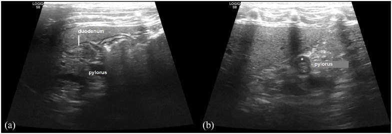
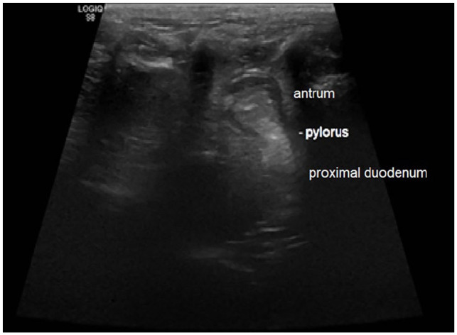
Ultrasonographic examination of the abdominal cavity showed no foreign material in the pylorus (Figure 1a) and a slightly thickened pyloric muscular layer (Figure 1b). A haematological profile and clinical chemistry panel, as well as feline pancreas-specific lipase and total thyroxine concentrations, were within the reference intervals, thus giving no evidence of any extragastrointestinal causes.
Figure 1.
(a) Longitudinal ultrasound image of the pyloric region. Examination of the pyloric antrum and proximal duodenum showed no foreign material. (b) The muscular layer appeared slightly thickened at the pyloroduodenal junction (asterisk)
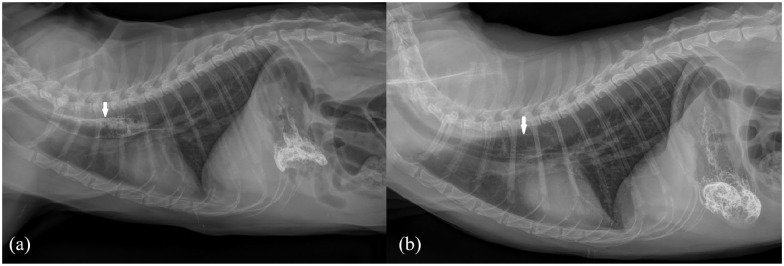
Based on the owner’s description of the cat’s problems, regurgitation could not be completely excluded. Fluoroscopy was unavailable, so an oral contrast study was conducted. Three minutes after barium sulfate 2 ml/kg (Micropaque; Guerbet) had been given orally, thoracic radiographs revealed some contrast medium in the oesophagus at the heart base (Figure 2a). After 10 mins, the whole of the contrast medium had passed into the stomach (Figure 2b). An oesophageal dilation or an oesophageal motility disorder were strongly suspected, but the clinical presentation and history were atypical and therefore it was decided to perform an oesophagogastroduodenoscopy.
Figure 2.
(a) Contrast study 5 mins after oral administration of barium sulfate. Moderate contrast medium retention (arrow) can be seen in the precordial part of the oesophagus. (b) After 10 mins the oesophagus was visualised as a dilated structure past the base of the heart (arrow)
Endoscopy revealed an oesophagus of normal appearance without any signs of megaoesophagus. The pyloric region showed a significantly narrowed pyloric lumen compared with the size of a 2.3 mm biopsy forceps (Figure 3). Several biopsies were taken from the pyloric region. Therapy with lansoprazole (1 mg/kg PO q12h [Lansoprazol AL; Aliud Pharma]) and sucralfate (60 mg/kg PO q8h [Sucrabest; Combustin]) was started. Histopathological examination of the pyloric biopsies revealed moderate chronic erosive lymphoplasmacytic and mild eosinophilic hyperplastic gastritis with mild-to-moderate mucosal fibrosis. No neoplastic process was found. In the pathologist’s report, it was suspected that the fibrosis could have contributed to the pyloric stenosis. As the inflammation was not pronounced and anti-inflammatory treatment was not effective, surgical treatment of the fibrosis was suggested.
Figure 3.
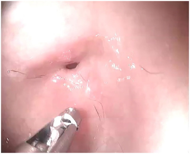
Endoscopic view of the pylorus. Note the narrowed lumen of the pylorus compared with the 2.3 mm endoscopic biopsy forceps
The severe pyloric obstruction was treated surgically by Y-U pyloroplasty (Figure 4).7 A full-thickness biopsy was taken from the pyloric sphincter. Histopathological examination of the pyloric sphincter biopsy showed focal hyperplasia of the pyloric smooth muscles (Figure 5). No inflammatory, fibrotic or neoplastic process was found. The biopsy taken from the duodenum had a normal appearance and there were no signs of any inflammatory process. No other biopsy samples were taken from the gastrointestinal tract.
Figure 4.
Y-U pyloroplasty. (a) The pyloric region was isolated. (b) Stay sutures were placed into the stomach and duodenum adjacent to the planned pyloroplasty site. (c,d) A Y-shaped full-thickness incision was performed – the main stem of the Y was made through the antimesenteric part of the duodenum and the pyloric sphincter and the arms of the Y were made on the pyloric antrum. The U-shaped flap was advanced forward to the base of the Y incision using a suture on its tip and was sutured into the aboral end of the incision with an absorbable monofilament synthetic suture
Figure 5.
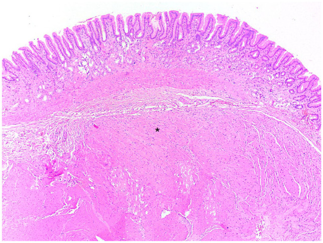
A photomicrograph (low-power magnification) of a full-thickness pyloric biopsy. The thickness of the smooth muscle layer is focally increased (asterisk). Haematoxylin and eosin. Image courtesy of Dr Wolf von Bomhard, Fachpraxis für Tierpathologie, Munich, Germany
The cat was hospitalised after surgery and discharged after 4 days. Treatment with lansoprazole (Lansoprazol AL; Aliud Pharma) was continued postoperatively and tapered out after 1 week. Three weeks postoperatively, the cat was presented for a follow-up appointment. The owner mentioned lethargic behaviour after feeding during the first week and three episodes of vomiting directly after food intake since discontinuing lansoprazole. The ultrasonographic examination showed normal peristalsis of the gastrointestinal tract, an open lumen of the pylorus and chymus in the most proximal duodenum (Figure 6).
Figure 6.
An ultrasonographic picture of the pyloroduodenal junction after surgery. A large opening was created between the antrum and the proximal duodenum
Lansoprazole was reinstituted and the owner reported no further vomiting when the cat was fed tinned food of one commercial brand based on various animal protein sources. According to the owner, hypoallergenic diets were not accepted by the cat. To document pyloric viability, a follow-up contrast study was conducted, which demonstrated prompt gastric emptying and good intestinal propulsion (Figure 7) after the oral administration of barium sulfate 10 ml/kg. The cat was discharged with lansoprazole (1 mg/kg PO q24h) for the next 6–12 weeks. The patient did not have further gastrointestinal signs during the 6-month follow-up period.
Figure 7.
Abdominal radiographs 8 weeks postoperatively. (a) Five minutes after barium sulfate administration. (b,c) Ten minutes after barium sulfate administration. Rapid gastric emptying and passage to the duodenum was seen
Discussion
This case is the first description of a Y-U pyloroplasty in a domestic cat (Felis catus). Y-U pyloroplasty was first described by Francesco Durantes.8 Y-U pyloroplasty has become a popular method owing to its simplicity, the short duration required to perform the procedure and maximal widening of the gastric outflow tract.9–11
A single case report of a Y-U pyloroplasty performed on a feline described a 4-year-old cheetah (Acinonyx jubatus), with chronic vomiting and spiral bacteria-associated gastritis.12 The preoperative contrast radiographs showed delayed gastric emptying. Y-U pyloroplasty was performed and no vomiting was observed for the following 3 years. The histological examination showed only mild superficial lymphocytic gastritis.
According to the literature, the median feline pyloric diameter is 9 mm (range 8–10),13 and the pyloroduodenal junction should be easily intubated with an endoscope with an external diameter <7.8 mm.14 The inability to do this could provide indirect evidence of pyloric narrowing.14 Freiche et al2 compared the pyloric diameter to the width of the open biopsy forceps. In this study, the cats in which the pylorus could not be passed during endoscopy also had a subjectively narrower pyloric sphincter than the control group. Most of the cats were treated with prednisolone (0.2–0.3 mg/kg q24h) and fed highly digestible food 3–5 times daily. Surgical intervention was needed in only two cases (balloon dilation and pyloroplasty, respectively, and not further specified). Vomiting occurred less frequently in all cats after medical or surgical therapy. In our patient, the pylorus could only be passed with 2.3 mm biopsy forceps. The free pyloric opening was estimated at about 2 mm.
The results of the endoscopic biopsy from our case confirmed long-standing gastritis with defects in the pyloric epithelial surface. This could be a possible cause of impaired gastric motility, a result of chronic delayed gastric emptying or both. The mucosal inflammation followed by mucosal fibrosis could have led to a lack of pyloric elasticity and a reduction in pyloric diameter, as suggested.2
On the contrary, the histopathological examination of the full-thickness pyloric wall sample showed only focal hyperplasia of the pyloric smooth muscles in our patient. This finding is more consistent with findings of hypertrophic pyloric gastropathy common in dogs with pyloric stenosis.15–17 In contrast to cats, dogs often show thick mucosal folds around the pylorus.18 The dissimilarity of our biopsy results could be explained by using different biopsy techniques (endoscopic vs full-thickness) and thus by samples of different quality and origin. The full-thickness biopsy was taken directly from the region of the pyloric sphincter. Endoscopic biopsies were obtained from the gastric part of the pyloric region and might not represent the narrowest part of the gastric outlet.
Pyloric stenosis has been rarely reported in cats.2–6 Pearson et al3 described pyloric stenosis in 13 cats with chronic vomiting. The contrast radiographs showed moderate gastric dilation and a delayed passage of the contrast medium. Additionally, in 7/13 cats, retention of the contrast medium within the oesophagus was seen, with mild oesophageal dilation in some cases, which is consistent with the case reported here. Siamese, Burmese and Tonkinese cats are suspected to have a narrower pyloric canal.14 Some authors report a high prevalence of young Siamese cats with congenital pyloric stenosis, which could suggest an inherited nature of the disease in the Siamese breed.3,5,6,19 Burmese cats with congenital pyloric stenosis have not yet been reported. In our patient, acquired narrowing of the gastric outlet due to chronic gastritis seems most probable, but a congenital cause cannot entirely be ruled out.
Eosinophilic infiltrates, lymphoid follicular hyperplasia, atrophy and fibrosis were found in the pyloric mucosa of cats infected with Helicobacter species,20,21 which is very similar to our histological results. In our case, a faecal sample had been examined for Helicobacter species by PCR, and no spiral bacteria were seen on histology, therefore Helicobacter species infection seems unlikely in our patient.
Lymphoplasmacytic infiltration of the gastric mucosa has been reported in a group of cats with pylorus sphincter narrowing, as well as in a control group.2 The significance of this finding is unclear.
Food intolerance or hypersensitivity could be a possible underlying cause of the pyloric stenosis in our patient, as suggested by eosinophilic infiltration, but it was not possible to prove this by a dietary trial.
Inflammatory bowel disease (IBD) was considered a possible underlying disorder in our case. However, the small sample of duodenal tissue examined histologically did not support a duodenal involvement. Diarrhoea or borborygmi were never reported as a problem. A purely gastric expression of IBD seems unlikely and the histological finding of mild-to-moderate inflammation and erosion could alternatively be explained by the chronic strain on the gastric mucosa by frequent vomiting. As the cat responded very well to the surgical intervention, further treatment with anti-inflammatory agents was not deemed necessary after surgery
Conclusions
Multifactor pathogenesis of feline acquired pyloric stenosis should be considered. Further investigation is required.
Acknowledgments
The authors wish to thank Dr Wolf von Bomhard for providing the microphotographic image.
Footnotes
Accepted: 15 October 2022
The authors declared no potential conflicts of interest with respect to the research, authorship, and/or publication of this article.
Funding: The authors received no financial support for the research, authorship, and/or publication of this article.
Ethical approval: The work described in this manuscript involved the use of non-experimental (owned or unowned) animals. Established internationally recognised high standards (‘best practice’) of veterinary clinical care for the individual patient were always followed and/or this work involved the use of cadavers. Ethical approval from a committee was therefore not specifically required for publication in JFMS Open Reports. Although not required, where ethical approval was still obtained it is stated in the manuscript.
Informed consent: Informed consent (verbal or written) was obtained from the owner or legal custodian of all animal(s) described in this work (experimental or non-experimental animals, including cadavers) for all procedure(s) undertaken (prospective or retrospective studies). No animals or people are identifiable within this publication, and therefore additional informed consent for publication was not required.
References
- 1. Batchelor DJ, Devauchelle P, Elliott J, et al. Mechanisms, causes, investigation and management of vomiting disorders in cats: a literature review. J Feline Med Surg 2013; 15: 237–265. [DOI] [PMC free article] [PubMed] [Google Scholar]
- 2. Freiche V, Da Riz F, Benchekroun G, et al. Endoscopic assessment of presumed acquired pyloric narrowing in cats: a retrospective study of 27 cases. Res Vet Sci 2021; 136: 408–415. [DOI] [PubMed] [Google Scholar]
- 3. Pearson H, Gaskell CJ, Gibbs C, et al. Pyloric and oesophageal dysfunction in the cat. J Small Anim Pract 1974; 15: 487–501. [DOI] [PubMed] [Google Scholar]
- 4. Syrcle JA, Gambino JM, Kimberlin WW. Treatment of pyloric stenosis in a cat via pylorectomy and gastroduodenostomy (Billroth I procedure). J Am Vet Med Assoc 2013; 242: 792–797. [DOI] [PubMed] [Google Scholar]
- 5. Twaddle AA. Pyloric stenosis in three cats and its correction by pyloroplasty. N Z Vet J 1970; 18: 15–17. [DOI] [PubMed] [Google Scholar]
- 6. Twaddle AA. Congenital pyloric stenosis in two kittens corrected by pyloroplasty. N Z Vet J 1971; 19: 26–27. [DOI] [PubMed] [Google Scholar]
- 7. Cornell K. Stomach. In: Johnston S, Tobias K. (eds). Veterinary surgery: small animals. 2nd ed. St Louis, MO: Elsevier Saunders, 2017, pp 1700–1730. [Google Scholar]
- 8. Durantes F. Plastica gastroduodenale. Bull Delia Real Accademia Di Roma 1892; 18: 526. [Google Scholar]
- 9. Moschel DM, Walske BR, Neumayer F. A new technique for pyloroplasty. Surgery 1958; 44: 813–816 [PubMed] [Google Scholar]
- 10. Randolph JG. Y-U advancement pyloroplasty. Ann Surg 1975; 181: 586–590. [DOI] [PMC free article] [PubMed] [Google Scholar]
- 11. Szauer JS, Vivas CA, Baylis SM. Y-U pyloroplasty. Am J Surg 1977; 134: 270–271. [DOI] [PubMed] [Google Scholar]
- 12. Neiffer DL, Pardo AD, Klein EC. Use of pyloroplasty (Y-U) to treat presumed delayed gastric emptying in a cheetah (Acinonyx jubatus). J Zoo Wildl Med 2000; 31: 552–557. [DOI] [PubMed] [Google Scholar]
- 13. Lamoureux A, Benchekroun G, German AJ, et al. An endoscopic method for semi-quantitatively measuring internal pyloric diameter in healthy cats: a prospective study of 24 cases. Res Vet Sci 2019; 122: 165–169. [DOI] [PubMed] [Google Scholar]
- 14. Tams TR. Gastroscopy. In: Tams TR, Rawlings CA. (eds). Small animal endoscopy. 3rd ed. St Louis, MO: Elsevier Saunders, 2011, pp 97–173. [Google Scholar]
- 15. Bellenger CR, Maddison JE, MacPherson GC, et al. Chronic hypertrophic pyloric gastropathy in 14 dogs. Aust Vet J 1990; 67: 317–320. [DOI] [PubMed] [Google Scholar]
- 16. Happé RP, van der Gaag I, Wolvekamp WT. Pyloric stenosis caused by hypertrophic gastritis in three dogs. J Small Anim Pract 1981; 22: 7–17. [DOI] [PubMed] [Google Scholar]
- 17. Walsh MH, Quigley PJ. Pyloric stenosis in the dog caused by hypertrophy of the circular muscle of the pylorus. Vet Rec 1966; 78: 13–15. [DOI] [PubMed] [Google Scholar]
- 18. Leib MS, Saunders GK, Moon ML, et al. Endoscopic diagnosis of chronic hypertrophic pyloric gastropathy in dogs. J Vet Intern Med 1993; 7: 335–341. [DOI] [PubMed] [Google Scholar]
- 19. Washabau RJ, Day MJ, Willard MD, et al. WSAVA International Gastrointestinal Standardization Group. Endoscopic, biopsy, and histopathologic guidelines for the evaluation of gastrointestinal inflammation in companion animals. J Vet Intern Med 2010; 24: 10–26. [DOI] [PubMed] [Google Scholar]
- 20. Scanziani E, Simpson KW, Monestiroli S, et al. Histological and immunohistochemical detection of different Helicobacter species in the gastric mucosa of cats. J Vet Diagn Invest 2001; 13: 3–12. [DOI] [PubMed] [Google Scholar]
- 21. Simpson KW, Strauss-Ayali D, Scanziani E, et al. Helicobacter felis infection is associated with lymphoid follicular hyperplasia and mild gastritis but normal gastric secretory function in cats. Infect Immun 2000; 68: 779–790. [DOI] [PMC free article] [PubMed] [Google Scholar]


