Abstract
Knee osteoarthritis (KOA), whose prevalence keeps rising, is still unsolved pathobiological/therapeutical problem. Historically, knee osteoarthritis was thought to be a “wear and tear” disease, while recent etiology hypotheses stressed it as a chronic, low-grade inflammatory disease. Inflammasomes mediated by the innate immunity systems have an important role in inflammatory diseases including KOA. A deluge of recent studies focused on the NLRP3 inflammasome with suggestions that its pharmacologic block would hinder degeneration. However, known inflammasomes are numerous and can also trigger IL-1β/IL-18 production and cells’ pyroptotic death. Among them, AIM2 inflammasome is involved in key aspects of various acute and chronic inflammatory diseases. Therefore, while presently leaving out little-studied inflammasomes in KOA, this review focuses on the AIM2 inflammasomes that participate in KOA’s complex mechanisms in conjunction with the activation of AIM2 inflammasomes in other diseases combined with the current studies on KOA mechanisms. Although human-specific data about it are relatively scant, we stress that only a holistic view including several inflammasomes including AIM2 inflammasome and other potential pathogenetic drivers will lead to successful therapy for knee osteoarthritis.
Keywords: knee osteoarthritis, inflammasome, absent in melanoma 2
Introduction
Knee osteoarthritis (KOA) is a progressive joint disease that primarily affects the middle-aged and elderly population, with progressive disease progression gradually affecting the patient’s mobility and quality of life.1 Its main pathological feature is the destruction and loss of articular cartilage, but all joint tissues are involved in some form, including subchondral bone remodeling, meniscal degeneration, ligament degeneration and laxity, inflammation and fibrosis of infrapatellar fat pad, synovial inflammation, and periarticular muscle atrophy.2,3 And in the advanced stages of the disease, eventually leads to the destruction of the entire joint, indicating that KOA is more of a disease that arises from the joint as an organ. Historically, KOA was thought to be a “wear and tear” disease, but it is now considered a multifactorial disease that is not only related to local biomechanical factors of the knee joint, but also involves biochemical factors, including obesity, aging, and gender.4–6 Among them, the infiltration of a number of proinflammatory cytokines in the joint microenvironment especially the synovial tissues, play an important role in the clinical and pathological progress of KOA.7–9 Along with the introduction of the concept of osteoimmunology in 2000,10 increasing studies have shown that immunological mechanisms play a crucial role in the pathogenesis of KOA, especially the innate immune system.11 Nowadays, KOA is considered a chronic, low-grade inflammatory disease.12 Anti-inflammatories (NSAIDs and corticosteroids) are typically used to reduce ongoing inflammation and relieve the pain induced by KOA. However, the current treatments can not prevent the sustained release of proinflammatory cytokines.13,14
The innate immune system is the first line of defense in the body. It can recognize various endogenous and exogenous stimuli in the body through Pattern Recognition receptors (PRRs), including Pathogen-Associated Molecular Patterns (PAMP) and Danger-Associated Molecular Patterns (DAMP), to activate further immune responses.15 Among them, NOD-like receptors (NLRs) and absent in melanoma 2 (AIM2-like receptors, ALRs) can form a multiprotein structure of the inflammasome in response to various stimuli.16 The inflammasome converts caspase-1 precursors into active caspase-1, which promotes the maturation and secretion of inflammatory factors: interleukin-1 beta (IL-1β) and interleukin-18 (IL-18), while the activated substances are also involved in cleavage formation and cleavage Gasdermin D (GSDMD), which directly mediates cell death (pyroptosis).17,18 Under normal conditions, the immune system-mediated inflammatory response is an important function to clear pathogens and maintain homeostasis, but when it is sustained, excessive accumulation and activation can lead to tissue damage in the body. A variety of inflammasomes has been shown to play a role in a variety of diseases, including various autoimmune diseases (rheumatoid arthritis, systemic lupus erythematosus, psoriasis, etc.), chronic inflammatory diseases (chronic hepatitis, neuroinflammation, atherosclerosis, etc.), and various cancers.19–21 Understanding how uncontrolled inflammation is triggered and continues to progress is a key aspect of treatment in KOA. Currently, the most studied inflammasome in KOA is NLRP3,22 but few studies have been conducted on AIM2 inflammasome.
Although a small number of studies have pointed out the presence of several different inflammasomes, including AIM2 inflammasome, can be detected in KOA. Chen et al found that AIM2 expression existed in synovial tissue of KOA knees, but the specific mechanisms involved have not been elucidated.23 They also found that AIM2 was upregulated in fibroblast-like synoviocytes of rheumatoid arthritis (RA), and suppressing its expression leads to inhibiting effect on fibroblast-like synoviocytes proliferation. As we know, there exist many similarities between the synovial tissue of OA and RA. However, it has not been reported whether the AIM2 has the same effect in OA as in RA. This may be related to the fact that the activation of AIM2 inflammasome is different from other inflammasomes. In the NLR family inflammasome, they can be regulated by a variety of PAMP and DAMP, including potassium efflux, mitochondrial dysfunction, and ROS production, altered intracellular calcium ion concentration, and other stimuli to assemble into inflammasome.24 While the activation of AIM2 inflammasome can only be activated by binding itself to the double-stranded DNA fragments. However, as a multifactorial disease, studying the effect of one inflammatory vesicle alone is not comprehensive in KOA. Simultaneously, AIM2 inflammasome is involved in key aspects of various acute and chronic inflammatory diseases such as atherosclerosis, fatty liver, and cerebral ischemia.19,20,25 Therefore, the purpose of this review is to propose several potential mechanisms for the involvement of AIM2 inflammasome in KOA and to stress the need to focus the experts’ attention on its multiple roles of it.
Composition and Activation of AIM2 Inflammasome
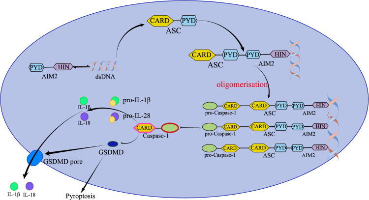
AIM2 inflammasomes consist of AIM2, apoptosis-associated speck-like protein containing a CARD (ASC), and cystatin protease 1 precursor (pro-caspase-1). One of the interferon-inducible proteins, AIM2, is a protein encoded by the AIM2 gene.16 It consists of two structural domains linked by long junctions: an N-terminal pyrin domain (PYD) and a C-terminal HIN (Hematopoietic, interferon-inducible, nuclear localization) domain. The PYD structural domain mediates homotypic protein-protein interactions, while the HIN structural domain binds to DNA through its two tandem oligonucleotide/oligosaccharide binding folds. DNA binding is the first step in a series of events in AIM2 inflammasome formation. The AIM2 HIN structural domain binds to both strands of a certain length of double-stranded DNA (viral, bacterial, or host) in a sequence-independent manner. The binding of the double-stranded DNA displaces the PYD structural domain, which then binds via homotypic PYD-PYD interactions to the downstream inflammatory vesicle bridging protein ASC, a dichotomous PYD-CARD containing protein. In turn, ASC interacts with no mature form of pro-caspase-1 through the CARD domain (CARD-CARD interaction) of the complex, thereby creating the basic structural element of the AIM2 inflammasome.26,27 Two main events occur after AIM2 inflammasome activation: the secretion of proinflammatory cytokines, IL-1β and IL-18, and the activation of pyroptosis (Figure 1). And these two events both are associated with the onset and development of KOA.4,22,28
Figure 1.
Composition and activation of AIM2 inflammasome.
Abbreviations: AIM2, absent in melanoma 2; dsDNA, double stranded DNA; PYD, N-terminal pyrin domain; HIN, hematopoietic, interferon-inducible, nuclear localization; ASC, apoptosis-associated speck‐like protein containing a CARD; GSDMD, gasdermin D; IL, interleukin. Created by Figdraw.
Probable Mechanisms for the Involvement of AIM2 Inflammasome in KOA
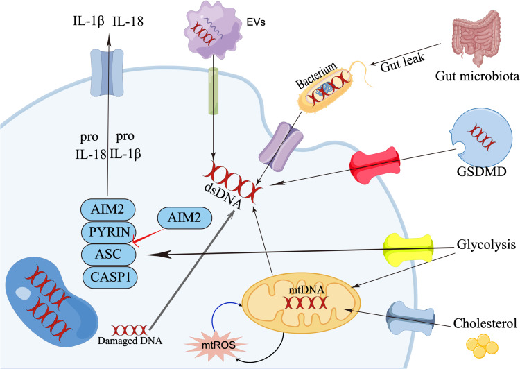
To study how AIM2 inflammasomes are involved in KOA pathogenesis, the primary question is how AIM2 inflammasomes are initiated in KOA, and it is this question that has limited the number of studies in this area. However, some endogenous or exogenous stimuli which have been shown to stimulate NLRP3 inflammasome can also activate the assembly of AIM2 inflammasome at the same time. The intensive study of AIM2 inflammasome in KOA may have been overlooked due to the fervor of NLRP3 inflammasome. Therefore, we propose the following possible ways of AIM2 inflammasome involvement in KOA in conjunction with the activation of AIM2 inflammasome in other diseases combined with the current studies on KOA mechanisms (Table 1, Figure 2).
Table 1.
Potential Mechanisms for the Involvement of AIM2 Inflammasome in KOA
| Factors Related to the KOA | The Activation of the AIM2 Inflammasome |
|---|---|
| Gut microbiota | The disorder of gut microbiota would invalidate intestinal barrier which lead to the leakage of intestinal contents including the gut microbiota into the circulatory system. The dsDNA of the bacterium can activate the AIM2 inflammasome. |
| Mitochondria | Mitochondria generate ROS radicals which can damage themselves. The release of the mtDNA activates AIM2 inflammasome. |
| Extracellular vesicles | EVs mediate intercellular communication via the contained biologically active molecules, including DNA which can be recognized by the AIM2. |
| EVs can also expell damaged DNA from cells, and when this process is impaired, the DNA which are damaged and carried by EVs can activate the AIM2 inflammasome. | |
| Metabolic factors | Elevated cholesterol causes impaired mitochondrial metabolism and mtDNA release, which in turn activate the AIM2 inflammasome. |
| The glycolysis can promote the regulation of AIM2 inflammasome activation. Glycolysis regulates the oligomerisation of ASC, which is required for AIM2 inflammasome formation. | |
| The glycolysis changes can also cause mitochondria dysfunction. The increased mitochondrial membrane potential loss and ROS production lead to the release of the mtDNA. | |
| Crosstalk between different inflammasomes | Pyroptosis caused by other inflammasomes could release the dsDNA of dead cells which in turn activate the AIM2 inflammasome in normal cells, and eventually lead to the uncontrolled cytokine storm. |
Abbreviations: KOA, knee osteoarthritis; AIM2, absent in melanoma 2; dsDNA, double stranded DNA; mtDNA, mitochondrial DNA; mROS, mitochondrial reactive oxygen species; EVs, extracellular vesicles.
Figure 2.
Potential mechanisms for the involvement of AIM2 inflammasome in KOA.
Abbreviations: AIM2, absent in melanoma 2; dsDNA, double stranded DNA; ASC, apoptosis-associated speck‐like protein containing a CARD; GSDMD, release of dsDNA caused by gasdermin D; IL, interleukin; EVs, extracellular vesicles; mtDNA, mitochondrial DNA; mROS, mitochondrial reactive oxygen species. Created by Figdraw.
Gut Microbiota -Knee-AIM2
Generally speaking, the presence of bacteria would not be detected in KOA knees, and a positive bacterial test usually implies the occurrence of an infection. However, with the current advances in detection technology and related theories, especially the gut microbiota theory, this concept is gradually changing.29,30 The gut microbiota is considered as a “microbial organ” whose functions are closely intertwined with host physiology and pathology, and which has an important role in nutrient absorption, intestinal mucosal barrier, hormone levels in the body, and regulation of immune/inflammatory responses.31,32 Among them, gut microbiota can influence intestinal barrier function, allowing intestinal metabolites, bacteria, and other substances to enter the human circulation.33 Numerous studies have shown that lipopolysaccharide, a major component of the cell wall of Gram-negative bacteria in the gut, can enter the human circulation and colonize the knee joint as PAMP to trigger the inflammatory response within the knee joint, and is a key factor in the current study of gut microbiota affecting KOA.34–36 As the lipopolysaccharide can pass through the intestinal mucosal barrier, bacterial DNA can also be present in the knee joint. In 2012, Témoin et al had already reported that bacterial DNA was found in the joint fluid of patients undergoing arthroplasty for KOA, and hip OA. They also found the same bacteria were detected in the periodontal tissue of some patients, and hypothesized that pathogenic bacteria could colonize the knee joint elsewhere in the body.37 More recently, in the study of Dunn et al, 16S ribosomal RNA gene sequencing detected a variety of bacterial DNA in all samples of human cartilage (hip and knee), with a predominance of gram-negative bacteria.38 None of the patients in these studies showed signs of knee infection, suggesting that bacterial DNA can be present in the KOA without causing infection. Jiang et al also found the gene expression of the bacteria in the synovial fluid of KOA.39 The presence of those bacteria in the joint may not be random and could play a physiological role. From the perspective of AIM2 inflammasome, these bacterial DNAs could then serve as a source of their activation and thus secrete downstream inflammatory factors to influence the occurrence and development of KOA, but further validation is still needed.
Mitochondria - AIM2
Mitochondria are important organelles of human cells that contain their own genetic material, mitochondrial DNA (mtDNA), and as the main energy supplier of cells, they have been closely associated with various diseases.40 In KOA, in vitro studies have demonstrated the presence of mitochondrial dysfunction in chondrocytes due to mtDNA mutations, which may affect several pathways related to cartilage degradation.41–43 In addition, mtDNA can also be involved in the activation of AIM2 inflammasome.44 While mitochondria generate ATP to meet the energy needs of the body, they also generate reactive oxygen species (ROS) radicals to damage themselves. When mitochondrial autophagy is impaired, damaged mitochondria accumulate in the cytoplasm and promote intracellular inflammatory responses, which may be due to the inability to remove mtDNA from damaged mitochondria in a timely manner, which in turn activates AIM2 inflammasome.45 In a study of a mouse model of nonalcoholic fatty liver, Xu et al found that the activation of AIM2 inflammasome was closely related to the damage of mtDNA, while the extracted mtDNA could activate the assembly of AIM2 inflammasome and trigger the pyroptosis of hepatocytes.46 On the other hand, ROS in human is mainly produced by mitochondria, and in addition to activating NLRP3 inflammasome,47 ROS are also closely linked to the activation of AIM2 inflammasome through the release of mtDNA.40,48,49 In the study of Wang et al, in the streptozotocin-induced diabetic rat model, AIM2 expression was significantly increased in heart tissue compared with the control. While the ROS inhibition reduced the level of AIM2.50 In Fibrotic Interstitial lung disease, Trachalaki et al also found that mtROS was significantly associated with heightened NLRP3 and AIM2 activation, and mitochondrial antioxidant treatment limited inflammasome activation.51 Therefore, activation of AIM2 inflammasome may also be a manner in which mitochondria influences KOA.
Extracellular Vesicles - AIM2
Extracellular vesicles (EVs) are a collective term for a variety of vesicular structures with membrane structures released by cells, which can be naturally released from almost all types of cells.52 These vesicles can be classified into endosomal types such as exosomes, microvesicles, and apoptotic vesicles according to their size and synthesis pathways. They carry a variety of biologically active molecules, including proteins, RNA, lipids, and DNA. EVs are nanoscale intercellular messengers that are secreted by cells to transmit biological signals.53 Current studies have shown that they can regulate host-pathogen interactions and participate in the pathological processes of many diseases such as infectious and inflammatory diseases, neurological diseases, and cancer, and play an important function in mediating intercellular communication in normal physiological processes.54 KOA is an inflammatory disease and the outer vesicles are important for its pathogenesis as well as for its treatment.55 Chondrocytes, synovial fibroblasts, osteoblasts, and tenocytes, can produce and secrete exosomes that influence the biological effects of targeted cells.56,57 In addition, exosomes from stem cells can protect the OA joint from damage by promoting cartilage repair, inhibiting synovitis, and mediating subchondral bone remodeling.58,59 However, most studies have focused on the RNA and protein content of EVs, while little has been reported on the DNA carried within them.60–63 Takahashi et al showed that EVs maintain cellular homeostasis and prevent aberrant activation of the innate immune response by expelling damaged DNA from cells, and when this process is impaired, the DNA they carry can activate inflammatory factors including the cGAS-STING pathway and the AIM2 inflammasome.64 In the study of the gastrointestinal response to the chemotherapeutic agent irinotecan, Lian et al found that chemotherapy-induced the release of large amounts of double-stranded DNA from exosomes in the gastrointestinal tract, and this “auto-DNA” entered the cytoplasm of innate immune cells and activated AIM2 inflammasome.65 This stimulated the secretion of mature IL-1β and IL-18 secretion and induced intestinal mucositis and delayed diarrhea. Whereas elimination of AIM2 signaling in AIM2-deficient mice or by pharmacological inhibitors significantly reduces the incidence of drug-induced diarrhea and does not affect the anticancer efficacy of the drugs. While in the KOA, the effects of DNA carried by EVs have not been studied.
Metabolic Phenotypes of KOA
Metabolic factors are associated with a variety of systemic diseases, including obesity, atherosclerosis, and type 2 diabetes. The dysfunction of these metabolic factors can cause a low-grade systemic inflammatory state, such as the systemic complications of diabetes.66,67 In OA, the most obvious one is obesity. Especially for weight-bearing joints such as knee and hip joints, obesity is one of the most likely risk factors for the development and progression of OA.68,69 Although obesity most notably changes physical factors such as increased joint loading, reduced muscle strength, and altered biomechanics, there is growing evidence that metabolic factors are also involved.70 For example, the relationship between obesity and hand OA and why not all obese individuals develop knee or hip OA.71 Although the mechanisms involved are unclear, other metabolic factors such as glucose and lipids have been shown to influence disease development through activation of AIM2 inflammasome.72,73 Wang et al found in a streptozotocin-induced diabetic rat model that diabetic rats exhibited severe left ventricular dysfunction, and AIM2 expression was significantly increased in heart tissue compared with the controls.50 While gene silencing of AIM2 alleviated cardiac dysfunction. Cho et al found that glucose transporter 1-dependent glycolysis could promote AIM2 inflammasome activation by regulating the oligomerisation of ASC, which is required for AIM2 inflammasome formation.72 Furthermore, the glycolysis changes can also cause mitochondria dysfunction, the increased mitochondrial membrane potential loss and ROS production lead to the release of the mtDNA which in turn activates the AIM2.73 As to the lipids, Pan et al found significantly increased expression of AIM2 in vascular smooth muscle cells and larger plaque lesion areas in mice fed a high-fat diet model of atherosclerosis compared with normally fed mice.74 Dang et al reported that the elevated cholesterol would cause impaired mitochondrial metabolism and mtDNA release, which in turn activate the AIM2 inflammasome.75 Although metabolic phenotype is one of the most common types of KOA in the clinic, no studies have reported the role of AIM2 inflammasome in it to date.
Crosstalk Between Different Inflammasomes
Although different inflammasomes have different initiation mechanisms, they share common downstream pathways.16 The presence of different inflammasomes can be detected in the same disease, and there must be an interconnection between them. In the study of Cunha et al, Legionella pneumophila infection first activated the formation of AIM2 inflammasome, after which activated caspase-1 induced cell membrane pore formation, which in turn caused K+ efflux-mediated NLRP3 activation, suggesting that different inflammasomes can regulate each other’s activities to ensure an effective immune response to infection.76 Witzenrath et al also found that in NLRP3 knockout mice, IL-1β secretion levels were not suppressed after Streptococcus pneumoniae infection, suggesting the activation of other NLR family or AIM2 inflammasome pathways.77 Heinisch et al found no effect on IL-1β levels after pharmacological inhibition of NLRP3 in microglia BV-2 cells in an acute ischemic stroke mouse model, where NLRC4 and AIM2 protein levels were increased, indicating the compensatory activity among inflammasomes, but the exact mechanism is unclear.78 The presence of a reciprocal regulatory relationship between inflammasomes in KOA has not been reported. However, based on the above studies, it appears that the factors that activate NLRP3 inflammasome can also activate AIM2 inflammasome, and when NLRP3 is blocked or silenced, various stimuli can further activate AIM2 inflammasome. In KOA, there should exist an interaction between different inflammasomes. However, it is curious that in the published articles, blocking the formation of NLRP3 inflammasome alone can also lead to a decrease in IL-β levels in KOA, it is possible that these drugs also block the activation of other inflammasomes at the same time,79 while the specific mechanism deserves further in-depth study for a comprehensive understanding of the therapeutic effects of these drugs.
Conclusion and Future Perspective
AIM2 inflammasome plays an important role in inflammatory diseases, but the role in KOA has not yet received attention. In this review, we have identified several possible mechanisms of action of AIM2 inflammasome by combining the action of AIM2 inflammasome in other inflammatory diseases with the latest research progress in KOA, to provide new perspectives on the role of AIM2 inflammasome in KOA. However, the expression of different inflammasomes is not the same in different organs, tissues, and cells, so the role of AIM2 inflammasome in KOA could be different in other diseases, while further in-depth study of the mechanism in key cells such as chondrocytes and synoviocytes in KOA will be more helpful to our understanding of the pathogenesis of KOA and the development of new therapeutic approaches. More importantly, as a multifactorial disease, it is not comprehensive to focus on only a single inflammasome in KOA, but should be studied as a whole. We should also note that although current animal studies have shown that silencing or inhibiting the formation of the inflammasome can reduce the severity of KOA, whether the immune capacity of other organs and tissues in the human body would be affected has not yet received attention, and focusing on local inhibition of excessive and sustained activation of various inflammasome may be the future direction of treatment.
Disclosure
The authors report no conflicts of interest in this work.
References
- 1.Hunter DJ, Schofield D, Callander E. The individual and socioeconomic impact of osteoarthritis. Nat Rev Rheumatol. 2014;10(7):437–441. doi: 10.1038/nrrheum.2014.44 [DOI] [PubMed] [Google Scholar]
- 2.Felson DT. Priorities for osteoarthritis research: much to be done. Nat Rev Rheumatol. 2014;10(8):447–448. doi: 10.1038/nrrheum.2014.76 [DOI] [PubMed] [Google Scholar]
- 3.Eymard F, Chevalier X. Inflammation of the infrapatellar fat pad. Joint Bone Spine. 2016;83(4):389–393. doi: 10.1016/j.jbspin.2016.02.016 [DOI] [PubMed] [Google Scholar]
- 4.Motta F, Barone E, Sica A, Selmi C. Inflammaging and osteoarthritis. Clin Rev Allerg Immu. 2022;1–17. doi: 10.1007/s12016-022-08941-1 [DOI] [PubMed] [Google Scholar]
- 5.Batushansky A, Zhu S, Komaravolu RK, South S, Mehta-D’souza P, Griffin TM; Fundamentals of OA. An initiative of osteoarthritis and cartilage. Chapter 9: obesity and metabolic factors in OA. Osteoarthr Cartilage. 2021;30(4):501–515. doi: 10.1016/j.joca.2021.06.013 [DOI] [PMC free article] [PubMed] [Google Scholar]
- 6.Coaccioli S, Sarzi-Puttini P, Zis P, Rinonapoli G, Varrassi G. Osteoarthritis: new insight on its pathophysiology. J Clin Med. 2022;11(20):6013. doi: 10.3390/jcm11206013 [DOI] [PMC free article] [PubMed] [Google Scholar]
- 7.de Lange-Brokaar BJE, Ioan-Facsinay A, van Osch GJVM, et al. Synovial inflammation, immune cells and their cytokines in osteoarthritis: a review. Osteoarthr Cartilage. 2012;20(12):1484–1499. doi: 10.1016/j.joca.2012.08.027 [DOI] [PubMed] [Google Scholar]
- 8.Sanchez-Lopez E, Coras R, Torres A, Lane NE, Guma M. Synovial inflammation in osteoarthritis progression. Nat Rev Rheumatol. 2022;18(5):258–275. doi: 10.1038/s41584-022-00749-9 [DOI] [PMC free article] [PubMed] [Google Scholar]
- 9.Griffin TM, Scanzello CR. Innate inflammation and synovial macrophages in osteoarthritis pathophysiology. Clin Exp Rheumatol. 2019;37(5):57–63. [PMC free article] [PubMed] [Google Scholar]
- 10.Arron JR, Choi Y. Bone versus immune system. Nature. 2000;408(6812):535–536. doi: 10.1038/35046196 [DOI] [PubMed] [Google Scholar]
- 11.Takayanagi H. Osteoimmunology as an intrinsic part of immunology. Int Immunol. 2021;33(12):dxab057. doi: 10.1093/intimm/dxab057 [DOI] [PubMed] [Google Scholar]
- 12.Robinson WH, Lepus CM, Wang Q, et al. Low-grade inflammation as a key mediator of the pathogenesis of osteoarthritis. Nat Rev Rheumatol. 2016;12(10):580–592. doi: 10.1038/nrrheum.2016.136 [DOI] [PMC free article] [PubMed] [Google Scholar]
- 13.Ghouri A, Conaghan PG. Prospects for Therapies in Osteoarthritis. Calcified Tissue Int. 2021;109(3):339–350. doi: 10.1007/s00223-020-00672-9 [DOI] [PMC free article] [PubMed] [Google Scholar]
- 14.Delplace V, Boutet MA, Visage CL, Maugars Y, Guicheux J, Vinatier C. Osteoarthritis: from upcoming treatments to treatments yet to come. Joint Bone Spine. 2021;88(5):105206. doi: 10.1016/j.jbspin.2021.105206 [DOI] [PubMed] [Google Scholar]
- 15.Li D, Wu M. Pattern recognition receptors in health and diseases. Signal Transduct Target Ther. 2021;6(1):291. doi: 10.1038/s41392-021-00687-0 [DOI] [PMC free article] [PubMed] [Google Scholar]
- 16.Schroder K, Tschopp J. The Inflammasomes. Cell. 2010;140(6):821–832. doi: 10.1016/j.cell.2010.01.040 [DOI] [PubMed] [Google Scholar]
- 17.Wang B, Yin Q. AIM2 inflammasome activation and regulation: a structural perspective. J Struct Biol. 2017;200(3):279–282. doi: 10.1016/j.jsb.2017.08.001 [DOI] [PMC free article] [PubMed] [Google Scholar]
- 18.Murakami T, Nakaminami Y, Takahata Y, Hata K, Nishimura R. Activation and function of NLRP3 inflammasome in bone and joint-related diseases. Int J Mol Sci. 2022;23(10):5365. doi: 10.3390/ijms23105365 [DOI] [PMC free article] [PubMed] [Google Scholar]
- 19.Uresti-Rivera EE, García-Hernández MH, Li H, Wang D. AIM2-inflammasome role in systemic lupus erythematous and rheumatoid arthritis. Autoimmunity. 2022;55(1):1–12. doi: 10.1080/08916934.2022.2103802 [DOI] [PubMed] [Google Scholar]
- 20.Lozano-Ruiz B, Bachiller V, García-Martínez I, et al. Absent in melanoma 2 triggers a heightened inflammasome response in ascitic fluid macrophages of patients with cirrhosis. J Hepatol. 2015;62(1):64–71. doi: 10.1016/j.jhep.2014.08.027 [DOI] [PubMed] [Google Scholar]
- 21.Chao-yang G, Peng C, Hai-hong Z. Roles of NLRP3 inflammasome in intervertebral disc degeneration. Osteoarthr Cartilage. 2021;29(6):793–801. doi: 10.1016/j.joca.2021.02.204 [DOI] [PubMed] [Google Scholar]
- 22.Roškar S, Hafner-Bratkovič I. The role of inflammasomes in osteoarthritis and secondary joint degeneration diseases. Life. 2022;12(5):731. doi: 10.3390/life12050731 [DOI] [PMC free article] [PubMed] [Google Scholar]
- 23.Chen Y, Fujuan Q, Chen E, et al. Expression of AIM2 in rheumatoid arthritis and its role on fibroblast-like synoviocytes. Mediat Inflamm. 2020;2020:1693730. doi: 10.1155/2020/1693730 [DOI] [PMC free article] [PubMed] [Google Scholar]
- 24.Huang Y, Xu W, Zhou R. NLRP3 inflammasome activation and cell death. Cell Mol Immunol. 2021;18(9):2114–2127. doi: 10.1038/s41423-021-00740-6 [DOI] [PMC free article] [PubMed] [Google Scholar]
- 25.Lammert CR, Frost EL, Bellinger CE, et al. AIM2 inflammasome surveillance of DNA damage shapes neurodevelopment. Nature. 2020;580(7805):647–652. doi: 10.1038/s41586-020-2174-3 [DOI] [PMC free article] [PubMed] [Google Scholar]
- 26.Wang B, Bhattacharya M, Roy S, Tian Y, Yin Q. Immunobiology and structural biology of AIM2 inflammasome. Mol Aspects Med. 2020;76:100869. doi: 10.1016/j.mam.2020.100869 [DOI] [PMC free article] [PubMed] [Google Scholar]
- 27.Jin T, Perry A, Smith P, Jiang J, Xiao TS. Structure of the absent in melanoma 2 (AIM2) pyrin domain provides insights into the mechanisms of aim2 autoinhibition and inflammasome assembly*. J Biol Chem. 2013;288(19):13225–13235. doi: 10.1074/jbc.m113.468033 [DOI] [PMC free article] [PubMed] [Google Scholar]
- 28.Chang X, Kang Y, Yang Y, et al. Pyroptosis: a novel intervention target in the progression of osteoarthritis. J Inflamm Res. 2022;15:3859–3871. doi: 10.2147/jir.s368501 [DOI] [PMC free article] [PubMed] [Google Scholar]
- 29.Berthelot JM, Sellam J, Maugars Y, Berenbaum F. Cartilage-gut-microbiome axis: a new paradigm for novel therapeutic opportunities in osteoarthritis. Rmd Open. 2019;5(2):e001037. doi: 10.1136/rmdopen-2019-001037 [DOI] [PMC free article] [PubMed] [Google Scholar]
- 30.Wei Z, Li F, Pi G. Association between gut microbiota and osteoarthritis: a review of evidence for potential mechanisms and therapeutics. Front Cell Infect Mi. 2022;12:812596. doi: 10.3389/fcimb.2022.812596 [DOI] [PMC free article] [PubMed] [Google Scholar]
- 31.Li XV, Leonardi I, Iliev ID. Gut mycobiota in immunity and inflammatory disease. Immunity. 2019;50(6):1365–1379. doi: 10.1016/j.immuni.2019.05.023 [DOI] [PMC free article] [PubMed] [Google Scholar]
- 32.Nicholson JK, Holmes E, Kinross J, et al. Host-gut microbiota metabolic interactions. Science. 2012;336(6086):1262–1267. doi: 10.1126/science.1223813 [DOI] [PubMed] [Google Scholar]
- 33.Morgan XC, Tickle TL, Sokol H, et al. Dysfunction of the intestinal microbiome in inflammatory bowel disease and treatment. Genome Biol. 2012;13(9):R79. doi: 10.1186/gb-2012-13-9-r79 [DOI] [PMC free article] [PubMed] [Google Scholar]
- 34.Loeser RF, Arbeeva L, Kelley K, et al. Association of increased serum lipopolysaccharide, but not microbial dysbiosis, with obesity‐related osteoarthritis. Arthritis Rheumatol. 2022;74(2):227–236. doi: 10.1002/art.41955 [DOI] [PMC free article] [PubMed] [Google Scholar]
- 35.Hernandez CJ, Guss JD, Luna M, Goldring SR. Links between the microbiome and bone. J Bone Miner Res. 2016;31(9):1638–1646. doi: 10.1002/jbmr.2887 [DOI] [PMC free article] [PubMed] [Google Scholar]
- 36.Collins KH, Paul HA, Reimer RA, Seerattan RA, Hart DA, Herzog W. Relationship between inflammation, the gut microbiota, and metabolic osteoarthritis development: studies in a rat model. Osteoarthr Cartilage. 2015;23(11):1989–1998. doi: 10.1016/j.joca.2015.03.014 [DOI] [PubMed] [Google Scholar]
- 37.Témoin S, Chakaki A, Askari A, et al. Identification of oral bacterial DNA in synovial fluid of patients with arthritis with native and failed prosthetic joints. J Clin Rheumatol. 2012;18(3):117–121. doi: 10.1097/rhu.0b013e3182500c95 [DOI] [PMC free article] [PubMed] [Google Scholar]
- 38.Dunn CM, Velasco C, Rivas A, et al. Identification of cartilage microbial DNA signatures and associations with knee and hip osteoarthritis. Arthritis Rheumatol. 2020;72(7):1111–1122. doi: 10.1002/art.41210 [DOI] [PMC free article] [PubMed] [Google Scholar]
- 39.Jiang P, Sun S, Zhang J, et al. RNA expression profiling from the liquid fraction of synovial fluid in knee joint osteoarthritis patients. Am J Transl Res. 2021;14(9):6782–6791. [PMC free article] [PubMed] [Google Scholar]
- 40.Schapira AH. Mitochondrial diseases. Lancet. 2012;379(9828):1825–1834. doi: 10.1016/s0140-6736(11)61305-6 [DOI] [PubMed] [Google Scholar]
- 41.Martel-Pelletier J, Pelletier JP. Is there a mitochondrial DNA haplogroup connection between osteoarthritis and elite athletes? A narrative review. Rmd Open. 2022;8(2):e002602. doi: 10.1136/rmdopen-2022-002602 [DOI] [PMC free article] [PubMed] [Google Scholar]
- 42.Crane DD, Bauler TJ, Wehrly TD, Bosio CM. Mitochondrial ROS potentiates indirect activation of the AIM2 inflammasome. Front Microbiol. 2014;5:438. doi: 10.3389/fmicb.2014.00438 [DOI] [PMC free article] [PubMed] [Google Scholar]
- 43.Blanco FJ, Rego I, Ruiz-Romero C. The role of mitochondria in osteoarthritis. Nat Rev Rheumatol. 2011;7(3):161–169. doi: 10.1038/nrrheum.2010.213 [DOI] [PubMed] [Google Scholar]
- 44.Bae JH, Jo SI, Kim SJ, et al. Circulating cell-free mtDNA contributes to AIM2 inflammasome-mediated chronic inflammation in patients with type 2 diabetes. Cells. 2019;8(4):328. doi: 10.3390/cells8040328 [DOI] [PMC free article] [PubMed] [Google Scholar]
- 45.Scherz-Shouval R, Elazar Z. Regulation of autophagy by ROS: physiology and pathology. Trends Biochem Sci. 2011;36(1):30–38. doi: 10.1016/j.tibs.2010.07.007 [DOI] [PubMed] [Google Scholar]
- 46.Xu L, Zhou J, Che J, et al. Mitochondrial DNA enables AIM2 inflammasome activation and hepatocyte pyroptosis in nonalcoholic fatty liver disease. Am J Physiolgastr L. 2021;320(6):G1034–G1044. doi: 10.1152/ajpgi.00431.2020 [DOI] [PubMed] [Google Scholar]
- 47.Chen Z, Zhong H, Wei J, et al. Inhibition of Nrf2/HO-1 signaling leads to increased activation of the NLRP3 inflammasome in osteoarthritis. Arthritis Res Ther. 2019;21(1):300. doi: 10.1186/s13075-019-2085-6 [DOI] [PMC free article] [PubMed] [Google Scholar]
- 48.Zorov DB, Juhaszova M, Sollott SJ. Mitochondrial Reactive Oxygen Species (ROS) and ROS-induced ROS release. Physiol Rev. 2014;94(3):909–950. doi: 10.1152/physrev.00026.2013 [DOI] [PMC free article] [PubMed] [Google Scholar]
- 49.Savu O, Sunkari VG, Botusan IR, Grünler J, Nikoshkov A, Catrina S. Stability of mitochondrial DNA against reactive oxygen species (ROS) generated in diabetes. Diabetes Metabol Res Rev. 2011;27(5):470–479. doi: 10.1002/dmrr.1203 [DOI] [PubMed] [Google Scholar]
- 50.Wang X, Pan J, Liu H, et al. AIM2 gene silencing attenuates diabetic cardiomyopathy in type 2 diabetic rat model. Life Sci. 2019;221:249–258. doi: 10.1016/j.lfs.2019.02.035 [DOI] [PubMed] [Google Scholar]
- 51.Trachalaki A, Tsitoura E, Mastrodimou S, et al. Enhanced IL-1β release following NLRP3 and AIM2 inflammasome stimulation is linked to mtROS in airway macrophages in pulmonary fibrosis. Front Immunol. 2021;12:661811. doi: 10.3389/fimmu.2021.661811 [DOI] [PMC free article] [PubMed] [Google Scholar]
- 52.Raposo G, Stoorvogel W. Extracellular vesicles: exosomes, microvesicles, and friends. J Cell Biol. 2013;200(4):373–383. doi: 10.1083/jcb.201211138 [DOI] [PMC free article] [PubMed] [Google Scholar]
- 53.Yáñez‐Mó M, Siljander PRM, Andreu Z, et al. Biological properties of extracellular vesicles and their physiological functions. J Extracell Vesicles. 2015;4(1):27066. doi: 10.3402/jev.v4.27066 [DOI] [PMC free article] [PubMed] [Google Scholar]
- 54.Sanwlani R, Gangoda L. Role of extracellular vesicles in cell death and inflammation. Cells. 2021;10(10):2663. doi: 10.3390/cells10102663 [DOI] [PMC free article] [PubMed] [Google Scholar]
- 55.Zhou QF, Cai YZ, Lin XJ. The dual character of exosomes in osteoarthritis: antagonists and therapeutic agents. Acta Biomater. 2020;105:15–25. doi: 10.1016/j.actbio.2020.01.040 [DOI] [PubMed] [Google Scholar]
- 56.Kato T, Miyaki S, Ishitobi H, et al. Exosomes from IL-1β stimulated synovial fibroblasts induce osteoarthritic changes in articular chondrocytes. Arthritis Res Ther. 2014;16(4):R163. doi: 10.1186/ar4679 [DOI] [PMC free article] [PubMed] [Google Scholar]
- 57.Wu X, Crawford R, Xiao Y, Mao X, Prasadam I. Osteoarthritic subchondral bone release exosomes that promote cartilage degeneration. Cells. 2021;10(2):251. doi: 10.3390/cells10020251 [DOI] [PMC free article] [PubMed] [Google Scholar]
- 58.Ni Z, Zhou S, Li S, et al. Exosomes: roles and therapeutic potential in osteoarthritis. Bone Res. 2020;8(1):25. doi: 10.1038/s41413-020-0100-9 [DOI] [PMC free article] [PubMed] [Google Scholar]
- 59.Noor NAM, Nurul AA, Zain MRAM, Aduni WKWN, Azlan M. Extracellular vesicles from mesenchymal stem cells as potential treatments for osteoarthritis. Cells. 2021;10(6):1287. doi: 10.3390/cells10061287 [DOI] [PMC free article] [PubMed] [Google Scholar]
- 60.Wu X, Bian B, Lin Z, et al. Identification of exosomal mRNA, lncRNA and circRNA signatures in an osteoarthritis synovial fluid-exosomal study. Exp Cell Res. 2022;410(1):112881. doi: 10.1016/j.yexcr.2021.112881 [DOI] [PubMed] [Google Scholar]
- 61.Wu Y, Li J, Zeng Y, et al. Exosomes rewire the cartilage microenvironment in osteoarthritis: from intercellular communication to therapeutic strategies. Int J Oral Sci. 2022;14(1):40. doi: 10.1038/s41368-022-00187-z [DOI] [PMC free article] [PubMed] [Google Scholar]
- 62.Fan WJ, Liu D, Pan LY, et al. Exosomes in osteoarthritis: updated insights on pathogenesis, diagnosis, and treatment. Front Cell Dev Biol. 2022;10:949690. doi: 10.3389/fcell.2022.949690 [DOI] [PMC free article] [PubMed] [Google Scholar]
- 63.Yu M, Wang D, Chen X, Zhong D, Luo J. BMSCs-derived mitochondria improve osteoarthritis by ameliorating mitochondrial dysfunction and promoting mitochondrial biogenesis in chondrocytes. Stem Cell Rev Rep. 2022;1–20. doi: 10.1007/s12015-022-10436-7 [DOI] [PubMed] [Google Scholar]
- 64.Matsuyama H, Shindo A, Shimada T, et al. Chronic cerebral hypoperfusion activates AIM2 and NLRP3 inflammasome. Brain Res. 2020;1736:146779. doi: 10.1016/j.brainres.2020.146779 [DOI] [PubMed] [Google Scholar]
- 65.Lian Q, Xu J, Yan S, et al. Chemotherapy-induced intestinal inflammatory responses are mediated by exosome secretion of double-strand DNA via AIM2 inflammasome activation. Cell Res. 2017;27(6):784–800. doi: 10.1038/cr.2017.54 [DOI] [PMC free article] [PubMed] [Google Scholar]
- 66.Kassi E, Pervanidou P, Kaltsas G, Chrousos G. Metabolic syndrome: definitions and controversies. Bmc Med. 2011;9(1):48. doi: 10.1186/1741-7015-9-48 [DOI] [PMC free article] [PubMed] [Google Scholar]
- 67.Chakraborty S, Zawieja S, Wang W, Zawieja DC, Muthuchamy M. Lymphatic system: a vital link between metabolic syndrome and inflammation. Ann Ny Acad Sci. 2010;1207:E94–E102. doi: 10.1111/j.1749-6632.2010.05752.x [DOI] [PMC free article] [PubMed] [Google Scholar]
- 68.Kulkarni K, Karssiens T, Kumar V, Pandit H. Obesity and osteoarthritis. Maturitas. 2016;89:22–28. doi: 10.1016/j.maturitas.2016.04.006 [DOI] [PubMed] [Google Scholar]
- 69.Misra D, Fielding RA, Felson DT, et al. Risk of knee osteoarthritis with obesity, sarcopenic obesity, and sarcopenia. Arthritis Rheumatol. 2019;71(2):232–237. doi: 10.1002/art.40692 [DOI] [PMC free article] [PubMed] [Google Scholar]
- 70.Mobasheri A, Rayman MP, Gualillo O, Sellam J, van der Kraan P, Fearon U. The role of metabolism in the pathogenesis of osteoarthritis. Nat Rev Rheumatol. 2017;13(5):302–311. doi: 10.1038/nrrheum.2017.50 [DOI] [PubMed] [Google Scholar]
- 71.Marshall M, Watt FE, Vincent TL, Dziedzic K. Hand osteoarthritis: clinical phenotypes, molecular mechanisms and disease management. Nat Rev Rheumatol. 2018;14(11):641–656. doi: 10.1038/s41584-018-0095-4 [DOI] [PubMed] [Google Scholar]
- 72.Cho SJ, Moon JS, Nikahira K, et al. GLUT1-dependent glycolysis regulates exacerbation of fibrosis via AIM2 inflammasome activation. Thorax. 2020;75(3):227. doi: 10.1136/thoraxjnl-2019-213571 [DOI] [PMC free article] [PubMed] [Google Scholar]
- 73.Xie M, Yu Y, Kang R, et al. PKM2-dependent glycolysis promotes NLRP3 and AIM2 inflammasome activation. Nat Commun. 2016;7(1):13280. doi: 10.1038/ncomms13280 [DOI] [PMC free article] [PubMed] [Google Scholar]
- 74.Pan J, Han L, Guo J, et al. AIM2 accelerates the atherosclerotic plaque progressions in ApoE−/− mice. Biochem Bioph Res Co. 2018;498(3):487–494. doi: 10.1016/j.bbrc.2018.03.005 [DOI] [PubMed] [Google Scholar]
- 75.Dang EV, McDonald JG, Russell DW, Cyster JG. Oxysterol restraint of cholesterol synthesis prevents AIM2 inflammasome activation. Cell. 2017;171(5):1057–1071.e11. doi: 10.1016/j.cell.2017.09.029 [DOI] [PMC free article] [PubMed] [Google Scholar]
- 76.Cunha LD, Silva ALN, Ribeiro JM, et al. AIM2 engages active but unprocessed caspase-1 to induce noncanonical activation of the NLRP3 inflammasome. Cell Rep. 2017;20(4):794–805. doi: 10.1016/j.celrep.2017.06.086 [DOI] [PubMed] [Google Scholar]
- 77.Witzenrath M, Pache F, Lorenz D, et al. The NLRP3 inflammasome is differentially activated by pneumolysin variants and contributes to host defense in pneumococcal pneumonia. J Immunol. 2011;187(1):434–440. doi: 10.4049/jimmunol.1003143 [DOI] [PubMed] [Google Scholar]
- 78.Heinisch O, Zeyen T, Goldmann T, et al. Erythropoietin abrogates post-ischemic activation of the NLRP3, NLRC4, and AIM2 inflammasomes in microglia/macrophages in a TAK1-dependent manner. Transl Stroke Res. 2022;13(3):462–482. doi: 10.1007/s12975-021-00948-8 [DOI] [PMC free article] [PubMed] [Google Scholar]
- 79.Kim J, Ahn H, Han BC, et al. Korean red ginseng extracts inhibit NLRP3 and AIM2 inflammasome activation. Immunol Lett. 2014;158(1–2):143–150. doi: 10.1016/j.imlet.2013.12.017 [DOI] [PubMed] [Google Scholar]


