


Abstract
Introduction:
The aim of the present study was to compare the amount of apical debris extrusion after preparation using hand files, reciprocating files, and full rotary nickel-titanium systems.
Methods and Materials:
One hundred extracted human mandibular molars with two separated canals in mesial root were divided into five groups and prepared using reciprocating systems (Reciproc file and Safesider endodontic reamers file), full rotary systems (Mtwo and Neoniti A1 files) and hand instrumentation systems. Endodontic access was prepared and a #15 K-file was passed beyond the apex of the mesiobuccal canal by 1 mm to ensure the canal patency. All mesiobuccal canals were prepared 1 mm shorter than the anatomic apex. In each case, extruded debris was collected in an Eppendorf tube and weighed after desiccation. The mean weight of extruded material was calculated in each group. The analysis was carried out using the Kruskal–Wallis test followed by two tailed and Mann-Whitney U test at a significance level of 0.05. The Bonferroni correction was also applied to correct multiple comparisons.
Results:
There was a statistically significant difference between the reciprocal and other techniques in debris extrusion (P<0.05). The order of groups ranked in terms of debris extrusion from the lowest to highest was as follows: 1) Hand instrumentation group (with crown down technique), 2) Mtwo group, 3) Neoniti A1 group, 4) Safesider endodontic reamer group, and 5) Reciproc group.
Conclusion:
Based on this in vitro study, all systems have some apical debris extrusion; however, using the hand instrumentation system resulted in extrusion of significantly less debris compared to the Reciproc group. It seems that hand and rotary instrumentation systems are better than reciprocating instrumentation systems in terms of the amount of debris extrusion.
Key Words: Endodontics, Root Canal Preparation, Rotary Instrumentation
Introduction
To achieve successful endodontic treatment, it is very important to clean the root canal system both mechanically and chemically using endodontic files and irrigant solutions. On the other hand, extrusion of irrigants, dental chips, necrotic tissues, pulp tissue remnants, and microorganisms and their products to the periapical area is possible during canal preparation, which is caused by instrumentation techniques and leads to inflammation and treatment failure [1]. In fact, instruments act as a piston and push debris out of the apical foramen [1]. Siqueira [2] mentioned that the apical extrusion of debris acts as an important cause of flare-up in endodontics. Extrusion of bacteria and their products in periradicular region leads to acute inflammation and pain, or flare-up and delayed healing. The intensity of the response depends on the number of bacteria (amount of debris extrusion), pathogenicity of bacteria (virulence) and host defense [3, 4].
Researches demonstrated that all instrumentation systems result in a bacteria extrusion beyond the apical foramen even if preparation be shorter than apical constriction [3, 5-9]. However, its amount is different in various preparation techniques. Therefore, minimizing the extrusion of debris from the apical area should be noticed in canal instrumentation.
According to the literature, it seems that extrusion of debris and the subsequent postoperative pain severity in the crown-down technique is less than other techniques [10, 11]. In crown-down technique the coronal area is prepared prior to the apical area. Enlarging the coronal third of the root canal and providing a path for the exit of debris from the root canals, results in the extrusion of less debris [12, 13].
On the other hand, it was shown that techniques involving a filing (linear) motion cause significantly more blockages and extrude significantly more apical dentin debris because of piston-like movements and extrusion of more debris and irrigation solutions compared with other instrumentation techniques [14, 15].
Neoniti (NEOLIX, Châtres-la-Forêt, France) is a nickel-titanium (NiTi) rotary system. The manufacturer claims that it has controlled memory and a rough surface, resulting in abrasive properties, satisfactory shaping and no screwing effect. This system is used in continuous rotation and consists of the A1 and C1 files. The latter can be used as an optional orifice shaper [16].
It was demonstrated that instrumentation systems with reciprocal motion lead to faster mechanical preparation of the root canal and a higher amount of dentinal chips and debris [5, 17]. There are several studies about the amount of apical extrusion of debris using these instruments [12, 18-20]. Two newer files with a reciprocal motion are Reciproc single file system (VDW, Munich, Germany) and Safesider endodontic reamers (multi-file system) (Essential Dental Systems, South Hackensack, NJ, USA). Reciproc is a single-file system which is used in a reciprocating motion. It has an S-shaped horizontal cross-section and 2 cutting edges [9]. The Safesider endodontic reamers is an instrumentation system that has recently been introduced. This system includes a non-interrupted flat-sided design for both reamers and files which reduces dentinal engagement and, consequently, the resistance of the instruments within the canal, as well as shortening the time for canal preparation [21]. Since just a few studies have evaluated comparison of the apical extrusion of debris in conventional reciprocating and rotary instrumentation systems and their results are inconsistent, thus, the purpose of this ex vivo experiment was to compare the apical extrusion of debris using Reciproc and Safesider endodontic reamer systems, Neoniti A1 (Neoniti A1, France) and Mtwo (VDW, Munich, Germany) rotary files, and also the manual instrumentation system (with crown-down technique).
Materials and Methods
Using a simple random sampling method, 100 mandibular molar teeth extracted in the surgery department of dentistry faculty of Zahedan University of Medical Science from 2013 to 2015 were selected. This in vitro study was reviewed and approved by the ethics committee of Zahedan University of Medical Sciences. The teeth were stored in 10% formalin until the experiment [22]. This research was verified and confirmed by Vice Chancellor for Research of Zahedan University of Medical Sciences. The teeth were examined both clinically and using radiographs. The inclusion criterion was mesial roots of mandibular molar teeth with two separate orifices, canals and apical foramens. Teeth with curvature less than 10° (according to Schneider’s technique) in the mesiobuccal (MB) canal were selected [23]. The crowns were adjusted so that all teeth had the same initial length, mature apices without previous endodontic treatment, and with the apical patency. The initial diameter of apical foramens was the same as the #15 K-file. Each tooth was cut in half buccolingually at the furcation area and the mesial half of the tooth was separated. Radiography was done proximally to confirm the existence of two separate canals in the mesial root.
First, root surfaces were cleaned and debrided using a periodontal scaler. Then, all caries and previous fillings were removed and standard access cavities were made using round diamond burs (#1014; KG Sorensen, Barueri, SP, Brazil) at a high speed and under air-water spray cooling.
This study was done on the mesiobuccal canal. A stable reference point was established on all teeth flattening of mesiobuccal cusp tip as well as the same working length for all specimens.
A #10 K-file (Maillefer, Dentsply, Switzerland) was used visually to control the apical patency and to determine the working length. The working length was measured one millimeter less than the point that was touched by the file tip in the apical foramen.
The teeth were positioned in the experimental setup proposed by Myers and Montgomery [24]. Eppendorf tubes were used to gather debris (Figure 1). Each tube was weighed three times using an electronic semi-micro balance with an accuracy of 0.1 mg (Mettler AC 100; Mettler Instruments, Greifensee, Switzerland) and the mean weight was recorded and summarized in a specified table. Samples were divided into five groups randomly. There were 20 teeth in each group (n=20). According to the manufacture guidelines, the teeth were prepared with VDW Silver electric motors (VDW, Munich, Germany) by one operator. To standardize root canal preparation and the amount of irrigating solution used for each sample, these steps were repeated until the working length was achieved and a total amount of 10 mL distilled water irrigating solution was used.
Figure 1.
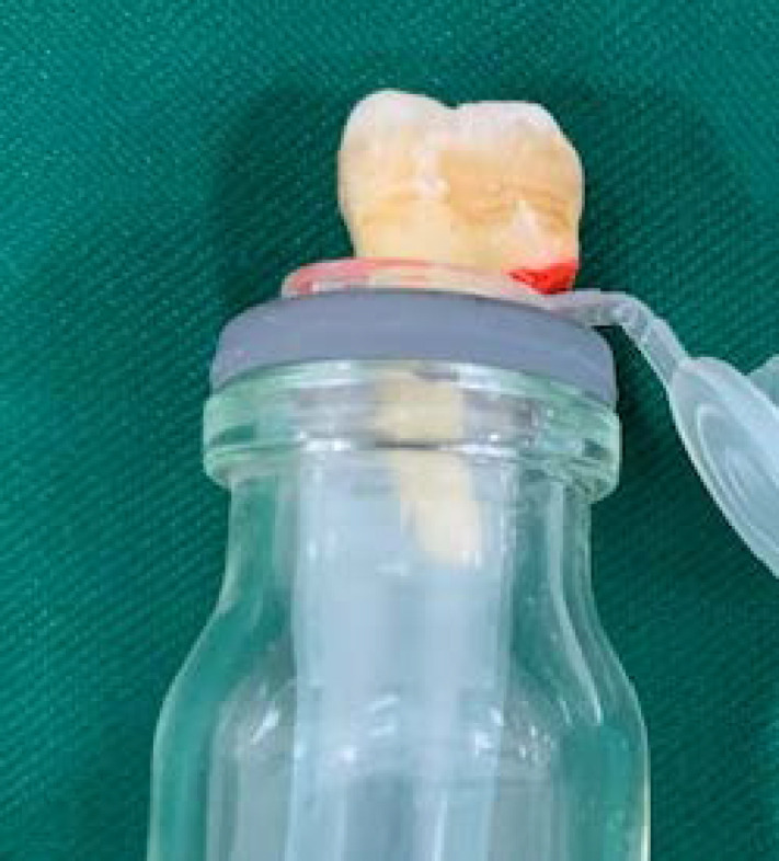
Experimental setup has been shown according to Myers and Montgomery
Canal preparation techniques/instruments
Neoniti A1 file : This system was used as a single-file technique. File #25/0.8 at a rotational speed of 350 rpm and a torque of 1.5N/cm was used over working length.
Mtwo files : The files of this system were used with gentle in-and-out motions and controlled torque. Sizes #10/0.04, #15/0.05, #20/0.06, and #25/0.06 files were used in order. Each instrument was used in the working length
Safesider endodontic reamers : According to the manufacturer's instruction, reamerswere used in reciprocating motion at a rotational speed of 2500 rpm with in-and-out tip motion. Sizes #15/0.02, #20/0.02, and #25/0.02 files were used in a sequence in the working length. Then, #2 Peeso reamer was used in coronal half of working length for flaring and direct access.
Reciproc file : According to the manufacturer's instruction, #25/0.8 file was used with gentle in-and-out motions. After every three pecking motions, instrument flutes were cleaned. The instrument was removed from the canal when it reached the working length freely.
Manual technique : Crown down technique was used with hand K-files up to a depth in the canal which required no pressure. Then, files #40, 35, 30, 25 and 20 were used in the canal. Afterwards, files #15, 20 and 25 were used in the working length, and then, the canal was flared with the step-back technique. Between any two files, patency was controlled with a #10 file.
For all groups, after each file and after every 3 pecks of reciprocating files, the canal was irrigated and a total volume of 10 mL double-distilled water (ddH2O) was used with #30 gauge needle during preparation. In the end, the stopper, needle, and tooth were removed from the Eppendorf tube and that attached debris was washed from the root surface in a tube using 1 mL ddH2O. To achieve dry debris, the tubes were removed from the setup and were put in an incubator at 70 °C for 5 days. The specimens were dried and weighed under the same condition. The debris were weighed three times by an operator who was totally blind about experimental groups. The mean of these measurements was considered to be the new weight of the tubes. Subtracting the weight of the empty tube, the net weight of debris was achieved.
Statistical analysis
The SPSS software (SPSS ver. 20, SPSS, Chicago, IL, USA) was used to analyze the data. In the descriptive statics part, the mean and standard deviations were calculated. The data were analyzed at a significance level of 0.05 using the Kruskal–Wallis test followed by two tailed Mann-Whitney U test. The Bonferroni correction was also applied to correct multiple comparisons.
Results
All systems cause extrusion of debris. The Kruskal-Wallis test showed that there were significant differences between the groups. A two by two comparison of files using Mann-Whitney U test showed that the amount of extruded debris was significantly larger using Reciproc and Safesider endodontic reamer files in comparison with Mtwo, Neoniti A1, and hand files (P<0.05) (Table 1). The highest and lowest amounts of extruded debris observed in Reciproc and crown down manual groups, respectively.
Table 1.
Amount of apically extruded debris using different instruments
| Debris extrusion (mg) | Neoniti A1 | Mtow | Safe sider | Reciproc | Hand K File |
|---|---|---|---|---|---|
| Mean (SD) | 0.21 (0.301) b | 0.17 (0.086) b | 0.42 (0.417) a | 0.50 (0.604) a | 0.08 (0.093) b |
| Min | 1.0 | 0.4 | 1.5 | 2.1 | 0.3 |
| Max | 0.35 | 0.21 | 0.61 | 0.78 | 0.13 |
| Median | 0.10 | 0.15 | 0.30 | 0.10 | 0.10 |
Values with the different letters were statistically different at P <0.05
The mean and standard deviations of extruded debris are presented in Table 1.
There was no significant difference in extruded debris between Mtwo, Neoniti A1, and hand K files (P>0.05).
Discussion
A significant complication which is undesirable for both the patient and the practitioner is the inter appointment flare-ups that occur as a consequence of apical extrusion during root canal procedures.
This study showed that the amount of extruded debris was significantly larger in reciprocal techniques including Reciproc and Safesider endodontic reamer in comparison with Mtwo rotary multi-files system, Neoniti A1, and also the manual instrumentation (with crown-down technique). On the other hand, the amount of extruded debris was significantly larger by Reciprocal systems compared to other techniques.
No significant difference was found in the amount of extruded debris between two studied reciprocating systems.
The Reciproc instrument has an S-shaped cross-sectional design with sharp cutting edges that increases the cutting ability and may enhance the transportation of debris toward the apex when used in combination with a reciprocating motion [25]. Continuous rotation may improve the coronal transportation of dentine chips and debris by acting like a screw conveyor [5].
In the crown-down technique, the coronal area is prepared first and then the apical area is prepared. Enlarging the coronal third of the root canal and providing a path for the exit of debris from the root canals, results in the extrusion of less debris [12, 13].
On the other hand, Safesider endodontic reamer with reciprocation and pecking motions results in more extruded debris. This is different from hand files with pull and push motions which may result in less extruded debris when used with the crown down technique and kept away from the apical foramen. Ferraz et al. [26] reported the same results. They mentioned that there was no significant difference in the amount of extruded debris using the balanced force manual technique compared to full rotary techniques. However, Zarrabi et al. [27] presented that there was more extruded debris using the step-back technique in comparison with the Flex master, RaCe, and Profile systems. In the crown-down technique, irrigant solution can better remove debris from the orifice area because it is firstly required to prepare the coronal part of the canal. On the other hand, the more taper of rotary files results in more dentin cut and specifically more extruded debris in comparison to hand files [28].
Burklein et al. [5, 25] also reported a lower amount of extruded debris using full rotary systems and also multi-files preparation in comparison with reciprocal systems. It seems that the amount of extruded debris of reciprocal systems is also because of their cross-section design, more cutting ability, and more dentin removal.
These results are similar to the results of the other studies that have been reported that instruments used with a reciprocating movement may cause a higher amount of extruded debris compared to files used with a continuous rotary movement [8, 29, 30].
However, Uzun et al. [31], Kocak et al. [7], and Dincer et al. [32] reported a lower amount of extruded debris for reciprocal systems which are different from our findings. It may because of using single canal teeth in their study which naturally is wider in apical area and less dentin cut is produced during preparation, and as a result, there is a less piston-like effect of the file on apical area. Previous studies also showed that the amount of extruded apical debris can be related to the root canal anatomy and/or the instrumentation technique [17, 19, 33]. This study was carried out on mesiobuccal roots of lower molars which probably and the more extruded debris may be because of the smaller diameter of the canal and application of more force during preparation. Increased level of instrumentation difficulty and because the highly variable anatomy and degree of curvature of the root canals in these teeth affect the extrusion of apical debris in this canal.
Neoniti A1 instruments have a non-homothetic rectangular cross-section with an abrasive surface [34]. There was no significant difference between Neoniti A1 and Mtwo rotary multi-files systems and the manual technique (crown down). De-Deus et al. [18] concluded that more extruded debris is associated with full sequence rotary files because of several times of irrigation and insertion of instruments in the canal. However, this study didn’t show any relationship between the number of instruments used and the amount of extruded debris.
The irrigate solution is of great importance. Impurities of different irrigants may affect the weight of dry debris and it is known as a confounding factor [14, 35]. In the present study, to avoid this problem, the double-distilled water was used. Adjusting the working length is also an important factor. In the present study, the working length was considered to be one millimeter shorter than the anatomic apex. This is different in each study. As the working length gets closer to the apical foramen, there is more debris extrusion [1]. In this study, the apical patency was established in all steps. Thus, there was no limit for the extrusion of debris. One of the disadvantages of this apparatus is that it doesn’t mimic the periapical tissues and their back pressures. It has been suggested to simulate the resistance of periapical tissues by using floral foam [36]. However, the foam may absorb some debris when used as a barrier, thus, just like Burklein et al. [25], no attempt was made in this study to simulate the resistance of periapical tissues. The change in the microhardness of extracted human teeth may affect the results. Additionally, the relationship between the amount of extruded debris and available pathogenic microorganisms remains unknown [8]. Thus, the extending of these results to the in vivo conditions should be done with caution. Additionally, it should be noted that other factors such as anatomic variation, type of irrigants, and multiple foramina are also effective.
Conclusions
All instrumentation systems cause extrusion of debris to the apical area. According to the results of the present study, the full rotation techniques produced less amount of extruded debris and yielded better results compared to reciprocal systems. It should be mentioned that because of variations in study designs, the direct comparison of different studies that addressed this problem is not possible and in vivo studies should be done to evaluate its clinical relevance.
Acknowledgment
The authors thank the vice-chancellery of Zahedan University of Medical Sciences for supporting this research.
Conflict of Interest:
‘None declared’.
References
- 1.Martin H, Cunningham WT. The effect of endosonic and hand manipulation on the amount of root canal material extruded. Oral Surg Oral Med Oral Pathol. 1982;53(6):611–3. doi: 10.1016/0030-4220(82)90350-4. [DOI] [PubMed] [Google Scholar]
- 2.Siqueira Jr J. Microbial causes of endodontic flare‐ups. Int Endod J. 2003;36(7):453–63. doi: 10.1046/j.1365-2591.2003.00671.x. [DOI] [PubMed] [Google Scholar]
- 3.Seltzer S, Naidorf IJ. Flare-ups in endodontics: I Etiological factors. J Endod. 1985;11(11):472–8. doi: 10.1016/S0099-2399(85)80220-X. [DOI] [PubMed] [Google Scholar]
- 4.Mollashahi NF, Saberi EA, Havaei SR, Sabeti M. Comparison of Postoperative Pain after Root Canal Preparation with Two Reciprocating and Rotary Single-File Systems: A Randomized Clinical Trial. Iran Endod J. 2017;12(1) doi: 10.22037/iej.2017.03. [DOI] [PMC free article] [PubMed] [Google Scholar]
- 5.Bürklein S, Benten S, Schäfer E. Quantitative evaluation of apically extruded debris with different single‐file systems: R eciproc, F 360 and O ne S hape versus M two. Int Endod J. 2014;47(5):405–9. doi: 10.1111/iej.12161. [DOI] [PubMed] [Google Scholar]
- 6.Saberi E, Zahedani SS, Ebrahimipour S. Apical extrusion of intracanal bacteria with single file and multifile rotary instrumentation systems. Int J Methods Psychiatr Res. 2017;7(5):292. doi: 10.4103/jispcd.JISPCD_199_17. [DOI] [PMC free article] [PubMed] [Google Scholar]
- 7.Koçak S, Koçak MM, Sağlam BC, Türker SA, Sağsen B, Er Ö. Apical extrusion of debris using self-adjusting file, reciprocating single-file, and 2 rotary instrumentation systems. J Endod. 2013;39(10):1278–80. doi: 10.1016/j.joen.2013.06.013. [DOI] [PubMed] [Google Scholar]
- 8.Uslu G, Özyürek T, Yılmaz K, Gündoğar M, Plotino G. Apically Extruded Debris during Root Canal Instrumentation with Reciproc Blue, HyFlex EDM, and XP-endo Shaper Nickel-titanium Files. J Endod. 2018;44(5):856–9. doi: 10.1016/j.joen.2018.01.018. [DOI] [PubMed] [Google Scholar]
- 9.Boijink D, Costa DD, Hoppe CB, Kopper PMP, Grecca FS. Apically Extruded Debris in Curved Root Canals Using the WaveOne Gold Reciprocating and Twisted File Adaptive Systems. J Endod. 2018 doi: 10.1016/j.joen.2018.04.011. [DOI] [PubMed] [Google Scholar]
- 10.Vaudt J, Bitter K, Neumann K, Kielbassa A. Ex vivo study on root canal instrumentation of two rotary nickel–titanium systems in comparison to stainless steel hand instruments. Int Endod J. 2009;42(1):22–33. doi: 10.1111/j.1365-2591.2008.01489.x. [DOI] [PubMed] [Google Scholar]
- 11.Fairbourn DR, McWalter GM, Montgomery S. The effect of four preparation techniques on the amount of apically extruded debris. J Endod. 1987;13(3):102–8. doi: 10.1016/S0099-2399(87)80174-7. [DOI] [PubMed] [Google Scholar]
- 12.Talebzadeh B, Nezafati S, Rahimi S, Shahi S, Lotfi M, Ghasemi N. Comparison of manual and rotary instrumentation on postoperative pain in teeth with asymptomatic irreversible pulpitis: A randomized clinical trial. Iran Endod J. 2016;11(4):273. doi: 10.22037/iej.2016.4. [DOI] [PMC free article] [PubMed] [Google Scholar]
- 13.Reddy SA, Hicks ML. Apical extrusion of debris using two hand and two rotary instrumentation techniques. J Endod. 1998;24(3):180–3. doi: 10.1016/S0099-2399(98)80179-9. [DOI] [PubMed] [Google Scholar]
- 14.Al-Omari M, Dummer P. Canal blockage and debris extrusion with eight preparation techniques. J Endod. 1995;21(3):154–8. doi: 10.1016/s0099-2399(06)80443-7. [DOI] [PubMed] [Google Scholar]
- 15.Ruiz-Hubard EE, Gutmann JL, Wagner MJ. A quantitative assessment of canal debris forced periapically during root canal instrumentation using two different techniques. J Endod. 1987;13(12):554–8. doi: 10.1016/S0099-2399(87)80004-3. [DOI] [PubMed] [Google Scholar]
- 16.Aminsobhani M, Meraji N, Khoshdel A, Ghorbanzadeh A. The effect of root canal preparation using single versus multiple endodontic rotary files on post-operative pain, a randomised clinical trial. Euro Endod J. 2017;2(1):1J–J. doi: 10.14744/eej.2017.17018. [DOI] [PMC free article] [PubMed] [Google Scholar]
- 17.Karataş E, Arslan H, Kırıcı D, Alsancak M, Çapar I. Quantitative evaluation of apically extruded debris with Twisted File Adaptive instruments in straight root canals: reciprocation with different angles, adaptive motion and continuous rotation. Int Endod J. 2016;49(4):382–5. doi: 10.1111/iej.12461. [DOI] [PubMed] [Google Scholar]
- 18.De-Deus G, Neves A, Silva EJ, Mendonça TA, Lourenço C, Calixto C, Lima EJM. Apically extruded dentin debris by reciprocating single-file and multi-file rotary system. Clin Oral Investig. 2015;19(2):357–61. doi: 10.1007/s00784-014-1267-5. [DOI] [PubMed] [Google Scholar]
- 19.Nevares G, Romeiro K, Albuquerque D, Xavier F, Fogel H, Freire L, Cunha R. Evaluation of Apically Extruded Debris during Root Canal Retreatment Using ProTaper Next and Reciproc in Severely Curved Canals. Iran Endod J. 2017;12(3):323. doi: 10.22037/iej.v12i3.15850. [DOI] [PMC free article] [PubMed] [Google Scholar]
- 20.Delvarani A, Akhlaghi NM, Aminirad R, Savadkouhi ST, Vahdati SA. In vitro comparison of apical debris extrusion using rotary and reciprocating systems in severely curved root canals. Iran Endod J. 2017;12(1) doi: 10.22037/iej.2017.07. [DOI] [PMC free article] [PubMed] [Google Scholar]
- 21.Musikant BL, Cohen BI, Deutsch AS. Comparison instrumentation time of conventional reamers and files versus a new, noninterrupted, flat-sided design. J Endod. 2004;30(2):107–9. doi: 10.1097/00004770-200402000-00011. [DOI] [PubMed] [Google Scholar]
- 22.Attam K, Talwar S, Yadav S, Miglani S. Comparative analysis of the effect of autoclaving and 10% formalin storage on extracted teeth: A microleakage evaluation. J Conserv Dent. 2009;12(1):26. doi: 10.4103/0972-0707.53338. [DOI] [PMC free article] [PubMed] [Google Scholar]
- 23.Balani P, Niazi F, Rashid H. A brief review of the methods used to determine the curvature of root canals. J Restor Dent. 2015;3(3):57. [Google Scholar]
- 24.Myers GL, Montgomery S. A comparison of weights of debris extruded apically by conventional filing and Canal Master techniques. J Endod. 1991;17(6):275–9. doi: 10.1016/S0099-2399(06)81866-2. [DOI] [PubMed] [Google Scholar]
- 25.Bürklein S, Schäfer E. Apically extruded debris with reciprocating single-file and full-sequence rotary instrumentation systems. J Endod. 2012;38(6):850–2. doi: 10.1016/j.joen.2012.02.017. [DOI] [PubMed] [Google Scholar]
- 26.Ferraz C, Gomes N, Gomes B, Zaia A, Teixeira F, Souza‐Filho F. Apical extrusion of debris and irrigants using two hand and three engine‐driven instrumentation techniques. Int Endod J. 2001;34(5):354–8. doi: 10.1046/j.1365-2591.2001.00394.x. [DOI] [PubMed] [Google Scholar]
- 27.Zarrabi MH, Bidar M, Jafarzadeh H. An in vitro comparative study of apically extruded debris resulting from conventional and three rotary (Profile, Race, FlexMaster) instrumentation techniques. Journal of oral science. 2006;48(2):85–8. doi: 10.2334/josnusd.48.85. [DOI] [PubMed] [Google Scholar]
- 28.Silva E, Carapiá M, Lopes R, Belladonna F, Senna P, Souza E, De‐Deus G. Comparison of apically extruded debris after large apical preparations by full‐sequence rotary and single‐file reciprocating systems. Int Endod J. 2016;49(7):700–5. doi: 10.1111/iej.12503. [DOI] [PubMed] [Google Scholar]
- 29.Toyoğlu M, Altunbaş D. Influence of different kinematics on apical extrusion of irrigant and debris during canal preparation using K3XF instruments. J Endod. 2017;43(9):1565–8. doi: 10.1016/j.joen.2017.05.001. [DOI] [PubMed] [Google Scholar]
- 30.Caviedes‐Bucheli J, Castellanos F, Vasquez N, Ulate E, Munoz H. The influence of two reciprocating single‐file and two rotary‐file systems on the apical extrusion of debris and its biological relationship with symptomatic apical periodontitis A systematic review and meta‐analysis. Int Endod J. 2016;49(3):255–70. doi: 10.1111/iej.12452. [DOI] [PubMed] [Google Scholar]
- 31.Uzun I, Güler B, Özyürek T, Tunc T. Apical extrusion of debris using reciprocating files and rotary instrumentation systems. Niger J Clin Pract. 2016;19(1):71–5. doi: 10.4103/1119-3077.173715. [DOI] [PubMed] [Google Scholar]
- 32.Dincer A, Er O, Canakci B. Evaluation of apically extruded debris during root canal retreatment with several NiTi systems. Int Endod J. 2015;48(12):1194–8. doi: 10.1111/iej.12425. [DOI] [PubMed] [Google Scholar]
- 33.Kirchhoff AL, Fariniuk LF, Mello I. Apical extrusion of debris in flat-oval root canals after using different instrumentation systems. J Endod. 2015;41(2):237–41. doi: 10.1016/j.joen.2014.09.023. [DOI] [PubMed] [Google Scholar]
- 34.Moazzami F, Khojastepour L, Nabavizadeh M, Habashi MS. Cone-beam computed tomography assessment of root canal transportation by neoniti and reciproc single-file systems. Iran Endod J. 2016;11(2) doi: 10.7508/iej.2016.02.004. [DOI] [PMC free article] [PubMed] [Google Scholar]
- 35.Beeson T, Hartwell G, Thornton J, Gunsolley J. Comparison of debris extruded apically in straight canals: conventional filing versus profile 04 Taper series 29. J Endod. 1998;24(1):18–22. doi: 10.1016/S0099-2399(98)80206-9. [DOI] [PubMed] [Google Scholar]
- 36.Altundasar E, Nagas E, Uyanik O, Serper A. Debris and irrigant extrusion potential of 2 rotary systems and irrigation needles. Oral Surg Oral Med Oral Pathol Endod. 2011;112(4):e31–e5. doi: 10.1016/j.tripleo.2011.03.044. [DOI] [PubMed] [Google Scholar]