Introduction
Bioresorbable scaffolds (BRS) were introduced into clinical practice to overcome long-term limitations of drug-eluting stents (DES), but they were instead associated with a high rate of target lesion revascularisation (TLR) and thrombosis. Whereas scaffold discontinuity was the most frequent mechanism for polymeric BRS TLR1, there are no studies on the causes of second-generation drug-eluting absorbable metal scaffold (MgBRS) TLR (Magmaris; Biotronik, Bülach, Switzerland). We sought to determine the optical coherence tomography (OCT) findings in patients who experienced an MgBRS TLR. Moreover, we performed a systematic review of the reported cases.
Methods
A retrospective screening was conducted to identify all the patients with an MgBRS TLR documented by OCT at six tertiary hospitals in Spain. Clinical data were anonymously collected with the approval of the local ethics committees. The patient selection process is shown in Supplementary Appendix 1. OCT and quantitative coronary angiography data were analysed offline in a core lab (BARCICORE-lab, Barcelona, Spain) using dedicated software (LightLab Imaging, Mountain View, CA, USA, and CAAS; Pie Medical Imaging, Maastricht, the Netherlands). A qualitative assessment of the entire OCT pullback was performed by two investigators (L. Ortega-Paz and S. Brugaletta) to determine the main OCT findings associated with TLR. These were predefined as scaffold discontinuity, malapposition, evagination, uncovered struts, neoatherosclerosis, restenosis without neoatherosclerosis, scaffold underexpansion/device collapse, edge dissection, or edge-related disease progression1. In case of disagreement, a third analyst (J. Gomez-Lara) was asked in order to reach a consensus. The complete OCT definitions are detailed in Supplementary Appendix 2.
CASE REPORTS FROM THE LITERATURE
Two independent reviewers (L. Ortega-Paz and S. Brugaletta) performed a systematic review of the literature applying the methodology detailed in Supplementary Appendix 3 and Supplementary Figure 1.
Results
Between December 2016 and October 2018, 100 patients received MgBRS at the participating institutions. No deaths were reported, and 12 cases of TLR (12%) were found (Table 1). OCT data were available in all the TLR cases. The patients were mainly male (92%) with a median age of 56 (49-61) years. At the index procedure, all patients received one scaffold with a median diameter of 3.5 (3.0-3.5) mm and length of 20 (15-25) mm. In 10 out of 12 patients the device was implanted in an off-label scenario, mainly ST-segment elevation myocardial infarction (STEMI). Regarding the predilation, sizing, and post-dilation (PSP) technique, in one patient predilation was not performed; post-dilation was carried out in eight patients but in seven of them with a maximal balloon-to-scaffold ratio of 1:1. The complete patient and procedural characteristics are shown in Supplementary Table 1. There were no significant differences between the included and not included patients.
Table 1. Patient data at the index procedure, at the time of TLR and OCT analysis.
| CASE | Time to failure (days) | INDEX PCI | TLR | OCT ANALYSIS | ||||||||
| Baseline presentation | Target vessel | BRS size (mm) | Predilation (mm) | RVD (mm) (RVD/BRS) | Post-dilation (mm) @atm | Clinical presentation | P2Y12 on top of ASA | Main finding | Neointimal pattern | Thrombus | ||
| 1 | 6 | NSTEMI (off-label) | LCX | 3.0×20 | 2.5×15 | 2.23 (0.74) | No | STEMI (definite scaffold thrombosis) | None | Distal dissection | Homogeneous | Yes |
| 2 | 8 | NSTEMI (off-label) | RCA | 3.5×15 | 2.5×10 | 2.58 (0.74) | No | NSTEMI | Ticagrelor | Discontinuity | Homogeneous | Yes |
| 3 | 57 | sCAD (on-label) | LAD | 3.5×15 | No | 3.25 (0.93) | 3.5×12; @16 | sCAD | Clopidogrel | Discontinuity | Homogeneous | Yes |
| 4 | 84 | NSTEMI (off-label) | LAD | 3.5×20 | 3.5×15 | 3.13 (0.89) | 3.5×10; @20 | UA | Ticagrelor | Discontinuity | Heterogeneous layered | Yes |
| 5 | 85 | NSTEMI (off-label) | LAD | 3.5×25 | 3.5×15 | 2.95 (0.84) | No | NSTEMI | Clopidogrel | Collapse | Heterogeneous | No |
| 6 | 139 | STEMI (off-label) | LAD | 3.0×20 | 2.5×15 | 2.86 (0.95) | 3.0×15; @26 | sCAD | Prasugrel | Discontinuity | Heterogeneous layered | Yes |
| 7 | 190 | STEMI (off-label) | RCA | 3.5×20 | 2.5×10 | 3.49 (0.99) | 3.5×15; @20 | sCAD | Prasugrel | Collapse | Heterogeneous layered | No |
| 8 | 203 | UA (on-label) | RCA | 3.0×15 | 3.0×10 | 2.82 (0.94) | 3.5×10; @20 | sCAD | Ticagrelor | Underexpansion/collapse | Heterogeneous layered | No |
| 9 | 224 | STEMI (off-label) | LAD | 3.0×15 | 2.5×8 | 2.77 (0.92) | 3.0×12; @18 | UA | Ticagrelor | Underexpansion/collapse | Heterogeneous layered | No |
| 10 | 248 | NSTEMI (off-label) | LAD | 3.5×15 | 2.5×10 | 3.16 (0.90) | No | NSTEMI | Ticagrelor | Underexpansion/collapse | Heterogeneous layered | No |
| 11 | 366 | STEMI (off-label) | LAD | 3.5×25 | 2.5×15 | 2.23 (0.64) | 3.5×15; @20 | sCAD | Ticagrelor | Underexpansion/collapse | Heterogeneous layered | No |
| 12 | 388 | STEMI (off-label) | RCA | 3.0×20 | 2.5×6 | 2.63 (0.87) | 3.0×15; @18 | sCAD | Ticagrelor | Underexpansion/collapse | Heterogeneous layered hyperplasia | No |
| ASA: aspirin; atm: atmospheres; BRS: bioresorbable scaffold; LAD: left anterior descending artery; LCX: left circumflex artery; NSTEMI: non-ST-segment elevation myocardial infarction; OCT: optical coherence tomography; RCA: right coronary artery; RVD: reference vessel diameter; sCAD: stable coronary artery disease; STEMI: ST-elevation myocardial infarction; TLR: target lesion revascularisation; UA: unstable angina | ||||||||||||
At the time of the TLR, six (50%) patients experienced an acute coronary syndrome (ACS), with one case of subacute definite scaffold thrombosis, due to abrupt interruption of dual antiplatelet treatment (DAPT), four days after the index procedure. The median occurrence of TLR was 164 days (IQR 63-242).
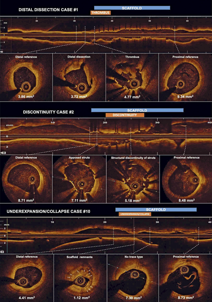
The main OCT findings were device underexpansion/collapse (n=7; 58%), scaffold discontinuity (n=4; 33%), and distal edge dissection (n=1; 9%) (Figure 1, Supplementary Figure 2, Supplementary Figure 3, Moving image 1-Moving image 3). Two cases of underexpansion (#5 and #7 in Table 1) also had OCT data at the index PCI, showing a well apposed and expanded scaffold, therefore confirming the occurrence of a device collapse. No other baseline OCT data were available. The scaffold discontinuity phenomenon was identified at an earlier stage than underexpansion/collapse (70 days [32-11] vs 224 days [190-366]), p=0.022).
Figure 1.
Second-generation drug-eluting absorbable metal scaffold target lesion revascularisation OCT findings. Minimum lumen area is shown per each cross-section. In case #2, predilation was performed before OCT assessment. Therefore, it cannot be ruled out that the scaffold structure was iatrogenically altered. A three-dimensional reconstruction of case #2 is shown in Supplementary Figure 3.
CASE REPORTS FROM THE LITERATURE
In the literature review, up to April 2019, we found six cases of MgBRS TLR with intravascular imaging assessment (Supplementary Table 2)2,3,4,5,6,7. The data relating to these cases are shown in Supplementary Appendix 4.
Discussion
The main findings of this case series study are the following. 1) The most frequent OCT findings were device underexpansion/collapse and scaffold discontinuity. 2) Scaffold discontinuity was found at an earlier stage than underexpansion/collapse. 3) The majority of the cases were off-label indications with a suboptimal PSP technique. 4) Only one case of scaffold thrombosis was observed.
In this retrospective analysis of patients treated with MgBRS who experienced TLR, the clinical characteristics and OCT findings resembled very closely those of the cases reported in the literature. Although both populations are very selected, the TLRs analysed may be related either to scaffold use in an off-label scenario or to a suboptimal PSP implantation technique8,9. Nevertheless, a recent study showed that the procedural characteristics do not impact on the MgBRS healing process10. It is worth mentioning that in first-in-man studies MgBRS were used in a very selected population, including only simple lesions and excluding ACS patients9. Notably, the off-label implantation in STEMI patients may be related to the high TLR rate because of the difficulties of accurate device sizing and worse clinical outcomes.
Of note is that, irrespective of adverse OCT findings, only one patient had definite scaffold thrombosis, in the context of early DAPT interruption, which is in line with previous studies showing a lower acute MgBRS thrombogenicity as compared to polymeric BRS11.
Eventually, prolongation of the scaffolding time and increasing the radial force could reduce the incidence of scaffold discontinuity or device collapse during follow-up. These hypotheses need to be confirmed in larger studies and should be taken into consideration for further improvements in the technology.
Limitations
This study has limitations that should be acknowledged. First, this is a retrospective study without a control group. However, to date, this is the largest cohort of MgBRS TLR analysed at a core lab facility. Second, only two cases had baseline and TLR OCT data. Third, for the assessment of MgBRS, the use of intravascular ultrasound (IVUS) and virtual histology may be better than OCT9, although the spatial resolution of OCT is higher than that of IVUS. Fourth, as the magnesium scaffold shadow disappears by six months, the struts can be ambiguously identified, making the discrimination of the OCT findings challenging. Fifth, the OCT definitions applied were derived from polymeric BRS studies. However, currently there are no standardised definitions for MgBRS failure assessment.
Conclusion
In patients who experienced an MgBRS TLR, the most frequent OCT finding was device underexpansion/collapse followed by scaffold discontinuity.
Impact on daily practice
The most common OCT findings in patients who experienced an MgBRS TLR were device underexpansion/collapse and scaffold discontinuity. The discontinuity cases occurred at an earlier stage compared to device underexpansion/collapse. However, a scaffold thrombosis conditioning a myocardial infarction was unusual. All these data should be considered for the clinical decision-making process and further improvements in the technology.
Supplementary data
Patient selection criteria.
Optical coherence tomography definitions.
Systematic literature review methodology.
Systematic literature review results.
Search strategy flow chart.
Second-generation drug-eluting absorbable metal scaffold target lesion revascularisation OCT findings.
OCT three-dimensional reconstruction of the scaffold discontinuity case number two.
Patient and procedural characteristics.
Case reports of magnesium-based bio-resorbable scaffold TLR found in the systematic literature review.
Distal dissection case #1.
Discontinuity case #2.
Underexpansion case #10.
Acknowledgments
Conflict of interest statement
The authors have no conflicts of interest to declare.
Contributor Information
Luis Ortega-Paz, Hospital Clínic i Provincial de Barcelona, IDIBAPS (Institut d’Investigacions Biomèdiques August Pi i Sunyer), Barcelona, Spain.
Salvatore Brugaletta, Hospital Clínic i Provincial de Barcelona, IDIBAPS (Institut d’Investigacions Biomèdiques August Pi i Sunyer), Barcelona, Spain.
Josep Gomez-Lara, Hospital Universitari de Bellvitge, Institut d’Investigació Biomèdica de Bellvitge (IDIBELL), Universitat de Barcelona, L’Hospitalet de Llobregat, Spain.
Juan Sanchis, Cardiology Department, Hospital Clínico Universitario de Valencia, INCLIVA, Departamento de Medicina, Universitat de València, València, Spain.
José Fernández-Díaz, Interventional Cardiology Unit, Hospital Universitario Puerta de Hierro, Majadahonda, Madrid, Spain.
Miquel Artaiz-Urdaci, Department of Cardiology, Clínica Universidad de Navarra, University of Navarra, Navarra, Spain.
Fernando Alfonso, Hospital Universitario de La Princesa, Madrid, Spain.
Hector Garcia-Garcia, Section of Interventional Cardiology, MedStar Cardiovascular Research Network, MedStar Washington Hospital Center, Washington, DC, USA.
Manel Sabaté, Hospital Clínic i Provincial de Barcelona, IDIBAPS (Institut d’Investigacions Biomèdiques August Pi i Sunyer), Barcelona, Spain.
References
- Yamaji K, Byrne RA, Windecker S, Karagiannis A, Amabile N, Lee M, Lee JKT, Christ G, Delarche N, Ielasi A, O’Sullivan CJ, Honton B, Boeder N, Meincke F, Abdel-Wahab M, Ueki Y, Hoppmann P, Motreff P, Timmers L, Gomez-Lara J, Wykrzykowska JJ, Lattuca B, Loh JP, Adriaenssens T, Nef H, Wiebe J, Daemen J, Souteyrand G, Räber L. Mechanisms of Very Late Bioresorbable Scaffold Thrombosis: The INVEST Registry. J Am Coll Cardiol. 2017;70:2330–44. doi: 10.1016/j.jacc.2017.09.014. [DOI] [PubMed] [Google Scholar]
- Mitomo S, Demir OM, Giannini F, Latib A, Colombo A. Magmaris Bioresorbable Scaffold - Possible Dismantling 2 Months After Implantation on Intravascular Ultrasound. Circ J. 2019;83:1418. doi: 10.1253/circj.CJ-18-0795. [DOI] [PubMed] [Google Scholar]
- Marynissen T, McCutcheon K, Bennett J. Early collapse causing stenosis in a resorbable magnesium scaffold. Catheter Cardiovasc Interv. 2018;92:310–2. doi: 10.1002/ccd.27676. [DOI] [PubMed] [Google Scholar]
- Roa-Garrido J, Cardenal-Piris RM, El Amrawy AM, Gomez-Menchero A, Camacho Freire S, Diaz-Fernandez JF. Optical Coherence Tomographic Image Pattern of Metallic Bioresorbable Vascular Scaffold Restenosis. JACC Cardiovasc Interv. 2018;11:707–8. doi: 10.1016/j.jcin.2017.11.025. [DOI] [PubMed] [Google Scholar]
- Barkholt TO, Neghabat O, Terkelsen CJ, Christiansen EH, Holm NR. Restenosis in a Collapsed Magnesium Bioresorbable Scaffold. Circ Cardiovasc Interv. 2017;10:e005677. doi: 10.1161/CIRCINTERVENTIONS.117.005677. [DOI] [PubMed] [Google Scholar]
- Yang H, Zhang F, Qian J, Chen J, Ge J. Restenosis in Magmaris Stents Due to Significant Collapse. JACC Cardiovasc Interv. 2018;11:e77–8. doi: 10.1016/j.jcin.2018.02.040. [DOI] [PubMed] [Google Scholar]
- McCutcheon K, Bennett J, Marynissen T, Adriaenssens T. Severe focal restenosis 15 months after implantation of a magnesium bioresorbable scaffold. Acta Cardiol. 2020;75:85–6. doi: 10.1080/00015385.2018.1548109. [DOI] [PubMed] [Google Scholar]
- Ortega-Paz L, Araszkiewicz A, Tamburino C, Sabate M, Munzel T, Colombo A, Naganuma T, Geraci S, Mattesini A, Pyxaras S, Mehilli J, Capodanno D, Wiebe J, Capranzano P, Lesiak M, Naber C, Di Mario C, Caramanno G, Latib A, Nef H, Gori T, Brugaletta S. Predilation, sizing and post-dilation scoring in patients undergoing everolimus-eluting bioresorbable scaffold implantation for prediction of cardiac adverse events: development and internal validation of the PSP score. EuroIntervention. 2017;12:2110–7. doi: 10.4244/EIJ-D-16-00974. [DOI] [PubMed] [Google Scholar]
- Haude M, Ince H, Kische S, Abizaid A, Tölg R, Alves Lemos P, Van Mieghem NM, Verheye S, von Birgelen C, Christiansen EH, Barbato E, Garcia-Garcia HM, Waksman R BIOSOLVE-II and III investigators. Safety and clinical performance of a drug eluting absorbable metal scaffold in the treatment of subjects with de novo lesions in native coronary arteries: Pooled 12-month outcomes of BIOSOLVE-II and BIOSOLVE-III. Catheter Cardiovasc Interv. 2018;92:E502–11. doi: 10.1002/ccd.27680. [DOI] [PMC free article] [PubMed] [Google Scholar]
- Ozaki Y, Garcia-Garcia HM, Hideo-Kajita A, Kuku KO, Haude M, Ince H, Abizaid A, Tölg R, Lemos PA, von Birgelen C, Christiansen EH, Wijns W, Escaned J, Dijkstra J, Waksman R. Impact of procedural characteristics on coronary vessel wall healing following implantation of second-generation drug-eluting absorbable metal scaffold in patients with de novo coronary artery lesions: an optical coherence tomography analysis. Eur Heart J Cardiovasc Imaging. 2019;20:916–24. doi: 10.1093/ehjci/jey210. [DOI] [PubMed] [Google Scholar]
- Waksman R, Lipinski MJ, Acampado E, Cheng Q, Adams L, Torii S, Gai J, Torguson R, Hellinga DM, Westman PC, Joner M, Zumstein P, Kolodgie FD, Virmani R. Comparison of Acute Thrombogenicity for Metallic and Polymeric Bioabsorbable Scaffolds: Magmaris Versus Absorb in a Porcine Arteriovenous Shunt Model. Circ Cardiovasc Interv. 2017;10(8) doi: 10.1161/CIRCINTERVENTIONS.116.004762. [DOI] [PubMed] [Google Scholar]
Associated Data
This section collects any data citations, data availability statements, or supplementary materials included in this article.
Supplementary Materials
Patient selection criteria.
Optical coherence tomography definitions.
Systematic literature review methodology.
Systematic literature review results.
Search strategy flow chart.
Second-generation drug-eluting absorbable metal scaffold target lesion revascularisation OCT findings.
OCT three-dimensional reconstruction of the scaffold discontinuity case number two.
Patient and procedural characteristics.
Case reports of magnesium-based bio-resorbable scaffold TLR found in the systematic literature review.
Distal dissection case #1.
Discontinuity case #2.
Underexpansion case #10.


