Abstract
Pertussis toxin is a member of the AB5 family of toxins and is composed of five subunits (S1 to S5) present in a 1:1:1:2:1 ratio. Secretion is a complex process. Each subunit has a secretion signal that mediates transport to the periplasm, where processing and assembly occur. Secretion of the assembled 105-kDa toxin past the outer membrane is mediated by the nine proteins encoded in the ptl operon. Previous studies have shown that S1, the catalytically active A subunit of pertussis toxin, is necessary for efficient secretion, suggesting that a domain on S1 may be required for interaction with the secretion apparatus. Previously, recombinant S1 from four different mutants (serine 54 to glycine, serine 55 to glycine, serine 56 to glycine, and arginine 57 to lysine) was shown to retain catalytic activity. We introduced these mutations into Bordetella pertussis and monitored pertussis toxin production and secretion. No pertussis toxin was detected in the serine 54-to-glycine mutant. The other S1 mutants produced periplasmic pertussis toxin, but little pertussis toxin secretion was observed. The arginine 57-to-lysine mutant had the most dramatic secretion defect. It produced wild-type levels of periplasmic pertussis toxin but secreted only 8% as much toxin as the wild-type strain. This phenotype was similar to that observed for strains with mutations in the ptl genes, suggesting that this region may have a role in pertussis toxin secretion.
Pertussis toxin is a major virulence factor of Bordetella pertussis, the gram-negative bacterium that is the causative agent of whooping cough. It is a member of the AB5 family of toxins, consisting of five subunits, S1, S2, S3, S4, and S5, present in a 1:1:1:2:1 ratio (18, 20, 27). S1 is the A or enzymatic subunit and catalyzes the ADP-ribosylation of G proteins in the target mammalian cell. Subunits S2 to S5 form the B pentamer, which delivers the S1 subunit to the target mammalian cell. Several of the mammalian cells targeted by pertussis toxin (including lymphocytes, macrophages, and neutrophils) are important effectors of the immune system, and toxin treatment compromises their ability to function, contributing to the severity of the disease (22, 23, 29).
Pertussis toxin assembly and secretion is a complex process. Each subunit is synthesized with a signal peptide (20), which mediates secretion to the periplasm via the equivalent of the Sec-mediated secretion machinery of Escherichia coli. Folding and assembly of the subunits occurs in the periplasm, and the ptl (pertussis toxin liberation) operon is required for efficient secretion of assembled toxin past the outer membrane (7, 9, 31). The ptl secretion machinery is specific for pertussis toxin, since secretion of other known virulence factors occurs normally in ptl mutant strains (31, 32). The secretion machinery appears to discriminate between assembled and unassembled pertussis toxin, since only assembled toxin is efficiently released from the bacteria. Since secretion involves substrate recognition, the Ptl secretion machinery must recognize some domain on pertussis toxin. In previous studies, S1 mutants were deficient for pertussis toxin secretion (26), implicating a role for S1 in pertussis toxin secretion. These mutations appeared to destabilize the molecule, and the strains were all deficient in S1 production, so no specific region of the S1 molecule was shown to play a role secretion. In this study we wanted to examine mutants capable of producing stable S1 and to define regions that may play a role in secretion.
The crystal structure of pertussis toxin has been determined. The active site in S1 appears to be in a cavity formed by an α-helix bent over a β-strand (termed the BA box) lined by β-strands containing the catalytic residues (8, 27). Several mutants with changes in the BA box region of S1 have been generated. The mutations were serine 54 to glycine (S54G), serine 55 to glycine (S55G), serine 56 to glycine (S56G), or arginine 57 to lysine (R57K). These changes did not appear to affect catalytic activity, since recombinant S1 overexpressed and purified from inclusion bodies in E. coli was shown to possess ADP-ribosylation activity (13). In this study we introduced these mutations into B. pertussis and examined their effect on pertussis toxin production and secretion. No S1 or pertussis toxin could be detected in mutant S54G. The other three mutants produced perplasmic pertussis toxin but secreted reduced levels of toxin compared to the wild-type strain. The R57K mutant expressed normal levels of periplasmic pertussis toxin but was defective in pertussis toxin secretion. These results suggest that arginine 57 and the adjacent regions of S1 may be involved in pertussis toxin secretion.
MATERIALS AND METHODS
Bacterial strains.
The B. pertussis strains used in this study are described in Table 1. B. pertussis were grown on Bordet-Gengou agar (BGA) medium (Difco, Detroit, Mich.) containing 15% sheep's blood (Colorado Serum, Denver, Colo.) or in Stainer Scholte minimal broth (SS broth) as previously described (30). E. coli strains were grown on L agar. When necessary, the following antibiotics at the indicated concentrations were added to the media: nalidixic acid, 30 μg/ml; gentamicin, 10 μg/ml (for maintenance of B. pertussis and E. coli strains) or 30 μg/ml (for selection of B. pertussis transconjugants); ampicillin, 100 μg/ml; and streptomycin, 300 μg/ml.
TABLE 1.
B. pertussis strains
| B. pertussis strain | Relevant characteristica | S1 expression | Source and/or reference |
|---|---|---|---|
| BP338 | Wild type; Tohama I background; Nalr | Wild type | 30 |
| BP338/cyc::WT | pKC113 conjugated into BP338; Nalr Genr | Wild type | 6, 7 |
| BPM3171 | Tn5 lac insertion into ptlC | Wild type | 31, 32 |
| BPM3171/cyc::WT | pKC113 conjugated into BPM3171 (Tn5 lac insertion into ptlC) | Wild type | This study; 6 |
| BPRA | Pertussis toxin deletion mutant; Nalr Strr | None | 1 |
| BPRA/cyc::WT | pKC113 conjugated into BPRA; Nalr Strr Genr | Wild type | 6, 7 |
| BPRA/cyc::S1-S54G | pS1-S54G conjugated into BPRA; Nalr Strr Genr | Serine 54 to glycine | This study; 6 |
| BPRA/cyc::S1-S55G | pS1-S55G conjugated into BPRA; Nalr Strr Genr | Serine 55 to glycine | This study; 6 |
| BPRA/cyc::S1-S56G | pS1-S56G conjugated into BPRA; Nalr Strr Genr | Serine 56 to glycine | This study; 6 |
| BPRA/cyc::S1-R57K | pS1-R57K conjugated into BPRA; Nalr Strr Genr | Arginine 57 to lysine | This study; 6 |
Nalr, nalidixic acid resistance; Strr, streptomycin resistance; Genr, gentamicin resistance.
Reagents.
Restriction enzymes and T4 DNA ligase were purchased from Gibco BRL (Gaithersburg, Md.) or New England BioLabs (Beverly, Mass.) and used according to the manufacturer's specifications. SeaKem and SeaPlaque (low-melting-point) agarose were obtained from FMC Bioproducts (Rockland, Maine). Antibiotics were purchased from Sigma Chemical Co. (St. Louis, Mo.). Sodium dodecyl sulfate-polyacrylamide gel electrophoresis (SDS-PAGE) reagents were obtained from Bio-Rad Laboratories (Hercules, Calif.). Tissue culture media, antibiotic supplements, and fetal bovine serum (FBS) were acquired from Gibco BRL. DNA and protein molecular weight markers were purchased from Gibco BRL or Bio-Rad Laboratories. Plasmids were isolated by using either the Midiprep Kit or the Miniprep Kit from Qiagen (Valencia, Calif.).
S1 mutant constructs.
The plasmids used in this study are described in Table 2. The original S1 site-directed mutations, S1/1E/pUC19 (S54G), S1/1F/pUC19 (S55G), S1/1G/pUC19 (S56G), and S1/1H/pUC19 (R57K), were generated by N. Burnette and V. Mar (13). In a previous study (7) we developed a suicide plasmid containing the pertussis toxin structural and secretion genes (the ptxptl operon). Plasmid pKC34 contains base pairs 0 to 13,025 of ptxptl operon in the EcoRI and PstI sites of pSP72. For these studies, the 307-bp NdeI/SphI fragment containing the S1 mutation was used to replace the corresponding wild-type sequences in pKC34 (6).
TABLE 2.
Plasmids
| Plasmid(s) | Descriptiona | Source or reference |
|---|---|---|
| pSP72 | Cloning vector; Ampr | Promega |
| pUC18, pUC19 | Cloning vector; Ampr | Gibco BRL |
| pBluescript | Cloning vector; Ampr | Stratagene |
| S1/1E/pUC19 | 307-bp NdeI/SphI fragment in pUC19; encodes the S1 S54G mutation | 13 |
| S1/1F/pUC19 | 307-bp NdeI/SphI fragment in pUC19; encodes the S1 S55G mutation | 13 |
| S1/1G/pUC19 | 307-bp NdeI/SphI fragment in pUC19; encodes the S1 S56G mutation | 13 |
| S1/1H/pUC19 | 307-bp NdeI/SphI fragment in pUC19; encodes the S1 R57K mutation | 13 |
| pKC34 | bp 0 to 13025 from ptxptl in the EcoRI and PstI sites of SP72 | 6, 7 |
| pUW2138 | Genr-oriT cassette in pBluescript SK(+) | 10 |
| pKC109 | Genr-oriT cassette and the 2.1-kb adenylate cyclase fragment (bp 4611 to 6740) in pUW2138 | 6, 7 |
| pKC113 | pKC34 with the 5.1-kb HindIII cyc/Genr/oriT fragment from pKC109 in the HindIII site | (6, 7 |
| pS1-S54G | pKC113 with the S1 bp 778 to 780 changed from AGC to GGT | This study; 6 |
| pS1-S55G | pKC113 with the S1 bp 781 to 783 changed from AGC to GGT | This study; 6 |
| pS1-S56G | pKC113 with the S1 bp 784 to 786 changed from AGC to GGT | This study; 6 |
| pS1-R57K | pKC113 with the S1 bp 787 to 789 changed from CGG to AAG | This study; 6 |
| pRK2013 | 6.3-kb EcoRI ColE1 replicon from pDF1 and 41.7-kb EcoRI fragment A of pRK212.2; Kanr | 11 |
Genr, gentamicin resistance; Ampr, ampicillin resistance; Kanr, kanamycin resistance.
A gene cassette was developed to facilitate introduction of genes into the chromosome of B. pertussis by mobilization (6, 7). A 2.1-kb PstI fragment internal to the adenylate cyclase toxin gene (bp 4611 to 6740) was cloned into the PstI site of pUW2138, resulting in pKC109. The gentamicin resistance gene, the origin of transfer (oriT) for the P-plasmid incompatibility group, and the adenylate cyclase toxin gene can be excised as a 5.1-kb HindIII fragment. The 5.1-kb HindIII fragment from pKC109 was cloned into the HindIII site of the resulting plasmids to create pS1-S54G, pS1-S55G, pS1-S56G, and pS1-R57K.
Conjugation.
Each ptl construct containing the S1 mutation was introduced into B. pertussis by a triparental mating as previously described (3). Transconjugants were selected on BGA plates containing nalidixic acid and gentamicin. Nonhemolytic colonies, indicative of plasmid integration into the adenylate cyclase toxin locus, were selected for characterization.
At a low frequency, integrated plasmids can recircularize and excise from the chromosome by homologous recombination. While this is a lethal event in the presence of antibiotic selection, it does allow recovery of the integrated plasmid. After the transconjugants were examined for pertussis toxin secretion, the plasmids were conjugated from B. pertussis into E. coli HB101 by triparental mating and selected on L agar plates containing ampicillin, gentamicin, and streptomycin. The presence of the mutation was verified by comparing the restriction pattern of the plasmid mobilized from B. pertussis to the parental plasmid propagated only in E. coli. Isolates that had undergone rearrangements were not included in the analysis.
PCR analysis of transconjugants.
The presence of the pertussis toxin deletion in the chromosome of the BPRA transconjugants was verified by PCR. Chromosomal DNA was prepared by suspending a few bacterial colonies in 500 μl of 50 mM Tris (pH 7.6) containing 0.2 mg of proteinase K (Sigma Chemical Co.) per ml and incubating at 65°C for 45 min, followed by boiling for 20 min. Then, 2 μl of this preparation was used per 25 μl of PCR reaction. PCR was performed by using the Advantage-GC cDNA PCR Kit (Clontech Laboratories, Palo Alto, Calif.) according to the manufacturer's instructions. Primers were designed to span the deletion: 5′-CAAGATAATCGTCCTGCTCAACCGC-3′ was used as the forward primer, and 5′-GTGAGGGCATAGGTCTGGAATGTGG-3′ was used as the reverse primer. The appropriate 795-bp band was observed in BPRA, and in BP338 the appropriate full-length 3,535-bp band was observed.
Secretion assay.
B. pertussis from 24-h BGA cultures were suspended to an optical density at 600 nm (OD600) of 0.1 in SS broth. Six milliliters of the suspension was plated on a BGA plate containing nalidixic acid and gentamicin. After 30 h at 37°C, the culture was harvested from the plate, and its volume was noted. The cells were pelleted at 7,000 rpm in an SS34 rotor for 10 min. The supernatant was filter sterilized and stored at −20°C for use in a pertussis toxin CHO cell assay. The bacteria were suspended in phosphate-buffered saline (PBS) to the volume harvested, and the OD600 values were measured. To assess intracellular toxin levels, a 1-ml aliquot of cells was pelleted at 6,000 rpm in a microcentrifuge and then suspended in 100 μl of 50 mM Tris and 50 mM EDTA containing 2 mg of lysozyme per ml. After 30 min at 37°C, 900 μl of PBS containing 0.05% Tween 20 was added. The cell debris was removed by centrifugation at 12,000 rpm in a microcentrifuge, and the supernatant filter was sterilized and stored at −20°C for use in a CHO cell assay.
To test the stability of mutant form of pertussis toxin, 5 μl of toxin solubilized from the wild type or the R57K mutant was added to 45 μl of a 30-h culture supernatant of BPRA grown in SS broth or to 45 μl of SS broth alone as a control. BPRA is a strain lacking pertussis toxin expression but still capable of secreting all other known virulence factors of B. pertussis. The samples were incubated for 1 h at 37°C and characterized for pertussis toxin activity in the CHO cell assay described below.
CHO cell assay.
The CHO cell assay was used to determine pertussis toxin activity (7, 12, 31). Pertussis toxin-treated CHO cells lose contact inhibition and clump together. Serial twofold dilutions of purified pertussis toxin (List Biologicals, Campbell, Calif.) and the unknowns were made in Ham's F-12 tissue culture medium containing 1% FBS, and 20 μl of the samples were added to 250 μl of CHO cells in 96-well plates. After 48 h, the cells were fixed and stained, and clumping was examined microscopically. The limit of detection for purified pertussis toxin was approximately 1 to 2 ng/ml, and the last positive well for an unknown sample was assigned that value. Each sample was assayed in duplicate. A student's t test was used to analyze the data.
SDS-PAGE and immunoblotting.
B. pertussis cells were grown and harvested as described for the secretion assay. SDS-PAGE was performed by the method of Laemmli (16) with the modifications of Peppler (21). Bacterial cells were centrifuged and concentrated to an OD600 of 16 in PBS, and samples were added to 3 volumes of Laemmli's solubilization buffer. Samples were boiled for 7 min, and 5 μl of each was subjected to electrophoresis on a Mini Protean II gel system by using 0.75-mm spacers (Bio-Rad) with 12.5% polyacrylamide in the separating gel and 4% polyacrylamide in the stacking gel. Electrophoresis was conducted for approximately 45 min at 200 V until the dye-front left the gels. Gels were blotted onto nitrocellulose membranes (0.45-μm pore size; Schleicher & Schuell, Dassel, Germany) in a submarine apparatus (Trans-Blot Tank; Bio-Rad) using a modified Towbin buffer (28) (15.6 mM Tris–120 mM glycine–0.02% SDS–20% methanol) at 4°C. Blotting was performed for 2 h at 100 V without further cooling. Proteins were detected by probing with monoclonal antibody C3X4 to S1 (14) and visualized by chemiluminescence using the Dupont Western blot Renaissance kit (NEN Research Products, Boston, Mass.). Peroxidase-conjugated goat anti-mouse secondary antibody was purchased from Cappel (West Chester, Pa.). Apparent molecular weights were determined by comparison with prestained molecular weight markers (Bio-Rad) and purified pertussis toxin controls.
RESULTS
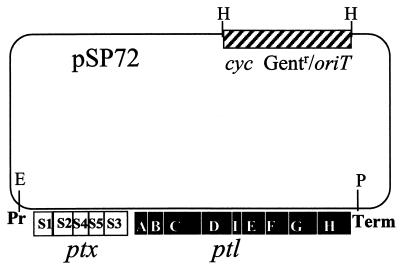
To facilitate genetic analysis, we constructed a shuttle vector that would allow us to integrate the entire ptxptl operon into a known region of the B. pertussis chromosome (7). The 13-kb ptxptl operon was cloned into a suicide shuttle plasmid capable of replicating in E. coli but not B. pertussis (Fig. 1A). A cassette containing the cis-acting region of the OriT (origin of transfer) of P-incompatibility plasmid (to allow for mobilization from E. coli into B. pertussis), a gentamicin resistance marker, and a 2.1-kb fragment of the adenylate cyclase toxin gene, cycA (Fig. 1), was cloned into the plasmid. The adenylate cyclase toxin gene was chosen because adenylate cyclase toxin causes hemolysis on blood plates. Integration of the plasmid into this locus results in a nonhemolytic phenotype making it easy to identify plasmids that had integrated into this region of the chromosome (7).
FIG. 1.
pKC113, containing the 13-kb ptxptl operon in pSP72. White boxes, pertussis toxin structural genes; black boxes, ptl genes; hatched box, cyc/Gentr/oriT region. Pr, ptxptl promoter; E, EcoRI; P, PstI; Term, terminator; cyc, adenylate cyclase toxin gene; Gentr, gentamicin resistance; oriT, P-plasmid origin of transfer; pSP72, plasmid vector.
The plasmid can integrate into either the pertussis toxin operon or into the adenylate cyclase toxin locus. We found that integration at the adenylate cyclase locus occurred less frequently than integration into the ptxptl operon, as determined by comparison of hemolytic to nonhemolytic transconjugants, probably due to its smaller size (2.1 kb for cycA versus 13 kb for ptxptl), but multiple independent transconjugants could be obtained. PCR analysis verified that integration into the adenylate cyclase toxin locus did not affect the chromosomal pertussis toxin locus. The appropriate 795-bp band was observed in transconjugants of BPRA (Table 1, deleted for part of the pertussis toxin operon), and in BP338 (Table 1, wild-type strain) the appropriate full-length 3,535-bp band was observed (data not shown).
The nonhemolytic transconjugants were stable when maintained under antibiotic selection. We did not detect any hemolytic revertants out of 20,000 colonies examined after the first passage in the absence of gentamicin selection. However, after a second passage without gentamicin, hemolytic, gentamicin-sensitive revertants were observed at a frequency of about 1/5,000, suggesting that they had arisen by excision of the plasmid. While integration at either site should allow pertussis toxin expression from the cloned copy, other studies characterizing transconjugants that had integrated into the adenylate cyclase toxin locus allowed us to generate stable merodiploid strains to perform complementation studies (7). In these studies, pertussis toxin production and secretion was examined in parallel for at least three independent nonhemolytic transconjugants containing mutant plasmids, and at least three independent nonhemolytic transconjugants containing the wild-type plasmid control.
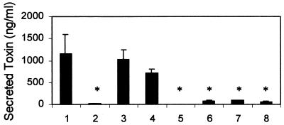
To determine whether the integrated ptxptl operon was capable of directing pertussis toxin production, the plasmid was introduced into the wild-type strain BP338 and into the pertussis toxin secretion mutant BPM3171, possessing a Tn5 lac insertion in the PtlC gene (Table 1). Pertussis toxin secretion was monitored. The wild-type strain secreted 1,162 ng of pertussis toxin per ml (Fig. 2, column 1) while the ptl secretion mutant only secreted 17 ng of pertussis toxin per ml (Fig. 2, column 2). Introduction of the wild-type ptxptl operon restored secretion to the ptl secretion mutant (Fig. 2, column 3); BPM3171 with the integrated plasmid produced 1,033 ng of pertussis toxin per ml, a statistically significant increase from BPM3171 lacking the integrated plasmid but not statistically different from the wild-type strain. These studies verify that integration of the ptxptl operon restores pertussis toxin production to Ptl secretion mutants. The presence of two intact copies of the ptxptl operon in the wild-type strain did not result in more pertussis toxin secretion. These results are similar to previous studies where increased copy number did not lead to increased pertussis toxin production (2, 7).
FIG. 2.
The amount of pertussis toxin secreted after 30 h as determined by CHO cell toxin assay. Columns: 1, BP338/cyc::WT (wild type with two copies of the ptxptl operon); 2, BPM3171 (Ptl mutant); 3, BPM3171/cyc::WT (Ptl mutant with one copy of the ptxptl operon); 4, BPRA/cyc::WT (ptx deletion with one copy of the ptxptl operon); 5, BPRA/cyc::S1-54G (ptx deletion with intact copy of the ptxptl operon); 6, BPRA/cyc::S1-S55G (ptx deletion with S1 mutation in the ptxptl operon); 7, BPRA/cyc::S1-S56G (ptx deletion with S1 mutation in the ptxptl operon); 8, BPRA/cyc::S1-R57K (ptx deletion with S1 mutation in the ptxptl operon). *, P < 0.05 for mutant compared to wild type.
The intact ptxptl operon was also introduced into BPRA, a virulent hemolytic strain with a deletion of the pertussis toxin promoter and the S1, S2, S4, and S5 genes. This strain fails to express pertussis toxin and the ptl secretion genes (1, 15). The introduction of the wild-type ptxptl operon restored pertussis toxin expression and secretion to BPRA (Fig. 2, column 4). BPRA containing the intact ptxptl operon appeared to secrete less pertussis toxin than BP338 or BPM3171 containing the same construct, but the difference was not statistically significant compared to BP338 (P < 0.052). In addition, BP338 and BPM3171 are isogenic, but BPRA possesses a chromosomal streptomycin resistance mutation that could affect protein synthesis.
Mutational analysis of pertussis toxin subunit S1 mutants.
Each of the S1 mutations was cloned into the ptxptl shuttle vector and introduced into BPRA, the pertussis toxin deletion strain. All of the mutants secreted statistically significantly less pertussis toxin than BPRA containing the wild-type ptxptl operon (Fig. 2, columns 5 to 8). One mutant, S54G, failed to secrete any detectable pertussis toxin (Fig. 2, column 5).
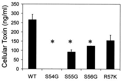
It has been shown previously that ptl secretion mutants are unable to efficiently secrete pertussis toxin past the outer membrane; however, they accumulate as much pertussis toxin in the periplasm as the wild-type strain (2, 6, 7, 31). To monitor intracellular pertussis toxin accumulation by the wild type and the S1 mutants, washed bacteria were lysed by treatment with lysozyme and EDTA, and the amount of functional pertussis toxin was determined by the CHO cell assay. BPRA with the wild-type ptxptl operon produced 266 ± 28 ng of cell-associated toxin per ml (Fig. 3, WT). No pertussis toxin was detected in the S54G mutant. In contrast, intracellular pertussis toxin was detected in the other three mutants. The S55G and S56G mutants produced a little less than half as much pertussis toxin activity as the control (Fig. 3), which was a statistically significant difference. In contrast, the R57K mutant produced about 60% as much cell-associated pertussis toxin as had the wild type, which was not a statistically significant difference.
FIG. 3.
The amount of cell associated pertussis toxin after 30 h as determined by CHO cell toxin assay. Columns: WT, BPRA/cyc::WT; S54G, BPRA/cyc::S1-S54G; S55G, BPRA/cyc::S1-S55G; S56G, BPRA/cyc::S1-S56G; R57K, BPRA/cyc::S1-R57K. *, P < 0.05 for mutant compared to the wild type.
Since the CHO cell assay is only able to detect the presence of functional pertussis toxin, Western blot analysis with a monoclonal antibody specific for S1 was used to assess the amount of antigenic S1 present in the periplasm. Full-length S1 (28 kDa, denoted by an arrow) and a smaller S1 breakdown product was detected in wild-type BP338 (Fig. 4, lane 1) and in BPRA containing the wild-type ptxptl plasmid (Fig. 4, lanes 3 and 8) but not in BPRA without the plasmid (Fig. 4, lane 2). Reduced levels of intact S1 were detected in the S54G mutant (Fig. 4, lane 4). The S55G mutant (Fig. 4, lane 5) and the S56G mutants (Fig. 4, lane 6) seemed to produce as much intact S1 as the wild-type strains, but less breakdown product was observed. In contrast, the R57K mutant produced wild-type levels of both S1 and the S1 breakdown product (Fig. 4, lane 7), suggesting that S1 production in this mutant is similar to that in the wild-type strain.
FIG. 4.
Western blot of intracellular S1. Lanes: 1, wild-type strain BP338; 2, pertussis toxin deletion strain BPRA; 3 and 8, BPRA/cyc::WT; 4, BPRA/cyc::S1-S54G; 5, S55G, BPRA/cyc::S1-S55G; 6, BPRA/cyc::S1-S56G; 7, BPRA/cyc::S1-R57K. The arrow denotes the migration point of intact 28-kDa S1 from purified pertussis toxin.
It could be hypothesized that while the R57K mutation does not affect the activity of periplasmic pertussis toxin, it could make the secreted form less stable; for example, it may be more susceptible to proteolysis. To test this hypothesis, three samples of periplasmic toxin solubilized from the strain expressing the wild-type ptxptl operon and the three samples of toxin solubilized from the strain expressing the 57K mutation (characterized in Fig. 3) were diluted 10-fold into culture supernatant from BPRA grown in SS broth or into SS broth as a negative control. BPRA is deficient in expression of pertussis toxin and the ptl genes, but it is capable of producing other secreted products. After 1 h at 37°C, all of the samples were tested for activity in the CHO cell assay. The wild-type toxin from BPRA/cyc::WT had a value of 176 ± 0 ng of toxin per ml when incubated with the BPRA supernatant and 260 ± 51 ng of toxin per ml when incubated with SS broth. The toxin produced by BPRA/cyc::S1-R57K had a value of 147 ± 29 ng of toxin per ml when incubated with the BPRA supernatant and 147 ± 30 ng of toxin per ml when incubated with SS broth. None of these values was statistically different from each other or the value or the value of 266 ± 28 ng/ml for the wild-type sample not incubated at 37°C. Together, these results suggest that the R57K mutant is as stable as the wild-type pertussis toxin.
DISCUSSION
Pertussis toxin has the most complex structure of any toxin, with five subunits coming together with an unusual 1:1:1:2:1 stoichiometry. Toxin assembly occurs in the periplasm and is uncoupled from secretion past the outer membrane, since equivalent amounts of intracellular toxin accumulate in the absence of a functional ptl secretion operon (2, 6, 7, 31). It has been suggested that the nine Ptl proteins produce a gated channel in the outer membrane that allows specific passage of pertussis toxin (33). In support of this model, Ptl mutants are not deficient for secretion of any of the other known B. pertussis virulence factors (32). Thus, the Ptl-secretion machinery must specifically recognize its substrate, assembled pertussis toxin, before opening the gate. The goal of this study was to identify domains on pertussis toxin that are required for secretion.
In the process of trying to genetically produce pertussis toxoid for vaccine purposes, several laboratories independently produced dozens of strains expressing mutant forms of S1 (4, 13, 17, 18, 24, 25). In many cases, only recombinant S1 produced by E. coli was characterized. In one of the few studies where the S1 mutations were introduced into B. pertussis and expressed in the context of the entire ptxptl operon, Pizza et al. implicated a role for the S1 subunit in secretion (24). However, these mutants all had the same phenotype in B. pertussis: a deficiency in S1 expression. As a result it was not possible to identify specific domains on S1 that were involved in secretion. In contrast, the same investigators isolated one S1 mutant (9K/129G) that was devoid of enzyme and toxin activity but was secreted at normal levels (25). This result suggests that secretion does not require enzymatic activity, and the amino acids at position 9 and 129 are not required for secretion.
To narrow the search for regions on pertussis toxin that may be required for secretion, we hypothesized that such a region would not be necessary for catalytic activity and should be exposed on the surface of pertussis toxin, where it would be accessible to the Ptl secretion machinery. The crystal structure of pertussis toxin has suggested that the catalytic site is in a cavity formed by an α-helix bent over a β-strand (termed the BA box), surrounded by β-strands containing the catalytic residues (8). Mutants in the BA box (S54G, S55G, S56G, and R57K) were of special interest to us because they retained ADP-ribosylating activity (13). This region is partially conserved among other AB5 toxins with ADP-ribosylating activity (8), suggesting that it may have some functional importance aside from toxicity. In examining the crystal structure of pertussis toxin, serine 54 is buried, but amino acids 55 through 58 project out from a cavity formed by the intersection of S1 with S3 and S4. The close proximity of this region of S1 to two different regions in the B subunit could provide a means to distinguish assembled toxin from free subunit.
We introduced these mutations into the pertussis toxin operon and examined secretion in B. pertussis. Like many of the previously characterized S1 mutants, the S54G mutant protein was unstable when expressed in B. pertussis. Low levels of antigenic S1, but no functional pertussis toxin could be detected in this mutant. In contrast, the other three mutants produced stable periplasmic pertussis toxin that retained toxin activity in the biological CHO cell assay following release from the periplasm. Slightly less biologically active toxin was recovered from the S55G and S56G mutants. The wild-type strain produced 266 versus 92 ng/ml for the S55G mutant and 122 ng/ml for the S56G mutant. The limit of detection is the CHO cell assay is about 1 ng/ml, so the phenotype of these two mutants is clearly different from the S54G mutant, for which no toxin activity was detected. When antigenic S1 was examined, the amount of full-length S1 produced by these mutants seemed similar to that produced with the wild type, but there was reduced expression of a breakdown product. The S1 mutants, like the Ptl secretion mutants characterized in previous studies (2, 6, 7, 31), accumulate about as much periplasmic pertussis toxin as the wild-type strain. However, when secreted toxin is also considered, the total amount of toxin expressed by the secretion mutants is much less than that produced by the wild-type strain. One explanation is that a feedback mechanism could shut down toxin synthesis when the periplasm becomes full. Alternatively, a more efficient periplasmic degradation pathway could be induced. The different phenotype of the mutants suggests that different mechanisms could be operating in each case.
In contrast, the R57K mutant expressed as much antigenic and functional toxin in the periplasm as the wild-type stain. However, the R57K mutant had a dramatic defect in secretion, and secreted only 8% as much toxin as the wild-type strain. For comparison, BPM3171, the first mutant we isolated in the ptl operon, also produced normal levels of periplasmic toxin; however, it secreted 2% as much toxin as the wild-type strain. These results suggest that arginine 57 and perhaps the adjacent regions of S1 may be involved in pertussis toxin secretion. Connell et al. (5) identified a mutation in the B subunit of cholera toxin (another member of the AB5 family of toxins) that affected toxin secretion. Since the secretion machinery can distinguish assembled toxin from unassembled subunits, it is likely that there are regions on the B subunit that are necessary for secretion in addition to the domain that we have identified on S1. In future studies we hope to identify B subunit domains needed for secretion and to select for the second site mutations in the Ptl proteins that recognize the R57K mutant and restore secretion.
ACKNOWLEDGMENTS
This work was supported by grant RO1 AI23695.
We would like to thank Neil Burnette and Vernon Mar for supplying us with the S1 mutants. We would also like to thank Paula Mobberley-Schuman for her technical expertise.
REFERENCES
- 1.Antoine R, Locht C. Roles of the disulfide bond and the carboxy-terminal region of the S1 subunit in the assembly and biosynthesis of pertussis toxin. Infect Immun. 1990;58:1518–1526. doi: 10.1128/iai.58.6.1518-1526.1990. [DOI] [PMC free article] [PubMed] [Google Scholar]
- 2.Baker, S. M., A. Masi, D.-F. L., B. K. Novitsky, and R. A. Deich. 1995. Pertussis toxin export genes are regulated by the ptx promoter and may be required for efficient translation of ptx mRNA in Bordetella pertussis. 63:3920–3926. [DOI] [PMC free article] [PubMed]
- 3.Barry E M, Weiss A A, Ehrmann I E, Gray M C, Hewlett E L, Goodwin M S M. Bordetella pertussis adenylate cyclase toxin and hemolytic activities require a second gene, cyaC, for activation. J Bacteriol. 1991;173:720–726. doi: 10.1128/jb.173.2.720-726.1991. [DOI] [PMC free article] [PubMed] [Google Scholar]
- 4.Burnette W N, Cieplak W, Mar V L, Kaljot K T, Sato H, Keith J M. Pertussis toxin S1 mutant with reduced enzyme activity and a conserved protective epitope. Science. 1988;242:72–74. doi: 10.1126/science.2459776. [DOI] [PubMed] [Google Scholar]
- 5.Connell T D, Metzger D J, Wang M, Jobling M G, Holmes R K. Initial studies of the structural signal for extracellular export of cholera toxin and other proteins recognized by Vibrio cholerae. Infect Immun. 1995;63:4091–4098. doi: 10.1128/iai.63.10.4091-4098.1995. [DOI] [PMC free article] [PubMed] [Google Scholar]
- 6.Craig K A. Characterization of pertussis toxin secretion from Bordetella pertussis. Ph.D. thesis. Cincinnati, Ohio: University of Cincinnati; 1999. [Google Scholar]
- 7.Craig-Mylius K A, Weiss A A. Mutants in the ptlA-H genes of Bordetella pertussis are deficient for pertussis toxin secretion. FEMS Microbiol Lett. 1999;179:479–484. doi: 10.1111/j.1574-6968.1999.tb08766.x. [DOI] [PubMed] [Google Scholar]
- 8.Domenighini M, Magagnoli C, Pizza M, Rappuoli R. Common features of the NAD-binding and catalytic site of ADP-ribosylating toxins. Mol Microbiol. 1994;14:41–50. doi: 10.1111/j.1365-2958.1994.tb01265.x. [DOI] [PubMed] [Google Scholar]
- 9.Farizo K M, Cafarella T G, Burns D L. Evidence for a ninth gene, ptlI, in the locus encoding the pertussis toxin secretion system of Bordetella pertussis and formation of a PtlI-PtlF complex. J Biol Chem. 1996;271:31643–31649. doi: 10.1074/jbc.271.49.31643. [DOI] [PubMed] [Google Scholar]
- 10.Fernandez R C, Weiss A A. Serum resistance in bvg-regulated mutants of Bordetella pertussis. FEMS Microbiol Lett. 1998;163:57–63. doi: 10.1111/j.1574-6968.1998.tb13026.x. [DOI] [PubMed] [Google Scholar]
- 11.Figurski D H, Helinski D R. Replication of an origin-containing derivative of plasmid RK2 dependent on a plasmid function provided in trans. Proc Natl Acad Sci USA. 1979;76:1648–1652. doi: 10.1073/pnas.76.4.1648. [DOI] [PMC free article] [PubMed] [Google Scholar]
- 12.Hewlett E L, Sauer K T, Myers G A, Cowell J L, Guerrant R L. Induction of a novel morphological response in Chinese hamster ovary cells by pertussis toxin. Infect Immun. 1983;40:1198–1203. doi: 10.1128/iai.40.3.1198-1203.1983. [DOI] [PMC free article] [PubMed] [Google Scholar]
- 13.Kaslow H R, Platler B W, Schlotterbeck J D, Mar V L, Burnette W N. Site-specific mutagenesis of the pertussis toxin S1 subunit gene: effects of amino acid substitutions involving residues 50–58. Vaccine Res. 1992;1:47–54. [Google Scholar]
- 14.Kim K J, Burnette W N, Sublett R D, Manclark C R, Kenimer J G. Epitopes on the S1 subunit of pertussis toxin recognized by monoclonal antibodies. Infect Immun. 1989;57:944–950. doi: 10.1128/iai.57.3.944-950.1989. [DOI] [PMC free article] [PubMed] [Google Scholar]
- 15.Kotob S I, Hausman S Z, Burns D L. Localization of the promoter for the ptl genes of Bordetella pertussis, which encode proteins essential for secretion of pertussis toxin. Infect Immun. 1995;63:3227–3230. doi: 10.1128/iai.63.8.3227-3230.1995. [DOI] [PMC free article] [PubMed] [Google Scholar]
- 16.Laemmli U K. Cleavage of structural proteins during the assembly of the head of bacteriophage T4. Nature (London) 1970;227:680–685. doi: 10.1038/227680a0. [DOI] [PubMed] [Google Scholar]
- 17.Locht C, Cieplak W, Marchitto K S, Sato H, Keith J M. Activities of complete and truncated forms of pertussis toxin subunits S1 and S2 synthesized by Escherichia coli. Infect Immun. 1987;55:2546–2553. doi: 10.1128/iai.55.11.2546-2553.1987. [DOI] [PMC free article] [PubMed] [Google Scholar]
- 18.Locht C, Keith J M. Pertussis toxin gene: nucleotide sequence and genetic organization. Science. 1986;232:1258–1264. doi: 10.1126/science.3704651. [DOI] [PubMed] [Google Scholar]
- 19.Loosmore S M, Zealey G R, Boux H A, Cockle S A, Radika K, Fahim R E F, Zobrist G J, Yacoob R K, Chong P C-S, Yao F-L, Klein M H. Engineering genetically detoxified pertussis toxin analogs for development of a recombinant whooping cough vaccine. Infect Immun. 1990;58:3653–3662. doi: 10.1128/iai.58.11.3653-3662.1990. [DOI] [PMC free article] [PubMed] [Google Scholar]
- 20.Nicosia A, Perugini M, Franzini C, Casagli M C, Borri M G, Antoni G, Almoni M, Neri P, Ratti G, Rappuoli R. Cloning and sequencing of the pertussis toxin genes: operon structure and gene duplication. Proc Natl Acad Sci USA. 1986;83:4631–4635. doi: 10.1073/pnas.83.13.4631. [DOI] [PMC free article] [PubMed] [Google Scholar]
- 21.Peppler M S. The isolation and characterization of isogenic pairs of domed hemolytic and flat nonhemolytic types of Bordetella pertussis. Infect Immun. 1982;25:840–851. doi: 10.1128/iai.35.3.840-851.1982. [DOI] [PMC free article] [PubMed] [Google Scholar]
- 22.Pittman M. Pertussis toxin: the cause of the harmful effects and prolonged immunity of whooping cough. A hypothesis. Rev Infect Dis. 1979;1:401–412. doi: 10.1093/clinids/1.3.401. [DOI] [PubMed] [Google Scholar]
- 23.Pittman M. The concept of pertussis as a toxin-mediated disease. Ped Infect Dis. 1984;3:467–486. doi: 10.1097/00006454-198409000-00019. [DOI] [PubMed] [Google Scholar]
- 24.Pizza M, Bartoloni A, Prugnola A, Silvestri S, Rappuoli R. Subunit S1 of pertussis toxin: mapping of the regions essential for ADP-ribosyltransferase activity. Proc Natl Acad Sci USA. 1988;85:7521–7525. doi: 10.1073/pnas.85.20.7521. [DOI] [PMC free article] [PubMed] [Google Scholar]
- 25.Pizza M, Covacci A, Bartoloni A, Perugini M, Nencioni L, De Magistris M T, Villa L, Nucci D, Manetti R, Bugnoli M, Giovannoni F, Olivieri R, Barbieri J T, Sato H, Rappuoli R. Mutants of pertussis toxin suitable for vaccine development. Science. 1989;246:497–246. doi: 10.1126/science.2683073. [DOI] [PubMed] [Google Scholar]
- 26.Pizza M, Bugnoli M, Manetti R, Covacci A, Rappuoli R. The subunit S1 important for pertussis toxin secretion. J Biol Chem. 1990;265:17759–17763. [PubMed] [Google Scholar]
- 27.Stein P E, Boodhoo A, Armstrong G D, Cockle S A, Klein M H, Read R J. The crystal structure of pertussis toxin. Structure. 1994;2:45–57. doi: 10.1016/s0969-2126(00)00007-1. [DOI] [PubMed] [Google Scholar]
- 28.Towbin H, Staehelin T, Gordon J. Electrophoretic transfer of proteins from polyacrylamide gels to nitrocellulose sheets: procedure and some applications. Proc Natl Acad Sci USA. 1979;76:4350–4354. doi: 10.1073/pnas.76.9.4350. [DOI] [PMC free article] [PubMed] [Google Scholar]
- 29.Weiss A A, Hewlett E L. Virulence factors of Bordetella pertussis. Annu Rev Microbiol. 1986;40:661–686. doi: 10.1146/annurev.mi.40.100186.003305. [DOI] [PubMed] [Google Scholar]
- 30.Weiss A A, Hewlett E L, Myers G A, Falkow S. Tn5-induced mutations affecting virulence factors of Bordetella pertussis. Infect Immun. 1983;42:33–41. doi: 10.1128/iai.42.1.33-41.1983. [DOI] [PMC free article] [PubMed] [Google Scholar]
- 31.Weiss A A, Johnson F D, Burns D L. Molecular characterization of an operon required for pertussis toxin secretion. Proc Natl Acad Sci USA. 1993;90:2970–2974. doi: 10.1073/pnas.90.7.2970. [DOI] [PMC free article] [PubMed] [Google Scholar]
- 32.Weiss A A, Melton A R, Walker K E, Andraos-Selim C, Meidl J J. Use of the promoter fusion transposon, Tn5 lac to identify Bordetella pertussis mutants in vir-regulated genes. Infect Immun. 1989;57:2674–2682. doi: 10.1128/iai.57.9.2674-2682.1989. [DOI] [PMC free article] [PubMed] [Google Scholar]
- 33.Winans S C, Burns D L, Christie P J. Adaptation of a conjugal transfer system for the export of pathogenic macromolecules. Trends Microbiol. 1996;4:64–68. doi: 10.1016/0966-842X(96)81513-7. [DOI] [PMC free article] [PubMed] [Google Scholar]


