Abstract
Binding of Pseudomonas aeruginosa strain PAK to mucin has been shown to be mediated by the flagellar cap protein, product of the fliD gene. Since the flagellar cap is very likely an exposed structure, the FliD polypeptide should be recognized by the host immune system, analogous to the recognition of dominant epitopes located in the exposed parts of the flagellin polypeptide within the assembled flagellum. In P. aeruginosa, a number of distinct flagellin variants are made, and these variable sequences presumably allow the newly infected P. aeruginosa to escape recognition by the antibody induced during a previous infection. Since similar mechanisms may direct the selection of FliD variants, we examined the extent of sequence heterogeneity among various FliD sequences among a selected group of P. aeruginosa. The results of PCR and nucleotide sequencing of the fliD region of eight different P. aeruginosa strains (laboratory strains PAK, PAO1, and PA103; clinical strains 1244, CS2, and CS32; cystic fibrosis strains CS29 and MDR) suggested that there were two distinct types of FliD in P. aeruginosa, which we named A type and B type. The results of Western blotting using the polyclonal antibodies raised against the purified FliD of A type (PAK) or B type (PAO1) further confirmed the existence of two distinct antigenic types of FliD proteins, with no cross-reactivity between the two serotypes. Further Western immunoblot analysis of the same strains using polyclonal FliC antibody showed that the strains with A-type FliD possessed a-type FliC and those with B-type FliD had b-type FliC. Similar Western blot analyses of 50 more P. aeruginosa strains obtained from varied sources revealed that all strains contained either A-type or B-type FliD, suggesting the existence of only two types of FliD in P. aeruginosa and indicating that fliC and fliD were coinherited. This limited diversity of FliC and FliD serotypes seems to be a unique feature of flagellar proteins. A chromosomal mutant having an insertion in the fliD gene of P. aeruginosa PAO1 was constructed. The motility defect of this mutant and a previously constructed PAK fliD mutant was better complemented with the fliD gene of the homologous types.
Pseudomonas aeruginosa carries a single polar flagellum that consists of a number of structural components. Some of these components, even when assembled into a structure, are exposed to environmental selective pressures and show sequence variation in response to immune selection in the host. Not surprisingly, flagellin subunits, the major structural components of the flagella, are variable between strains. The flagellin protein of P. aeruginosa has been classified into two major types, a and b, based on its reactions with specific polyclonal antibodies (2, 20) and molecular weight (1). Comparisons of the deduced amino acid sequences of the fliC genes, encoding flagellins, from different strains with either a or b type supports the division of flagellins into two major groups, with the group a flagellins being significantly more heterogeneous than those belonging to group b (23). Another structural component, the flagellar cap protein FliD, is found at the tip of the flagellar filament and was recently shown to be the mucin-specific adhesin (5). The exposed location of FliD encouraged us to explore the possibility that FliD proteins could represent a heterogeneous group similar to the flagellins.
One obvious benefit of expressing different types of flagellar cap proteins is the ability of P. aeruginosa to escape immune recognition by presenting different variants of a dominant surface epitope. Numerous examples of antigenic diversity of surface molecules of bacteria exist in the literature. Surface structures, e.g., pilus proteins (10, 19), flagellin (1), certain outer membrane proteins (11, 8), and lipopolysaccharides (14), present antigenic diversity in several bacteria. A variety of mechanisms by which bacteria cause antigenic variation have been reported: by phase variation (17), frameshift and compensatory mutations (21), recombination of pseudogenes (25), programmed gene arrangement (7), or simply sequence variations caused by nucleotide substitutions (23). Since the P. aeruginosa genomes carry only single copies of the fliC and fliD genes, there is little possibility for the existence of a phase or of antigenic variation mechanisms by selective expression or intragenic rearrangements. Instead, the type of antigenic diversity observed in P. aeruginosa has very likely evolved to enable an infecting strain to escape immune recognition when the immune system has been primed by prior exposure to a different flagellar variant.
With the availability of the PAO1 genome sequence on the World Wide Web, we compared the sequence of the PAK fliD gene with the PAO1 fliD gene sequence in the Pseudomonas genome database (www.pseudomonas.com). Surprisingly, the two sequences were widely different, suggesting a possibility of extensive polymorphism of the fliD genes among different isolates. Based on this observation, we analyzed eight different P. aeruginosa strains from diverse sources to obtain information about their FliD proteins. In this report, we present evidence that FliD proteins of P. aeruginosa belong to two groups that have two distinct antigenic types, A and B. The results of Western blotting demonstrated that the two serotypes of FliD are coinherited with the two serotypes of FliC. Furthermore, the A-type fliD mutant was better complemented with an A-type fliD and was only partially complemented with B-type fliD, and vice versa. The presence of only two serotypes of FliD proteins among different P. aeruginosa strains may have a limited role in avoiding host defenses but may be responsible for differential binding of strains to respiratory mucins.
MATERIALS AND METHODS
Bacterial strains, plasmids, and media.
All bacterial strains, plasmid vectors, and their derivatives are described in Table 1. The bacteria were propagated in liquid Luria broth or on L-agar (1.7%) plates with or without antibiotics. The antibiotics used were as follows: for Escherichia coli, ampicillin (200 μg/ml) and gentamicin (10 μg/ml); for P. aeruginosa, carbenicillin (300 μg/ml) and gentamicin (100 μg/ml).
TABLE 1.
Bacterial strains and plasmids used
| Strain or plasmid | Relevant characteristics | Source or reference |
|---|---|---|
| Escherichia coli | ||
| DH5α | hsdR recA lacZYA φ80 lacZ ΔM15 | GIBCO-BRL |
| BL21(DE3) | F−ompT hsdSB (rB− mB−) gal dcm (DE3) | Novagen |
| Pseudomonas aeruginosa | ||
| PAK | Laboratory strain, motile | D. Bradley |
| PAO1 | Laboratory strain, motile | M. Vasil |
| PA103 | Laboratory strain, nonmotile | 17 |
| CS2 | Clinical strain, motile | This study |
| CS29 | Cystic fibrosis strain, motile, nonmucoid | This study |
| CS32 | Cystic fibrosis strain, motile, nonmucoid | This study |
| 1244 | Clinical strain | |
| MDR | Clinical strain, motile | This study |
| PAK-D | PAK fliD::Gmr | 5 |
| PAO-D | PAO1 fliD::Gmr | This study |
| Plasmids | ||
| pLysS | Plasmid containing the T7 lysozyme gene | Novagen |
| pBluescriptKS (+) | E. coli vector, Ampr | Stratagene |
| pET15BVP | Expression vector, T7 promoter, His tag coding sequence, Ampr, pBR322 origin, contains broad-host-range origin of replication oriV | 4 |
| pET15BVPDb | PAO1 fliD gene inserted as a PCR product into the NdeI/BamHI sites of pET15BVP | This study |
| pET15BVPDa | PAK fliD gene inserted as a PCR product into the NdeI/BamHI sites of pET15BVP | This study |
| pBS55XBAM | 2-kb PCR product containing the complete PAO1 fliD gene and flanking sequence cloned into the EcoRI/BamHI sites of pBluescript | This study |
| pUC7G | pUC7 plasmid containing a Gmr cassette excisable with restriction enzymes PstI, SalI, BamHI, and EcoRI | S. Lory |
| pBS55XBAMG | Gmr cassette inserted into the unique SmaI site of pBS55XBAM; plasmid used for fliD knockout in PAO1 | This study |
Enzymes and chemicals.
T4 DNA ligase, Taq DNA polymerase, and all restriction enzymes were purchased from GIBCO-BRL Inc., Gaithersburg, Md. The chemicals were purchased either from Sigma Chemical Co., St. Louis, Mo., or Amresco, Inc., Solon, Ohio. The prestained molecular weight markers were purchased from Bio-Rad Laboratories, Hercules, Calif.
PCR amplification and primers.
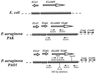
PCR was used for specific amplification of the fliDST regions of eight different P. aeruginosa strains and to obtain specific amplification products that were cloned into different vectors. PCRs were performed in a DNA Thermal Cycler 480 (Perkin-Elmer Cetus) in 100-μl volumes using Taq polymerase. Each reaction mixture contained 50 ng of DNA template, 2.5 U of Taq polymerase, 1.5 mM MgCl2, 0.1 mM deoxynucleoside triphosphate mix, and 0.2 μM primers (final concentrations). For certain amplifications, dimethyl sulfoxide was added to 5% (vol/vol; final concentration). Thirty-five cycles were run, each consisting of incubations for 1 min at 94°C, 1 min at 55°C, and 5 min at 72°C. The primers used for PCR were purchased from GIBCO-BRL. Restriction enzyme recognition sites were added at the ends of primers to facilitate subsequent cloning of the PCR products if desired. Six or more additional nucleotides were added 5′ to the restriction sites to ensure efficient cleavage. Primers used to amplify the fliDSS′ regions of different P. aeruginosa strains were RER55 (with an EcoRI site) and RER56 (with a BamHI site). These primers were made in the regions flanking the fliD gene where the nucleotide sequence was conserved in P. aeruginosa strains PAK and PAO1 (Fig. 1). In addition to primers RER55 and RER56, which were used in all P. aeruginosa strains for sequencing the fliD gene, three additional primers were used for sequencing. RER58 was used specifically for sequencing the PAO1-type strains, and RER59 and RER60 were used for sequencing the PAK-type strains. Primer RER57 (with a NdeI site) was used as the 5′ primer and primer RER56 was used as the 3′ primer to amplify the complete fliD gene from P. aeruginosa PAO1, which was cloned into the broad-host-range vector pET15BVP (4). Primers RER55 and XBAM (with a BamHI site) were used for the amplification of the fliD gene from PAO1 DNA, and the product was used for the subsequent construction of a mutation in this gene (Fig. 1).
FIG. 1.
Schematic representation of the fliDST regions of E. coli and P. aeruginosa strains PAK and PAO1. Locations and orientations of various primers used in PCR are shown. The deletion of ca. 300 bp in P. aeruginosa PAO1 is represented by the open triangle. The unique SmaI site used to insert the Gmr cassette in the PAO1 fliD gene and BamHI cleavage sites are marked.
DNA sequencing.
The PCR products containing the fliDST region of eight different P. aeruginosa strains were purified from agarose gels and used directly in sequencing reactions. DNA sequencing was performed using Taq DyeDeoxy terminator and dye primer cycle sequencing protocols developed by Applied Biosystems (Perkin-Elmer Corp., Foster City, Calif.). Fluorescently labeled dideoxynucleotides and primers respectively, were used. The labeled extension products were analyzed on an Applied Biosystems model 373A DNA sequencer. Double-stranded sequences were aligned and assembled using programs in the Sequencer software package (Gene Codes Corp., Ann Arbor, Mich.).
Plasmid constructions.
A 2.8-kb amplification product was obtained by PCR using primers RER55 and XBAM (Fig. 1), with PAO1 genomic DNA as the template. When this fragment was cut with EcoRI and BamHI, it was reduced to a 2-kb fragment because of an internal BamHI site in the insert. This 2-kb insert containing the PAO1 fliD gene and flanking DNA was cloned into the EcoRI and BamHI sites of pBluescriptKS (+), to give pBS55XBAM. Plasmid pBS55XBAM was linearized at the unique SmaI site in the PAO1 fliD gene, and a gentamicin resistance gene (Gmr) cassette excised from pUC7G and blunt ended by a fill-in reaction was inserted at that site, leading to the construction of pBS55XBAMG. This construct was used to generate a chromosomal mutation in the PAO1 fliD gene by marker exchange. The plasmid (pET15BVPDb) used for overexpression of PAO1 fliD and for complementation of the fliD mutation in PAO-D was obtained by cloning a 1.6-kb PCR fragment carrying the complete PAO1 fliD gene into the NdeI and BamHI sites of the expression vector pET15BVP (4). Plasmid pET15BVPDa, used for complementation of the fliD mutation in PAK-D, was constructed by cloning a 1.6-kb DNA fragment containing the PAK fliD gene into the NdeI and BamHI sites of the expression vector pET15BVP. This fragment was excised from pET15BD (5) with NdeI and BamHI.
Electroporations.
Electroporations were performed using a modification of the protocol of Smith and Iglewski (22). The plasmid DNA used for the electroporations was prepared by the alkaline lysis procedure (6). For gene replacement experiments involving chromosomal recombinations, the plasmid DNA was linearized by a restriction enzyme recognizing a site in the vector, and DNA was gel purified. About 1 μg of linear DNA fragment was electroporated into the electrocompetent P. aeruginosa cells. For complementation experiments, 50 to 100 ng of supercoiled, covalently closed circular plasmid DNA was electroporated into the target strains.
Motility assay.
To study the motility of P. aeruginosa, different strains were grown overnight at 37°C on fresh L-agar plates with or without antibiotics. The cells were then transferred with a sterile toothpick to 0.3% agar plates with or without antibiotics. These plates were incubated at 37°C for 16 h, and motility was assessed qualitatively by examining the circular swarm formed by the growing motile bacterial cells.
Expression and purification of FliD.
The complete PAO1 fliD coding sequence was inserted as a 1.6-kb PCR product into the NdeI/BamHI sites of plasmid pET15BVP. The resulting plasmid, pET15BVPDb, was introduced into the E. coli BL21(pLysS) (Novagen), which contains the T7 polymerase gene on the chromosome under the control of lacUV5 promoter. The fliD gene was overexpressed in the E. coli host, and the gene product was purified on a nickel affinity column as described previously (5).
Antibodies.
Polyclonal antibodies against the PAK and PAO1 FliD proteins were produced in New Zealand White rabbits by Cocalico Biologicals Inc., Reamstown, Pa. The antigen used in these immunizations was purified PAK or PAO1 FliD protein tagged with six histidines. Polyclonal antibodies against the PAK FliC were used as described earlier (26).
SDS-PAGE and Western Blotting.
Sodium dodecyl sulfate-polyacrylamide gel electrophoresis (SDS-PAGE) and Western blotting were used to determine the FliD and FliC serotypes in a series of P. aeruginosa strains. Proteins from whole cell lysates were separated by SDS PAGE (10% gel) (16) using Bio-Rad's mini-Protean II system. Proteins were then electrophoretically transferred to polyvinylidene difluoride membrane (Millipore) by using the mini-Transblot Western blotting system. The filters were blocked overnight in 1% solution of blocking reagent (Genius kit; Boehringer Mannheim) prepared in Tris-buffered saline. Subsequently the filters were incubated with 1:5,000 dilution of the PAK anti-FliD antibody or 1:4,000 dilution of the PAO1 anti-FliD antibody and 1:2,000 dilution of the PAK anti-FliC antibody, followed by washes with Tris-buffered saline containing 0.2% Tween 20. Finally, the secondary antibody (alkaline phosphatase-conjugated immunoglobulin G whole molecule from Sigma) was added, and the alkaline phosphatase activity was detected by color reaction.
Nucleotide sequence accession numbers.
The sequences reported here have been assigned GenBank accession no. AF139819 to AF139825.
RESULTS
Nucleotide sequence of the fliDST region.
In P. aeruginosa strain PAK, the open reading frame following the fliDS genes, previously called orf126 (5), has more homology to the fliS gene than the fliT gene of other bacteria. We therefore called it fliS′. The nucleotide sequence of the fliDSS′ (2.0-kb) region of PAK (accession no. L81176) was compared with that of strain PAO1 (contig 54; release date, March 15, 1999) obtained from the Pseudomonas genome project. Alignment of the two sequences using the GAP program of the Genetics and Computer Group showed that the two sequences were only 56% identical at the nucleotide level. Moreover, there was a major size difference between the two sequences. The PAO1 sequence had a deletion of 329 bp, in three different blocks, immediately following the fliD gene that included most of the sequence encoding the fliS gene. As a result, the operon structure in PAO1 is fliDS′, as opposed to fliDST in E. coli (12, 15, 27) and fliDSS′ in PAK (5) (Fig. 1).
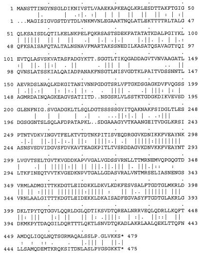
GAP analysis of the deduced amino acid sequences of the PAK and PAO1 fliD gene products showed that the two flagellar caps were different throughout the entire sequence (43% identity and 51% similarity), except for small stretches of homologous amino acids present predominantly in the C-terminal half of FliD (Fig. 2). Furthermore, PAK FliD was more homologous to E. coli FliD (34% identity and 42% similarity) than to PAO1 FliD (30% identity and 38% similarity).
FIG. 2.
Alignment of deduced amino acid sequences of the PAK (upper row) and PAO1 (lower row) fliD genes, using the GAP program of the Genetics and Computer Group package.
PCR and nucleotide sequence analysis of the fliDSS′ region of diverse P. aeruginosa strains.
Based on the differences between the fliD genes of the P. aeruginosa strains PAK and PAO1, we analyzed the fliD gene sequences of several additional P. aeruginosa strains. A total of eight different P. aeruginosa strains were analyzed: three laboratory strains (PAK, PAO1, and PA103), three clinical strains (CS2, MDR, and 1244), and two cystic fibrosis strains (CS29 and CS32). The fliD region of these strains was amplified by PCR using primers RER55 and RER56 and the corresponding chromosomal DNA templates as described in Materials and Methods. An amplification product of 2.0 kb was obtained from PAK, PA103, CS2, and CS32, while a smaller product (1.7 kb) was obtained from PAO1, CS29, MDR, and 1244 (data not shown). These PCR products were gel purified, and the nucleotide sequence of each DNA fragment was determined by direct sequencing of the PCR products. All eight sequences were submitted to GenBank by using the Sequin software available from the National Center for Biotechnology Information. All strains that were similar to PAO1 as judged by the size of the PCR product carried also a similar deletion, eliminating one of the fliS homologues seen in the corresponding region of PAK such that all strains yielded the 2.0-kb product. Moreover, the deduced amino acid sequences of FliD of PAK, PA103, CS2, and CS32 were more than 99% identical but different from those of PAO1, CS29, MDR, and 1244, which were also more than 99% identical. Based on these results, it appears there are two distinct types of FliD proteins in P. aeruginosa.
Are there two serotypes of FliD?
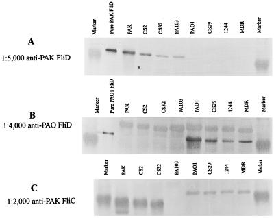
To ascertain whether the FliD proteins of the PAK and PAO1 groups represent two distinct serotypes in a larger group of P. aeruginosa isolates, polyclonal antibodies were raised against both PAK and PAO1 FliD proteins. PAK FliD was purified earlier (5); PAO1 FliD was purified using the same procedures and used as an antigen to generate polyclonal antibodies in rabbits. These antibodies were used to probe the whole cell extracts of different strains of P. aeruginosa in Western blots. As shown in Fig. 3, only strains belonging to the PAK group reacted with the PAK FliD antibody (Fig. 3A), while strains belonging to the PAO1 group reacted only with the PAO1 FliD antibody (Fig. 3B). Both PAK and PAO1 immunoreactive FliD proteins showed mobilities of ca. 53 kDa. However, the PAO1 FliD antisera also cross-reacted nonspecifically with a protein slightly heavier than FliD (Fig. 3B), which was also seen in the preimmune serum. These results suggest that there are two serotypes of FliD in P. aeruginosa. To further confirm that there are only two serotypes of FliD, we tested 50 other P. aeruginosa strains of diverse origins for reactivity with PAK or PAO1 FliD polyclonal antibodies. All of these strains reacted with either PAK FliD antibody or PAO1 FliD antibody (data not shown), confirming that there are only two serotypes of FliD in a large sampling of P. aeruginosa strains which we call A type (PAK type) and B type (PAO1 type).
FIG. 3.
Western blot analysis of different P. aeruginosa strains using specific polyclonal antibodies against A- and B-type FliD and a-type FliC proteins. Whole cell extracts of P. aeruginosa strains in panel A were probed with the PAK (A-type) FliD antibodies. The identical samples in panels B and C were probed with the PAO1 (B-type) FliD antibodies and a-type FliC antibodies, respectively.
Coinheritance of fliC and fliD genes.
The fact that there are two types of flagellins and the new finding that there are also only two types of flagellar cap proteins raised the interesting possibility that these two flagellar gene types may be coinherited. To explore this possibility, we probed four P. aeruginosa strains of A-type FliD serotype and 4 of B type by Western blotting using the polyclonal PAK FliC antibody. This antibody, raised against purified PAK flagella, reacts strongly with the a-type flagellin and weakly with the b-type flagellin. As shown in Fig. 3C, strains of the A-type FliD serotype (PAK, CS2, and CS32) showed a diffuse, strongly immunoreactive band of 45 kDa, while strains of the B-type FliD serotype (PAO1, 1244, CS29, and MDR) showed a single band of 53 kDa that reacted weakly with this antibody. PA103 did not show FliC antibody-reactive band as expected, since PA103 is naturally nonmotile (17). To further confirm this observation, the 50 P. aeruginosa strains of diverse origin were analyzed by Western blotting using the same antibodies. Again, each strain with an A-type FliD also had a-type FliC, and all strains with B-type FliD had b-type FliC (data not shown). These results are consistent with the hypothesis that the fliD and fliC genes were coinherited from an ancestral P. aeruginosa. The proximity of the two genes on the bacterial chromosome and their juxtaposition in the flagellar structure support this finding. The sequences of the flagellar regions from fliC to fliR (Fig. 1) from these two prototype strains were then compared. The gene between fliC and fliD, which we have named fleL, and the gene following fliS′, which we now call fliP, was also dissimilar in these strains; however, the regulatory genes fleQ, fleS, and fleR were identical in sequence. Thus, the entire region encoding these terminal flagellar proteins necessary for their transport may have been inherited as a block.
Complementation of fliD mutants.
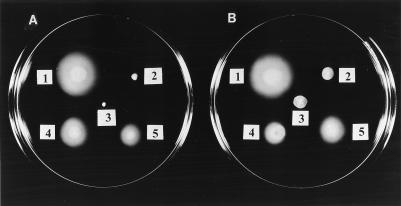
It is conceivable that the sequence conservation of the FliC and FliD proteins within a group is the reflection of an optimal match between these flagellar components for their proper function in motility. We therefore assessed the ability of the fliD gene of one strain type to complement the fliD mutant of the other type, and vice versa. To perform these experiments, a chromosomal mutant of PAO1 fliD was constructed by gene replacement. The P. aeruginosa PAO1 fliD gene located on a 2.0-kb EcoRI/BamHI PCR-generated fragment was inactivated by inserting a Gmr cassette into the unique SmaI site in the PAO1 fliD gene. The insertionally inactivated fliD gene on a nonreplicating, multicopy plasmid (pBS55XBAMG) was introduced into PAO1 by electroporation, where it replaced the corresponding chromosomal copy of the fliD gene by double reciprocal recombination, giving rise to a fliD mutant strain, PAO-D. Replacement of the wild-type fliD in PAO-D was confirmed by PCR (data not shown). This strain was nonmotile on 0.3% agar plates (Fig. 4).
FIG. 4.
Soft L-agar (0.3%) plates showing the motility phenotypes of PAK and its derivatives (A) and of PAO1 and its derivatives (B). (A) 1, wild-type PAK; 2, A-type fliD mutant PAK-D; 3, PAK-D containing vector control pET15BVP; 4, PAK-D complemented with pET15BVPa (A-type fliD); 5, PAK-D containing pET15BVPDb (B-type fliD). (B) 1, wild-type PAO1; 2, B-type fliD mutant PAO-D; 3, PAO-D containing vector control pET15BVP; 4, PAO-D complemented with pET15BVPDa (A-type fliD); 5, PAK-D containing pET15BVPDb (B-type fliD).
The plasmids carrying PAK fliD (pET15BVPDa) and PAO1 fliD (pET15BVPDb), and their vector controls without the fliD insert, were electroporated into two fliD mutant strains, PAK-D and PAO-D. The resulting strains were tested for motility on 0.3% agar plates. In both cases, full complementation was not achieved with the homologous type of fliD gene, as suggested by smaller motility zones of PAK-D(pET15BVPDa) and PAO-D(pET15BVPDb) compared to the motility zones of wild-type PAK and PAO1. Interestingly, PAK fliD (pET15BVPDa) complemented PAK-D better than PAO1 fliD (pET15BVPDb) (Fig. 4). Similarly, the PAO1 fliD (pET15BVPDb) complemented PAO-D better than PAK fliD (pET15BVPDa) (Fig. 4B). The vector controls in both cases did not complement the motility defects of either PAK-D or PAO-D, as expected. The explanation for partial complementation with the pET15BVP constructs is probably the weak expression of fliD from the T7 promoter in the absence of T7 polymerase or possibly the polar effect of the insertion. The reason for poorer complementation by the heterologous fliD type is possibly its abnormal export and/or assembly.
Phylogenetic analysis of FliD sequences.
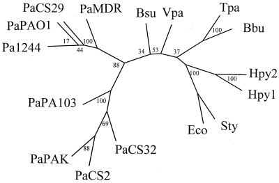
Sequences of various FliD proteins from this work and those obtained from databases were compared using the PHYLIP analysis program, version 3.5c (9) (Fig. 5). The P. aeruginosa sequences clearly show clustering into two distinct families, in complete agreement with their assignment to A type and B type, as determined by related immunogenicity and by direct comparisons of deduced amino acid sequences.
FIG. 5.
Phylogenetic analysis of the FliD sequences. The tree was constructed using P. aeruginosa (Pa) sequences from this work and additional sequences retrieved from GenBank: Bsu, Bacillus subtilis (U56901); Hly1 and Hly2, Helicobacter pylori (accession no. U82981) and H. pylori J99 (accession no. AE001500); Eco, E. coli K-12 MG1655 (accession no. AE000285); Tpa, Treponema pallidum (accession no. AE001257); Bbu, Borrelia burgdorferi (accession no. U66699); PaPAK, P. aeruginosa strain PAK (accession no. L81176); Vpa, Vibrio parahaemolyticus (accession no. U52957); and Sty, Salmonella enterica serovar Typhimurium (accession no. M33541). Bootstrap values represent 100 replications as indicated at branch points.
DISCUSSION
In this report, we demonstrate the existence of two distinct sequence variants of the flagellar cap proteins, FliD, in P. aeruginosa, which we refer to as A type and B type. The results of DNA sequencing and PCR analysis showed that eight different P. aeruginosa strains clearly fall into two groups (PAK and PAO1) based on the DNA sequence of the fliD locus. Western blot analysis using antibodies against A-type (PAK-type) or B-type (PAO1-type) FliD and a-type FliC show that the A- and B-type FliD proteins are immunologically different and suggest that the fliD and fliC genes are coinherited. In spite of their common role of serving as an important structural component of the flagellum to prevent the continued export of flagellin subunits from the cell, the two types of FliD proteins are not interchangeable, since the A-type fliD poorly complements the B-type fliD mutant, and vice versa. This lack of complementation reflects differences in the primary amino acid sequences of the two proteins which result in distinct conformation in the flagellum and a highly specific interaction with the rest of the flagellar components, most notably with the flagellin subunits. Moreover, since FliD has been shown to function as a mucin-specific bacterial adhesin, the sequence variation may lead to quantitative or qualitative differences in adherence to mucin between strains expressing the A-type or B-type variant of FliD.
Most strains of P. aeruginosa also express one of two antigenically distinct types of flagellins, a type or b type, which are distinguishable by their size and reaction with type-specific flagellin antibodies (Siadak, Oncogen). Both a-type (26) and b-type (13) flagellin genes have been cloned and sequenced; alignment of the amino acid sequences of the two types of flagellins showed that the amino- and carboxy-terminal domains were more conserved while the central regions were more divergent (26). A survey of a larger group of flagellin sequences revealed that b-type flagellins are highly conserved, with only few amino acid substitutions, while the a-type flagellins show greater variation, with majority of the substitutions clustered in the central region (23). The antibodies to the a-type flagellin cross-react with the b-type FliC, and vice versa (24). Strain-specific variants of FliD resemble FliC in that they apparently occur only as two types, A and B. However, there are significant differences between these two flagellar components. Alignment of the deduced amino acid sequences of the two types of the FliD proteins (Fig. 2) revealed the variation between the two sequences throughout the entire polypeptide sequence. Moreover, the antibodies raised against the A-type FliD did not cross-react with the B-type FliD, and vice versa. The reason for this specificity of the two antibodies could be that the two proteins present completely different array of epitopes. The lack of long stretches of homologous amino acids between the two types of FliD proteins (Fig. 2) is consistent with this observation.
It appears that the heterogeneity in the nucleotide sequence of the flagellar genes is probably limited to the more exposed structural proteins such as FliC and FliD since the nucleotide sequences of the PAK fliF gene (3), coding for the membrane and supramembrane ring, and PAK flgE gene (data not shown; submitted to GenBank), coding for the flagellar hook, were found to be identical to the corresponding PAO1 gene sequences. This would suggest that a need for antigenic diversity in the exposed terminal parts of the flagellum accounts for the existence of different serotypes. However, it is surprising that there are only two types of FliC and FliD proteins in P. aeruginosa. If the region under discussion was inherited as a block, it is not obvious why flagellins would allow some variation in structure even within one flagellar type, but the cap has very strict structural requirements allowing little or no variation between strains of a serotype.
The division of the FliD and FliC sequences into two distinct and coinheritable groups provides potentially important clues about the evolution of P. aeruginosa as a human pathogen and its adaptability during specific infections ranging from acute bacteremia to chronic respiratory infections of cystic fibrosis patients. To fully understand the selection process which leads to the coevolution of the flagellin subunit and the cap and adhesin polypeptides, the phylogenetic analysis will have to be greatly extended to include a larger number of strains associated with specific disease as well as strains from various environmental reservoirs.
ACKNOWLEDGMENTS
We acknowledge the Interdisciplinary Center for Biotechnology Research computer facilities of the University of Florida for use of the VAX computers for DNA sequence analyses. We thank Gita Bangera and William Hughes for assistance with the phylogenetic analysis.
This work was supported by NIH grant HL33622 (R.R.).
REFERENCES
- 1.Allison J S, Dawson M, Drake D, Montie T C. Electrophoretic separation and molecular weight characterization of Pseudomonas aeruginosa H-antigen flagellins. Infect Immun. 1985;49:770–774. doi: 10.1128/iai.49.3.770-774.1985. [DOI] [PMC free article] [PubMed] [Google Scholar]
- 2.Ansorg R. Flagella specific H antigenic schema of Pseudomonas aeruginosa. Zentbl Bakteriol Parasitenkd Infektkrankh Hyg Abt 1. 1978;224:228–238. [PubMed] [Google Scholar]
- 3.Arora S K, Ritchings B W, Almira E C, Lory S, Ramphal R. Cloning and characterization of Pseudomonas aeruginosa fliF necessary for flagellar assembly and bacterial adherence to mucin. Infect Immun. 1996;64:2130–2136. doi: 10.1128/iai.64.6.2130-2136.1996. [DOI] [PMC free article] [PubMed] [Google Scholar]
- 4.Arora S K, Ritchings B W, Almira E C, Lory S, Ramphal R. A transcriptional activator, FleQ, regulates mucin adhesion and flagellar gene expression in Pseudomonas aeruginosa in a cascade manner. J Bacteriol. 1997;179:5574–5581. doi: 10.1128/jb.179.17.5574-5581.1997. [DOI] [PMC free article] [PubMed] [Google Scholar]
- 5.Arora S K, Ritchings B W, Almira E C, Lory S, Ramphal R. The Pseudomonas aeruginosa flagellar cap protein, FliD, is responsible for mucin adhesion. Infect Immun. 1998;66:1000–1007. doi: 10.1128/iai.66.3.1000-1007.1998. [DOI] [PMC free article] [PubMed] [Google Scholar]
- 6.Birnboim H C, Doly J. A rapid alkaline extraction procedure for screening recombinant plasmid DNA. Nucleic Acids Res. 1979;7:1513–1523. doi: 10.1093/nar/7.6.1513. [DOI] [PMC free article] [PubMed] [Google Scholar]
- 7.Borst P, Greaves D R. Programmed gene rearrangements altering gene expression. Science. 1989;235:658–667. doi: 10.1126/science.3544215. [DOI] [PubMed] [Google Scholar]
- 8.Feavers I M, Fox A J, Gray S, Jones D M, Maiden M C. Antigenic diversity of meningococcal outer membrane protein PorA has implications for epidemiological analysis and vaccine design. Clin Diagn Lab Immunol. 1996;3:444–450. doi: 10.1128/cdli.3.4.444-450.1996. [DOI] [PMC free article] [PubMed] [Google Scholar]
- 9.Felsenstein J. Inferring phylogenies from protein sequences by parsimony, distance, and likelihood methods. Methods Enzymol. 1996;266:418–427. doi: 10.1016/s0076-6879(96)66026-1. [DOI] [PubMed] [Google Scholar]
- 10.Hanley J, Salit I E, Hofmann T. Immunochemical characterization of P pili from invasive Escherichia coli. Infect Immun. 1985;49:581–586. doi: 10.1128/iai.49.3.581-586.1985. [DOI] [PMC free article] [PubMed] [Google Scholar]
- 11.Hongyo H, Kokeguchi S, Kurihara H, Miyamoto M, Maeda H, Takashiba S, Murayama Y. Comparative study of two outer membrane protein genes from Porphyromonas gingivalis. Microbios. 1998;95:91–100. [PubMed] [Google Scholar]
- 12.Kawagishi I, Muller V, Williams A W, Irikura V M, Macnab R M. Subdivision of flagellar region III of the Escherichia coli and Salmonella typhimurium chromosomes and identification of two additional flagellar genes. J Gen Microbiol. 1992;138:1051–1065. doi: 10.1099/00221287-138-6-1051. [DOI] [PubMed] [Google Scholar]
- 13.Kelly-Winterberg K, Montie T C. Cloning and expression of Pseudomonas aeruginosa flagellin in Escherichia coli. J Bacteriol. 1989;171:6357–6362. doi: 10.1128/jb.171.11.6357-6362.1989. [DOI] [PMC free article] [PubMed] [Google Scholar]
- 14.Knirel Y A, Kochetkov N K. The structure of lipopolysaccharides from gram negative bacteria. III. Structure of O-specific polysaccharides. Biochemistry (Moscow) 1994;59:1784–1851. [PubMed] [Google Scholar]
- 15.Kutsukake K, Ohya Y, Yamaguchi S, Iino T. Operon structure of flagellar genes in Salmonella typhimurium. Mol Gen Genet. 1988;214:11–15. doi: 10.1007/BF00340172. [DOI] [PubMed] [Google Scholar]
- 16.Laemmli U K. Cleavage of structural proteins during the assembly of the head of bacteriophage T4. Nature (London) 1970;227:680–685. doi: 10.1038/227680a0. [DOI] [PubMed] [Google Scholar]
- 17.Liu P V. The roles of various fractions of Pseudomonas aeruginosa in its pathgenesis. II. Effects of lecithinase and protease. J Infect Dis. 1966;116:112–116. doi: 10.1093/infdis/116.1.112. [DOI] [PubMed] [Google Scholar]
- 18.Macnab R M. Flagella. In: Neidhardt F C, Ingraham J L, Magasanik B, Low K B, Schaechter M, Umbarger H E, editors. Escherichia coli and Salmonella typhimurium: cellular and molecular biology. Vol. 1. Washington, D.C.: American Society for Microbiology; 1987. pp. 70–83. [Google Scholar]
- 19.McCrea K W, Sauver J L, Marrs C F, Clemans D, Gilsdorf J R. Immunologic and structural relationships of the minor pilus subunits among Haemophilus influenza isolates. Infect Immun. 1998;66:4788–4796. doi: 10.1128/iai.66.10.4788-4796.1998. [DOI] [PMC free article] [PubMed] [Google Scholar]
- 20.Montie T C, Anderson T R. Enzyme-linked immunosorbent assay for detection of Pseudomonas aeruginosa H (flagellar) antigen. Eur J Clin Microbiol. 1988;7:256–260. doi: 10.1007/BF01963097. [DOI] [PubMed] [Google Scholar]
- 21.Relf W A, Martin D R, Sriprakash K S. Antigenic diversity within a family of M proteins from group A streptococci: evidence for the role of frameshift and compensatory mutations. Gene. 1994;144:25–30. doi: 10.1016/0378-1119(94)90198-8. [DOI] [PubMed] [Google Scholar]
- 22.Smith A W, Iglewski B H. Transformation of Pseudomonas aeruginosa by electroporation. Nucleic Acids Res. 1989;17:10509. doi: 10.1093/nar/17.24.10509. [DOI] [PMC free article] [PubMed] [Google Scholar]
- 23.Spangenberg C, Heuer T, Burger C, Tummler B. Genetic diversity of flagellins of Pseudomonas aeruginosa. FEBS Lett. 1996;396:213–217. doi: 10.1016/0014-5793(96)01099-x. [DOI] [PubMed] [Google Scholar]
- 24.Spangenberg C, Montie T C, Tummler B. Structural and functional implications of sequence diversity of Pseudomonas aeruginosa genes, oriC, ampC, and fliC. Electrophoresis. 1998;19:545–550. doi: 10.1002/elps.1150190414. [DOI] [PubMed] [Google Scholar]
- 25.Thon G, Baltz T, Eisen H. Antigenic diversity by the recombination of pseudogenes. Genes Dev. 1989;3:1247–1254. doi: 10.1101/gad.3.8.1247. [DOI] [PubMed] [Google Scholar]
- 26.Totten P A, Lory S. Characterization of the type a flagellin gene from Pseudomonas aeruginosa PAK. J Bacteriol. 1990;172:7188–7199. doi: 10.1128/jb.172.12.7188-7199.1990. [DOI] [PMC free article] [PubMed] [Google Scholar]
- 27.Yokoseki T, Kutsukake K, Ohnishi K, Iino T. Functional analysis of the flagellar genes in the fliD operon of Salmonella typhimurium. Microbiology. 1995;141:1715–1722. doi: 10.1099/13500872-141-7-1715. [DOI] [PubMed] [Google Scholar]


