Abstract
Simple Summary
Migratory birds have an enormous potential for dispersing pathogenic microorganisms. Ducks can host coronaviruses (CoVs), which have a high pathogenic expression and economic impacts, given their ability to migrate exceptional distances, facilitating the dispersal of microorganisms. This study aimed to identify and characterize the diversity of CoVs in migratory ducks from Portugal (Anas platyrhynchos, Anas acuta, and Anas crecca). Among the samples tested, 23 were characterized as gammacoronavirus and one as deltacoronavirus. The present study aimed to assess the circulation of CoVs in wild ducks from Portugal, being the first description of CoVs for these animals in Portugal.
Abstract
Coronaviruses (CoVs) are part of the Coronaviridae family, and the genera Gamma (γ) and Delta (δ) are found mostly in birds. Migratory birds have an enormous potential for dispersing pathogenic microorganisms. Ducks (order Anseriformes) can host CoVs from birds, with pathogenic expression and high economic impact. This study aimed to identify and characterize the diversity of CoVs in migratory ducks from Portugal. Duck stool samples were collected using cloacal swabs from 72 individuals (Anas platyrhynchos, Anas acuta, and Anas crecca). Among the 72 samples tested, 24 showed amplicons of the expected size. Twenty-three were characterized as Gammacoronavirus and one as Deltacoronavirus (accession numbers ON368935-ON368954; ON721380-ON721383). The Gammacoronaviruses sequences showed greater similarities to those obtained in ducks (Anas platyrhynchos) from Finland and Poland, Anas crecca duck from the USA, and mute swans from Poland. Birds can occupy many habitats and therefore play diverse ecological roles in various ecosystems, especially given their ability to migrate exceptional distances, facilitating the dispersal of microorganisms with animal and/or human impact. There are a considerable number of studies that have detected CoVs in ducks, but none in Portugal. The present study assessed the circulation of CoVs in wild ducks from Portugal, being the first description of CoVs for these animals in Portugal.
Keywords: coronavirus, migratory ducks, Gammacoronaviruses, Deltacoronavirus
1. Introduction
Coronaviruses (CoVs) belong to the Coronaviridae family, subfamily Orthocoronavirinae, having a positive-sense single-stranded RNA genome and an envelope equipped with protruding structures on their surface called spikes [1]. Its genome is one of the largest viral RNA genomes, with approximately 25–32 kb [2]. CoVs show a high genetic diversity that can be the result of their large genomes, high rates of mutation, infidelity of the RNA-dependent RNA polymerase, and high frequency of homologous RNA recombination [3].
The Orthocoronavirinae subfamily is divided into four genera based on genetic differences and serological cross-reactivity. Alpha and Betacoronaviruses might have a common ancestor, a CoV originating from bats [4]. For this reason, the viruses belonging to these two genera are found in bats [5] and other mammals, such as swine acute diarrhea syndrome coronavirus (SADS-CoV), transmissible gastroenteritis virus (TGEV), feline coronavirus (FCoV), bovine CoVs (BCoV), and rat coronavirus (RtCoV) [6,7]. On the other hand, Gamma and Deltacoronavirus evolved from a CoV originating in birds, with the majority of them causing diseases in birds, such as avian CoV infectious bronchitis virus (IBV), turkey CoV (TCoV), goose CoV, and duck CoV [8,9]. Both Gamma and Deltacoronavirus have been isolated and detected in wild and domestic birds in orders such as Anseriformes, Pelecaniformes, Ciconiiformes, Galliformes, Columbiformes. and Charadriformes [10,11,12]. However, Gammacoronavirus tends to be detected in domestic birds, while Deltacoronavirus infects both domestic and wild birds [13,14]. Two CoVs isolated from cetaceans have also been placed in the genus Gammacoronavirus [15,16], and Deltacoronavirus has also been found in pigs (porcine deltacoronavirus -PDCoV), causing acute diarrhea and dehydration [17]. Coronaviral infections have received significant attention from both the public and researchers [18].
CoVs have been identified in almost 15 orders of Aves, especially Charadriiformes (seagulls, plovers, sandpipers) and Anseriformes (ducks, geese, swans) [1]. Migratory birds have a huge potential for the transport and dispersal of a large number of pathogenic microorganisms [19]. Ducks, species from the Anseriformes order, can host a number of RNA viruses, including avian CoVs, and avian paramyxovirus type 1, with emerging evidence that suggests they may also be hosts of an array of avian astroviruses [20]. Infections by avian CoVs are characterized by acute, highly contagious, and economically important diseases in domesticated poultry [21]. However, the genetic diversity, evolution, distribution, and taxonomy of some CoVs dominant in birds still remain enigmatic [22]. Therefore, to add knowledge to this specific topic and to understand the role of circulation and the potential transboundary introduction of exotic avian CoVs, this study aimed to identify and characterize the diversity of CoVs in migratory ducks from Portugal.
2. Materials and Methods
2.1. Sample Collection
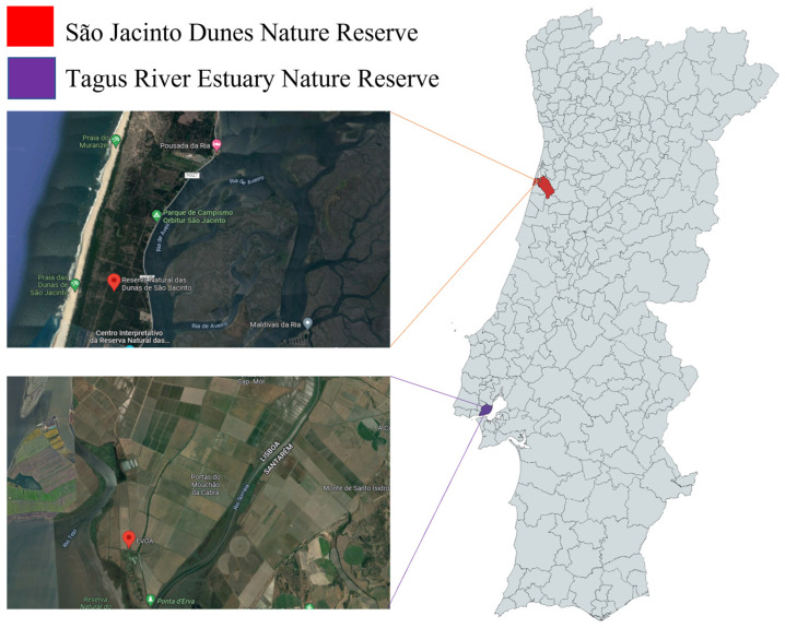
Samples of duck feces were collected on duck cloaca using cotton swabs and conserved at −23 ℃ from ducks captured for marking within duck ecology and migration studies. The species sampled were Mallard Anas platyrhynchos, Pintail Anas acuta, and Teal Anas crecca. Captures were performed in São Jacinto Dunes Nature Reserve (Aveiro) and at EVOA (in Tagus River Estuary Nature Reserve, Vila Franca de Xira) since these are areas of high concentration of ducks where long-term duck ecology studies have been performed (see Figure 1). Ducks were visually marked with nasal saddles and could be followed on the field. A license to capture and mark ducks was obtained from Instituto da Conservação da Natureza e das Florestas (ICNF), Portugal (permit number 40/2021).
Figure 1.
Selected sampling locations (city of Porto and Aveiro) in Portugal used in this study.
Fecal swabs were thoroughly mixed by vortexing in 500 µL of phosphate-buffered saline (PBS) pH 7.2. RNA was extracted from the fecal suspension using the QIAamp viral mini kit (Qiagen, Hilden, Germany) according to the manufacturer’s instructions using 140 µL of the clarified supernatants. Eluted RNA was then kept at −80 °C until further processing.
2.2. Screening for Coronaviruses
Extracted nucleic acids were tested for CoVs using a broad-spectrum pan-CoV nested RT-PCR assay targeting the RNA-dependent RNA polymerase (RdRp)-conserved region with a final product size of 440 bp [23]. The sensitivity of the nested pan-CoV primers has been recently compared with different protocols by combining existing primers from different studies showing high performance and combining the chances of detecting known and unknown CoVs from all matrices [23].
We employed the one-step RT-PCR kit from GRiSP®, Porto, Portugal, for the initial round of PCR. The following conditions were used in the Veriti 96-well thermal cycler (Thermo Fisher) for amplification reactions with positive and negative controls: an initial cycle of 3 min at 95 °C, followed by 40 cycles of 95 °C for 15 s, 50 °C for 15 s, and 72 °C for 2 s, with a final elongation at 72 C for 10 min. Then, 2 μL of the first round’s products was utilized as a template for the second round using the Xpert Fast Hotstart Mastermix (2 x) with dye (GRiSP®, Porto, Portugal). A final amount of 25 μL was used for the PCR. The same thermal cycler was used to perform the amplification reactions with the positive and negative controls. The following conditions were used: an initial cycle of 3 min at 95 °C, 40 cycles of 95 °C for 15 s, 52 °C for 15 s, and 72 °C for 2 s, followed by an elongation at 72 °C for 10 min.
In order to identify the target DNA fragments, PCR amplification products were electrophoresed at 120 V for 30 min on a 1% agarose gel stained with Xpert Green Safe DNA gel stain (Grisp, Porto, Portugal). Molecular weights were assessed using a DNA weight comparison (100 bp DNA ladder; Grisp, Porto, Portugal).
2.3. Sanger Sequencing and Phylogenetic Analysis
Amplicons of the expected size were purified using the GRS PCR Purification Kit (Grisp, Porto, Portugal). Bidirectional sequencing was then performed using the target gene’s specific primers by the Sanger method. Sequence alignment was performed using the Bi-oEdit Sequence Alignment Editor v7.1.9 software package, version 2.1 (Ibis Biosciences, Carlsbad, CA, USA). The obtained sequences were trimmed, and consensus sequences were compared with the sequences found online in the nucleotide database NCBI (Gen-Bank, Carlsbad, CA, USA).
The viral sequences obtained in this study were submitted to GenBank under the accession numbers ON368935-ON368954 and ON721380-ON721383. These sequences, together with 36 reference strains from the 4 genera (Alpha-, Beta-, Gamma-, and Deltacoronavirus) obtained from GenBank, were aligned using MEGA 11 software [24]. Models function on MEGA 11 was used to opt for the model with the smallest Bayesian information criterion (BIC) score [25] using the maximum likelihood method, based on the general time reversible model using a discrete Gamma distribution and assuming evolutionarily invariable sites, 1000 bootstraps replicated, followed by editing with the Interactive Tree of Life (iTOL) platform [26].
3. Results
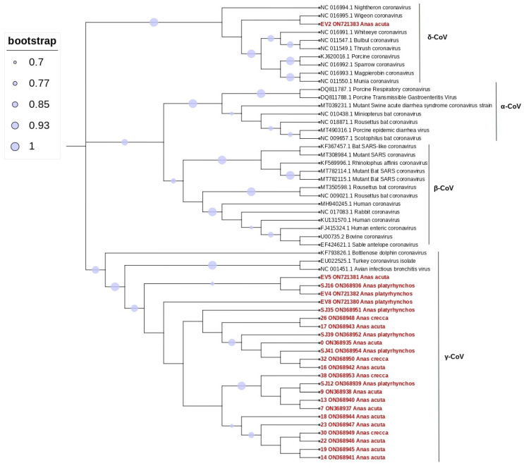
Among the total 72 samples tested, 24 presented amplicons of the expected size (33.3%; 95% confidence interval [CI]: 22.7–45.4). Bidirectional sequencing of these 24 products, followed by nucleotide BLAST analysis, showed that the majority (n = 23) were characterized as Gammacoronavirus (31.4%; 95% CI: 21.4–44.0), and one was characterized as Deltacoronavirus (1.4%; 95% CI: 0.03–7.5), as shown in Figure 2. Pintail showed a higher prevalence, with 12 of 24 samples positive (48%). Mallard, a resident species in Portugal (Rodrigues et al. 2000), also had positive samples.
Figure 2.
Phylogenetic tree constructed for the alpha, beta, gamma, and delta coronavirus, using 36 reference strains and 24 strains identified in this study. Phylogenetic analysis was based on a 406 nt partial region of the RdRp. The tree was constructed using MEGA 10 using the maximum likelihood based on the GTR + G model, and 1000 bootstraps were replicated. Samples from this study are indicated in red.
Sequence analysis within the obtained CoV sequences showed identities ranging from 94% and 100%. Characterization by BLAST indicated that the sequences showed the highest hits (97.76–99.44%) to those sequences obtained from ducks from Finland, Poland, and the USA and mute swans from Poland. Phylogenetic analysis using the obtained 24 CoV sequences and 36 reference strains confirmed the classification as Gammacoronaviruses and Deltacoronavirus (Figure 2).
The obtained phylogenetic tree showed that the retrieved sequences in this study clustered together with those obtained from a bottlenose dolphin (bottlenose dolphin coronavirus), from a chicken (avian infectious bronchitis virus), and a turkey (turkey coronavirus), with all of them clustering in Gammacoronavirus, except for one pintail sample, which clustered with a Wigeon Anas penelope (Wigeon coronavirus HKU20) Deltacoronavirus. Details of the samples from this study can be found in Table 1.
Table 1.
Details of the samples from this study.
| Collection Site | Sample ID | Host Species | Accession Number | CoV Genera |
|---|---|---|---|---|
| Evoa | #0 | Anas acuta | ON368935 | Gammacoronavirus |
| #7 | Anas acuta | ON368937 | Gammacoronavirus | |
| #9 | Anas acuta | ON368938 | Gammacoronavirus | |
| #13 | Anas acuta | ON368940 | Gammacoronavirus | |
| #14 | Anas acuta | ON368941 | Gammacoronavirus | |
| #16 | Anas acuta | ON368942 | Gammacoronavirus | |
| #17 | Anas acuta | ON368943 | Gammacoronavirus | |
| #18 | Anas acuta | ON368944 | Gammacoronavirus | |
| #19 | Anas acuta | ON368945 | Gammacoronavirus | |
| #22 | Anas acuta | ON368946 | Gammacoronavirus | |
| #23 | Anas acuta | ON368947 | Gammacoronavirus | |
| #26 | Anas crecca | ON368948 | Gammacoronavirus | |
| #30 | Anas crecca | ON368949 | Gammacoronavirus | |
| #32 | Anas crecca | ON368950 | Gammacoronavirus | |
| #38 | Anas crecca | ON368953 | Gammacoronavirus | |
| #EV2 | Anas acuta | ON721383 | Deltacoronavirus | |
| #EV4 | Anas platyrhynchos | ON721382 | Gammacoronavirus | |
| #EV5 | Anas acuta | ON721381 | Gammacoronavirus | |
| S São Jacinto Dunes Nature Reserve | ##EV8 | Anas platyrhynchos | ON721380 | Gammacoronavirus |
| #SJ12 | Anas platyrhynchos | ON368939 | Gammacoronavirus | |
| #SJ16 | Anas platyrhynchos | ON368936 | Gammacoronavirus | |
| #SJ35 | Anas platyrhynchos | ON368951 | Gammacoronavirus | |
| #SJ39 | Anas platyrhynchos | ON368952 | Gammacoronavirus | |
| #SJ41 | Anas platyrhynchos | ON368954 | Gammacoronavirus |
4. Discussion
The present study assessed the circulation of CoVs in wild ducks from Portugal, being the first description of CoVs for these animals in Portugal. We have also shown that the CoV strains found in the duck population under study are closely related to the gammacoronavirus strains retrieved from duck species Anas platyrhynchos from Poland and Finland [1,2,27], mute swans from Poland [1], the duck species Anas crecca from Hong Kong [11] and the USA (accession number: KJ741882).
As it is known, the Alphacoronavirus and Betacoronavirus infect mostly mammals, while gammacoronavirus and deltacoronavirus mainly infect birds [8,9]. While there is a growing number of studies focusing on the presence of coronavirus in bats, there is a variety of CoVs known to circulate among wild and domestic birds as well [2], which, upon entering poultry production premises, may cause severe morbidity and mortality to the birds [28], causing substantial economic losses [29].
Several viruses, including zoonotic and economically significant pathogens, are known to circulate among wild birds [2,30]. The main representative of avian coronavirus is the infectious bronchitis virus (IBV) [31], which is a Gammacoronavirus, a highly contagious viral disease that is considered responsible for significant economic losses in the poultry industry worldwide (Moreno et al., 2017). IBV control has been hampered by the intricate IBV evolution over the years, by the emergence of many different antigenic or genotypic types, commonly referred to as variants, that are likely to be facilitated by the spillover through migratory birds [32]; hence, continued monitoring of CoVs in migratory birds is key to mitigating potential outbreaks.
Birds can occupy many habitats and therefore serve diverse ecological roles in various ecosystems [33], especially because of their ability to migrate exceptional distances [34]. The most frequently proposed reason for birds’ migration is to benefit from a seasonal availability of resources, to breed, and also to avoid harsh winters [35]. This ability of dispersion can also, unfortunately, facilitate the dispersion of microorganisms with animal and/or human impact [2]. Hence alerts should be made towards surveillance of potentially pathogenic viruses in wild birds.
Gammacoronavirus and Deltacoronavirus were detected in a large variety of birds from different countries, such as Sweden [36], Norway [37], England [38], South Korea [39], and Australia [40] in either aquatic or nonaquatic variety, which highlights the potential for viral spillover to domestic species. There is a considerable number of studies that detected CoVs in ducks, but none in Portugal. Ducks are able to occupy diverse ecological niches and are either migratory or resident [1]. The strains of CoV found in our study are closely related to the strains from Finland, and Finnish mallards are strongly migratory [41]. Some species have adapted to urbanized landscapes, increasing their chances of being in contact with humans. The potential for transmissibility between bird species, even between wild and captive members of the same species (e.g., Anas platyrhynchos), or between other species, could trigger a captive poultry outbreak, especially in extensively farmed birds, leading to large economic losses in a wide geographical area [42].
Wildlife has been under epidemiological surveillance to identify its possible roles as a reservoir for emerging viruses that may pose a risk to humans and threaten wildlife [21]. Our investigation showed the presence of Gammacoronavirus and Deltacoronavirus in ducks, indicating that they harbor CoVs and possibly spread it. It is important to continue surveillance of wild ducks for the presence of avian coronavirus to understand better population flyways and ducks’ migration, especially the ones with close contact with humans, to understand better the evolution and ecology of coronaviruses, and to monitor emerging new strains that can possibly cause more losses to the poultry industry and transmit to humans [43]. The use of nasal saddles on sampled ducks can also allow the study of possible impacts of CoVs on wild duck species’ survival.
5. Conclusions
This study identified Gammacoronavirus and Deltacoronaviruses in migratory ducks from Portugal, and it is the first report of CoVs for these animals in the country. Migratory birds can occupy many habitats, primarily because of their ability to migrate exceptional distances. This ability of dispersion can also, unfortunately, facilitate the dispersion of microorganisms with animal and/or human impact. Hence, efforts should be made towards the surveillance of potentially pathogenic viruses in wild birds and for monitoring emerging new strains that can possibly cause more losses to the poultry industry and transmit to humans.
Acknowledgments
Mahima Hemnani thanks Fundação para a Ciência e a Tecnologia (FCT) for the financial support of her Ph.D. work under the Maria de Souza scholarship contract number 2021.09380.BD. Sérgio Santos-Silva thanks FCT for the financial support of his Ph.D. work under the Maria de Souza scholarship contract number 2021.09461.BD. This work was financed by national funds through FCT—Fundação para a Ciência e a Tecnologia, I.P., under the projects UIDB/04750/2020 and LA/P/0064/2020. Fieldwork was sponsored by the Association Nationale des Chasseurs de Gibier d’Eau (ANCGE, France) within the project with ESAC « Étude de la migration des canards hivernants au Portugal».
Author Contributions
Conceptualization, M.H. and J.R.M.; methodology, M.H., S.S.-S. and J.F.-e.-S.; formal analysis, M.H., D.R., N.S., M.E.F., P.H., H.R., P.P., G.T. and J.R.M.; investigation, M.H., S.S.-S., J.F.-e.-S. and J.R.M.; resources, D.R., N.S. and M.E.F.; sample collection, D.R., N.S. and M.E.F.; writing—original draft preparation, M.H.; writing—review and editing, D.R., N.S., M.E.F., P.H., H.R., P.P., G.T. and J.R.M.; supervision, J.R.M.; project administration, H.R., P.P., G.T. and J.R.M.; funding acquisition, M.H. and J.R.M. All authors have read and agreed to the published version of the manuscript.
Institutional Review Board Statement
The license to capture and mark ducks was obtained from Instituto da Conservação da Natureza e das Florestas (ICNB), Portugal (permit number 40/2021).
Informed Consent Statement
Not applicable.
Data Availability Statement
The data presented in this study are available on request from the corresponding author.
Conflicts of Interest
The authors declare no conflict of interest.
Funding Statement
This research was funded by Fundação para Ciência e Tecnologia (FCT), grant number 2021.09380.BD.
Footnotes
Publisher’s Note: MDPI stays neutral with regard to jurisdictional claims in published maps and institutional affiliations.
References
- 1.Domańska-Blicharz K., Miłek-Krupa J., Pikuła A. Diversity of Coronaviruses in Wild Representatives of the Aves Class in Poland. Viruses. 2021;13:1497. doi: 10.3390/v13081497. [DOI] [PMC free article] [PubMed] [Google Scholar]
- 2.Hepojoki S., Lindh E., Vapalahti O., Huovilainen A. Prevalence and genetic diversity of coronaviruses in wild birds, Finland. Infect. Ecol. Epidemiol. 2017;7:1408360. doi: 10.1080/20008686.2017.1408360. [DOI] [PMC free article] [PubMed] [Google Scholar]
- 3.Jackwood M.W., Hall D., Handel A. Molecular evolution and emergence of avian gammacoronaviruses. Infect. Genet. Evol. 2012;12:1305–1311. doi: 10.1016/j.meegid.2012.05.003. [DOI] [PMC free article] [PubMed] [Google Scholar]
- 4.Han Y., Du J., Su H., Zhang J., Zhu G., Zhang S., Wu Z., Jin Q. Identification of Diverse Bat Alphacoronaviruses and Betacoronaviruses in China Provides New Insights Into the Evolution and Origin of Coronavirus-Related Diseases. Front. Microbiol. 2019;10:1900. doi: 10.3389/fmicb.2019.01900. [DOI] [PMC free article] [PubMed] [Google Scholar]
- 5.Monchatre-Leroy E., Boué F., Boucher J.-M., Renault C., Moutou F., Gouilh M.A., Umhang G. Identification of Alpha and Beta Coronavirus in Wildlife Species in France: Bats, Rodents, Rabbits, and Hedgehogs. Viruses. 2017;9:364. doi: 10.3390/v9120364. [DOI] [PMC free article] [PubMed] [Google Scholar]
- 6.Alluwaimi A.M., Alshubaith I.H., Al-Ali A.M., Abohelaika S. The Coronaviruses of Animals and Birds: Their Zoonosis, Vaccines, and Models for SARS-CoV and SARS-CoV2. Front. Veter- Sci. 2020;7:582287. doi: 10.3389/fvets.2020.582287. [DOI] [PMC free article] [PubMed] [Google Scholar]
- 7.Lin C.-N., Chan K., Ooi E., Chiou M.-T., Hoang M., Hsueh P.-R., Ooi P. Animal Coronavirus Diseases: Parallels with COVID-19 in Humans. Viruses. 2021;13:1507. doi: 10.3390/v13081507. [DOI] [PMC free article] [PubMed] [Google Scholar]
- 8.Woo P.C.Y., Lau S.K.P., Lam C.S.F., Lau C.C.Y., Tsang A.K.L., Lau J.H.N., Bai R., Teng J.L.L., Tsang C.C.C., Wang M., et al. Discovery of Seven Novel Mammalian and Avian Coronaviruses in the Genus Deltacoronavirus Supports Bat Coronaviruses as the Gene Source of Alphacoronavirus and Betacoronavirus and Avian Coronaviruses as the Gene Source of Gammacoronavirus and Deltacoronavirus. J. Virol. 2012;86:3995–4008. doi: 10.1128/jvi.06540-11. [DOI] [PMC free article] [PubMed] [Google Scholar]
- 9.Cui J., Li F., Shi Z.-L. Origin and evolution of pathogenic coronaviruses. Nat. Rev. Microbiol. 2019;17:181–192. doi: 10.1038/s41579-018-0118-9. [DOI] [PMC free article] [PubMed] [Google Scholar]
- 10.Muradrasoli S., Bálint Á., Wahlgren J., Waldenström J., Belák S., Blomberg J., Olsen B. Prevalence and Phylogeny of Coronaviruses in Wild Birds from the Bering Strait Area (Beringia) PLoS ONE. 2010;5:e13640. doi: 10.1371/journal.pone.0013640. [DOI] [PMC free article] [PubMed] [Google Scholar]
- 11.Chu D.K.W., Leung C.Y.H., Gilbert M., Joyner P.H., Ng E.M., Tse T.M., Guan Y., Peiris J.S.M., Poon L.L.M. Avian Coronavirus in Wild Aquatic Birds. J. Virol. 2011;85:12815–12820. doi: 10.1128/JVI.05838-11. [DOI] [PMC free article] [PubMed] [Google Scholar]
- 12.Honkavuori K.S., Briese T., Krauss S., Sanchez M.D., Jain K., Hutchison S.K., Webster R.G., Lipkin W.I. Novel Coronavirus and Astrovirus in Delaware Bay Shorebirds. PLoS ONE. 2014;9:e93395. doi: 10.1371/journal.pone.0093395. [DOI] [PMC free article] [PubMed] [Google Scholar]
- 13.Woo P.C.Y., Lau S.K.P., Lam C.S.F., Lai K.K.Y., Huang Y., Lee P., Luk G.S.M., Dyrting K.C., Chan K.-H., Yuen K.-Y. Comparative Analysis of Complete Genome Sequences of Three Avian Coronaviruses Reveals a Novel Group 3c Coronavirus. J. Virol. 2009;83:908–917. doi: 10.1128/JVI.01977-08. [DOI] [PMC free article] [PubMed] [Google Scholar]
- 14.Ma M., Ji L., Ming L., Xu Y., Zhao C., Wang T., He G. Co-circulation of coronavirus and avian influenza virus in wild birds in Shanghai, 2020–2021. Transbound. Emerg. Dis. 2022:1–7. doi: 10.1111/tbed.14694. [DOI] [PMC free article] [PubMed] [Google Scholar]
- 15.Mihindukulasuriya K., Wu G., Leger J.S., Nordhausen R.W., Wang D. Identification of a Novel Coronavirus from a Beluga Whale by Using a Panviral Microarray. J. Virol. 2008;82:5084–5088. doi: 10.1128/JVI.02722-07. [DOI] [PMC free article] [PubMed] [Google Scholar]
- 16.Woo P.C.Y., Lau S.K.P., Lam C.S.F., Tsang A.K.L., Hui S.-W., Fan R.Y.Y., Martelli P., Yuen K.-Y. Discovery of a Novel Bottlenose Dolphin Coronavirus Reveals a Distinct Species of Marine Mammal Coronavirus in Gammacoronavirus. J. Virol. 2014;88:1318–1331. doi: 10.1128/JVI.02351-13. [DOI] [PMC free article] [PubMed] [Google Scholar] [Retracted]
- 17.Zhang H., Han F., Shu X., Li Q., Ding Q., Hao C., Yan X., Xu M., Hu H. Co-infection of porcine epidemic diarrhoea virus and porcine deltacoronavirus enhances the disease severity in piglets. Transbound. Emerg. Dis. 2022;69:1715–1726. doi: 10.1111/tbed.14144. [DOI] [PubMed] [Google Scholar]
- 18.Łukaszuk E., Dziewulska D., Stenzel T. Occurrence and Phylogenetic Analysis of Avian Coronaviruses in Domestic Pigeons (Columba livia domestica) in Poland between 2016 and 2020. Pathogens. 2022;11:646. doi: 10.3390/pathogens11060646. [DOI] [PMC free article] [PubMed] [Google Scholar]
- 19.Hubálek Z. An annotated checklist of pathogenic microorganisms associated with migratory birds. J. Wildl. Dis. 2004;40:639–659. doi: 10.7589/0090-3558-40.4.639. [DOI] [PubMed] [Google Scholar]
- 20.Wille M., Lindqvist K., Muradrasoli S., Olsen B., Järhult J.D. Urbanization and the dynamics of RNA viruses in Mallards (Anas platyrhynchos) Infect. Genet. Evol. 2017;51:89–97. doi: 10.1016/j.meegid.2017.03.019. [DOI] [PMC free article] [PubMed] [Google Scholar]
- 21.Rohaim M.A., El Naggar R.F., Helal A.M., Bayoumi M.M., El-Saied M.A., Ahmed K.A., Shabbir M.Z., Munir M. Genetic Diversity and Phylodynamics of Avian Coronaviruses in Egyptian Wild Birds. Viruses. 2019;11:57. doi: 10.3390/v11010057. [DOI] [PMC free article] [PubMed] [Google Scholar]
- 22.Zhuang Q.-Y., Wang K.-C., Liu S., Hou G.-Y., Jiang W.-M., Wang S.-C., Li J.-P., Yu J.-M., Chen J.-M. Genomic Analysis and Surveillance of the Coronavirus Dominant in Ducks in China. PLoS ONE. 2015;10:e0129256. doi: 10.1371/journal.pone.0129256. [DOI] [PMC free article] [PubMed] [Google Scholar]
- 23.Drzewnioková P., Festa F., Panzarin V., Lelli D., Moreno A., Zecchin B., De Benedictis P., Leopardi S. Best Molecular Tools to Investigate Coronavirus Diversity in Mammals: A Comparison. Viruses. 2021;13:1975. doi: 10.3390/v13101975. [DOI] [PMC free article] [PubMed] [Google Scholar]
- 24.Tamura K., Stecher G., Kumar S. MEGA11: Molecular Evolutionary Genetics Analysis Version 11. Mol. Biol. Evol. 2021;38:3022–3027. doi: 10.1093/molbev/msab120. [DOI] [PMC free article] [PubMed] [Google Scholar]
- 25.Zhang D., Kan X., Huss S.E., Jiang L., Chen L.-Q., Hu Y. Using Phylogenetic Analysis to Investigate Eukaryotic Gene Origin. J. Vis. Exp. 2018;2018:e56684. doi: 10.3791/56684. [DOI] [PMC free article] [PubMed] [Google Scholar]
- 26.Letunic I., Bork P. Interactive Tree Of Life (iTOL) v4: Recent updates and new developments. Nucleic Acids Res. 2019;47:256–259. doi: 10.1093/nar/gkz239. [DOI] [PMC free article] [PubMed] [Google Scholar]
- 27.Miłek J., Blicharz-Domańska K. Coronaviruses in avian species—Review with focus on epidemiology and diagnosis in wild birds. J. Veter- Res. 2018;62:249–255. doi: 10.2478/jvetres-2018-0035. [DOI] [PMC free article] [PubMed] [Google Scholar]
- 28.Erasmus Medical Centre (NL) Istituto Zooprofilattico Sperimentale delle Venezie (IT) Richard M., Fouchier R., Monne I., Kuiken T. Mechanisms and risk factors for mutation from low to highly pathogenic avian influenza virus. EFSA Support. Publ. 2017;14:1287E. doi: 10.2903/sp.efsa.2017.en-1287. [DOI] [Google Scholar]
- 29.Cavanagh D. Coronavirus avian infectious bronchitis virus. Veter- Res. 2007;38:281–297. doi: 10.1051/vetres:2006055. [DOI] [PubMed] [Google Scholar]
- 30.Wille M., Holmes E.C. Wild birds as reservoirs for diverse and abundant gamma- and deltacoronaviruses. FEMS Microbiol. Rev. 2020;44:631–644. doi: 10.1093/femsre/fuaa026. [DOI] [PMC free article] [PubMed] [Google Scholar]
- 31.Domanska-Blicharz K., Sajewicz-Krukowska J. Recombinant turkey coronavirus: Are some S gene structures of gammacoronaviruses especially prone to exchange? Poult. Sci. 2021;100:101018. doi: 10.1016/j.psj.2021.101018. [DOI] [PMC free article] [PubMed] [Google Scholar]
- 32.Decaro N., Lorusso A. Novel human coronavirus (SARS-CoV-2): A lesson from animal coronaviruses. Vet. Microbiol. 2020;244:108693. doi: 10.1016/j.vetmic.2020.108693. [DOI] [PMC free article] [PubMed] [Google Scholar]
- 33.Şekercioğlu H., Daily G.C., Ehrlich P.R. Ecosystem consequences of bird declines. Proc. Natl. Acad. Sci. USA. 2004;101:18042–18047. doi: 10.1073/pnas.0408049101. [DOI] [PMC free article] [PubMed] [Google Scholar]
- 34.Maurice M.E., Fuashi N.A., Mbua R.L., Mendzen N.S., Okon O.-A., Ayamba N.S. The Environmental Influence on the Social Activity of Birds in Buea University Campus, Southwest Region, Cameroon. Interdiscip. J. Environ. Sci. Educ. 2020;16:e2210. doi: 10.29333/ijese/6446. [DOI] [Google Scholar]
- 35.Somveille M., Rodrigues A.S.L., Manica A. Why do birds migrate? A macroecological perspective. Glob. Ecol. Biogeogr. 2015;24:664–674. doi: 10.1111/geb.12298. [DOI] [Google Scholar]
- 36.Wille M., Muradrasoli S., Nilsson A., Järhult J.D. High Prevalence and Putative Lineage Maintenance of Avian Coronaviruses in Scandinavian Waterfowl. PLoS ONE. 2016;11:e0150198. doi: 10.1371/journal.pone.0150198. [DOI] [PMC free article] [PubMed] [Google Scholar]
- 37.Jonassen C.M., Kofstad T., Larsen I.-L., Løvland A., Handeland K., Follestad A., Lillehaug A. Molecular identification and characterization of novel coronaviruses infecting graylag geese (Anser anser), feral pigeons (Columbia livia) and mallards (Anas platyrhynchos) J. Gen. Virol. 2005;86:1597–1607. doi: 10.1099/vir.0.80927-0. [DOI] [PubMed] [Google Scholar]
- 38.Hughes L.A., Savage C., Naylor C., Bennett M., Chantrey J., Jones R. Genetically Diverse Coronaviruses in Wild Bird Populations of Northern England. Emerg. Infect. Dis. 2009;15:1091–1094. doi: 10.3201/eid1507.090067. [DOI] [PMC free article] [PubMed] [Google Scholar]
- 39.Kim H.-R., Oem J.-K. Surveillance of Avian Coronaviruses in Wild Bird Populations of Korea. J. Wildl. Dis. 2014;50:964–968. doi: 10.7589/2013-11-298. [DOI] [PubMed] [Google Scholar]
- 40.Chamings A., Nelson T., Vibin J., Wille M., Klaassen M., Alexandersen S. Detection and characterisation of coronaviruses in migratory and non-migratory Australian wild birds. Sci. Rep. 2018;8:5980. doi: 10.1038/s41598-018-24407-x. [DOI] [PMC free article] [PubMed] [Google Scholar]
- 41.Gunnarsson G., Waldenström J., Fransson T. Direct and indirect effects of winter harshness on the survival of Mallards Anas platyrhynchos in northwest Europe. Ibis. 2012;154:307–317. doi: 10.1111/j.1474-919X.2011.01206.x. [DOI] [Google Scholar]
- 42.Blagodatski A., Trutneva K., Glazova O., Mityaeva O., Shevkova L., Kegeles E., Onyanov N., Fede K., Maznina A., Khavina E., et al. Avian Influenza in Wild Birds and Poultry: Dissemination Pathways, Monitoring Methods, and Virus Ecology. Pathogens. 2021;10:630. doi: 10.3390/pathogens10050630. [DOI] [PMC free article] [PubMed] [Google Scholar]
- 43.Suryaman G.K., Soejoedono R.D., Setiyono A., Poetri O.N., Handharyani E. Isolation and characterization of avian coronavirus from healthy Eclectus parrots (Eclectus roratus) from Indonesia. Veter-World. 2019;12:1797–1805. doi: 10.14202/vetworld.2019.1797-1805. [DOI] [PMC free article] [PubMed] [Google Scholar]
Associated Data
This section collects any data citations, data availability statements, or supplementary materials included in this article.
Data Availability Statement
The data presented in this study are available on request from the corresponding author.


