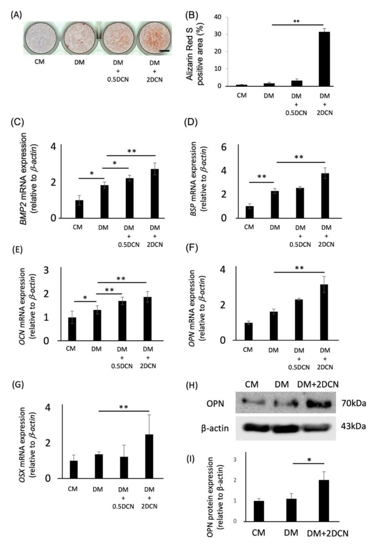
Figure 2.
Effects of DCN on the osteoblastic differentiation of HPDLSCs. (A) HPDLSCs were cultured in 10% FBS/α-MEM (CM), CM with 1.5 mM CaCl2 (DM), DM on a 0.5 μg/mL DCN coating (DM + 0.5 DCN), or DM on a 2 μg/mL DCN coating (DM + 2 DCN) for 2 weeks. Alizarin red S staining was performed to evaluate the mineralization of HPDLSCs. Bar = 500 µm. (B) The graph shows the quantification of Alizarin red S-positive areas. (C–G) Quantitative RT-PCR was performed to analyze the gene expression of BMP2 (C), BSP (D), OCN €, OPN (F), and OSX (G) in HPDLSCs cultured under the same conditions for 7 days (C–F) or 12 h (G). (H,I) HPDLSCs were cultured in CM, DM, or DM + 2 DCN for 7 days. (H) Western blot analysis was performed to determine the expression of OPN. β-actin was used as a loading control. (I) The graph shows the quantification of OPN expression. Normalization of protein expression was performed against β-actin expression. Values are the mean ± SD of three indepenDent. experiments. ** p < 0.01, * p < 0.05.

