Abstract
To study the postoperative visualisation of the electrode array insertion angle through transcanal Veria approach in both round window and cochleostomy techniques. Retrospective study. Tertiary care centre. 26 subjects aged 2–15 years implanted with a MED-EL STANDARD electrode array (31.5 mm) through Veria technique were selected. 16 had the electrode insertion through the round window, 10 through anteroinferior cochleostomy. DICOM files of postoperative computer tomography (CT) scans were collected and analysed using the OTOPLAN 3.0 software. Examined parameters were cochlear duct length, average angle of insertion depth. Pearson’s Correlation Test was utilized for statistical analysis. Average cochlear duct length was 38.12 mm, ranging from 34.2 to 43 mm. Average angle of insertion depth was 666 degrees through round window insertion and 670 degrees through cochleostomy insertion. Pearson’s correlation showed no significant difference in average angle of insertion depth between subjects with cochleostomy and round window insertion. Detailed study on the OTOPLAN software has established that there remains no difference between round window insertion or cochleostomy insertion when it comes to electrode array position and placement in the scala tympani. It is feasible to perform round window insertion and cochleostomy insertion through transcanal Veria approach as this technique provides good visualisation.
Supplementary Information
The online version contains supplementary material available at 10.1007/s12070-022-03228-5.
Keywords: Veria technique, Cochleostomy, Round window insertion, Otoplan, Cochlear Duct length, Angle of Insertion
Introduction
Cochlear implantation is a widely accepted medical and surgical option to treat sensorineural hearing loss in individuals with limited benefit from hearing aids. Cochlear implants (CIs) consist of an internal component and an external audio processor. The internal component consists of a receiver/stimulator and an intracochlear electrode array. The implant is surgically placed over the temporal bone and the electrode array is inserted into the scala tympani of the cochlear duct either through round window (RW) insertion or through cochleostomy (CS).
After Dr House introduced the mastoidectomy with posterior tympanotomy approach (MPTA) for cochlear implantation in 1961 [1], several studies describing cochlear implantation with modified surgical approaches have been published. The steps commonly described include mastoidectomy, posterior tympanotomy, and electrode array insertion through the RW or through CS. In the posterior tympanotomy approach, cortical mastoidectomy is followed by the opening of the facial recess. This step is considered crucial as the RW can be visualised through the facial recess [8]. This technique has been modified to reduce the risk of injury to the essential structures in and around the surgical site—the facial nerve and the chorda tympani. Both RW and CS proved to be surgically feasible through posterior tympanotomy but rely on the large facial recess approach to identify both the facial nerve and the chorda tympani [2].
Another approach for CI surgery, known as the Veria technique, was described by Dr. Trifone Kiratzidis in 2002. The Veria technique is a non-mastoidectomy surgical technique using a transcanal approach to the middle ear and the cochlea where a direct tunnel is drilled through the posterosuperior bony canal wall for electrode array insertion [3, 4]. Here, a tympanomeatal flap is elevated and the middle ear is visualised. A special “Trifone-perforator” is used to make the posterior canal wall tunnel. Unlike the posterior tympanotomy approach, the facial recess is not opened directly. The facial nerve is protected by the special perforator where a 1.6 mm drill bit is paired with a straight guide. The tip of the guide is very close to the burr allowing the drilling to be less than 0.5 mm under the surface of the bone. On the right side, the entry to the well is in the 11 o’clock position and the exit in the 9 o’clock position just above the chorda tympani exit and below the tympanic annulus. Here, the middle ear is already exposed and thus the RW is visible. This surgical approach allows both CS and RW insertion of the electrode array.
RW insertion entails various challenges that require careful consideration, including the extent to which the RW membrane is exposed in the middle ear and variations in morphology of the RW opening that may affect electrode array insertion [5]. The RW is the site of electrode array insertion as described in the conventional posterior tympanotomy technique while CS has been described as the method of electrode array insertion by proponents of the Veria technique [4, 6].
CI surgery either through posterior tympanotomy or transcanal approach involves a series of steps that require precise preoperative planning. Preoperative radiological imaging using computed tomography (CT) and/or magnetic resonance imaging (MRI) provides essential information about the structures of the temporal bone that are essential to be taken care of during CI surgery. Further, postoperative radiological imaging allows visualisation of full insertion or partial insertion of the electrode array, correct site of insertion, angle of insertion depth (AID) via the RW or CS, position of the electrode array in the cochlear duct, and any visible anomaly of the intracochlear electrode array.
Radiological imaging provides the benefit of visualising the electrode array position, however without the feasibility of 3-dimensional visualisation and without the frequency place information of the electrode contacts. OTOPLAN is a multifunctional otological planning software developed by CAScination AG, a medical technology company from Bern (Switzerland), in association with MED-EL, a hearing implant company from Innsbruck (Austria). It facilitates presurgical planning for otological surgery. This software allows 3D visualisation of the CT and MRI scans of the anatomical areas of interest. For a cochlear implant surgery, preoperative analysis of cochlea, Internal auditory canal, Vestibular structures, facial nerve and chorda tympani can be done using OTOPLAN software. Postoperative analysis is an efficient tool of the OTOPLAN software that can be used to evaluate the positioning of the electrode array and its AID in degrees. Along with the position, the OTOPLAN software provides frequency place information of individual electrode contacts.
Various studies described electrode array insertions through the RW in a posterior tympanotomy technique. However, there is a dearth of studies that compare the electrode position and placement between RW and CS insertions using the transcanal Veria approach. In this study, we aimed to visualise the electrode insertion angle postoperatively in transcanal Veria approach cases with either RW or CS insertion and to compare them for similarities and differences using the OTOPLAN software. We also aimed to analyse the intracochlear electrode array position in both approaches of electrode array insertion.
Objectives
Using OTOPLAN software to perform postoperative evaluation of intracochlear electrode array position following both RW and CS insertion in transcanal Veria technique
To study the parameters of electrode array position (angle/depth of insertion, scala tympani insertion etc.) following RW insertion and CS insertion in transcanal Veria technique
To look for any significant differences in intracochlear electrode array position between two different insertion routes – RW and CS in transcanal Veria technique
Materials and Methods
Subjects
26 Subjects aged 2–15 years who had bilateral severe to profound sensorineural hearing loss and who underwent CI surgery were selected (Tables 1, 2). Subjects were excluded if they were affected by active middle ear disease or cochlear malformation.
Table 1.
Demographic detail of patients undergoing round window insertion of the cochlear electrode
| Subject number | Age | Gender | RW/CS |
|---|---|---|---|
| S1 | 6 | M | RW |
| S2 | 5 | M | RW |
| S3 | 6 | F | RW |
| S4 | 3 | M | RW |
| S10 | 3 | M | RW |
| S11 | 4 | M | RW |
| S12 | 3 | F | RW |
| S13 | 3 | F | RW |
| S14 | 4 | M | RW |
| S15 | 4 | M | RW |
| S16 | 3 | F | RW |
| S17 | 3 | M | RW |
| S18 | 4 | F | RW |
| S19 | 6 | F | RW |
| S20 | 7 | F | RW |
| S29 | 3 | F | RW |
Table 2.
Demographic details of patients undergoing insertion of the cochlear electrode via the cochleostomy
| Subject number | Age | Gender | RW/CS (Round window/Cochleostomy) |
|---|---|---|---|
| S5 | 5 | M | CS |
| S6 | 4 | M | CS |
| S7 | 10 | M | CS |
| S8 | 9 | M | CS |
| S9 | 14 | M | CS |
| S22 | 11 | F | CS |
| S23 | 9 | F | CS |
Ethics
Study conducted after written informed consent of participants and ethical approval.
Study Conduct
In this retrospective study, intracochlear positioning of the electrode array was analysed using the OTOPLAN software in two different surgical approaches for cochlear implantation—RW insertion via transcanal technique and CS via transcanal technique.
DICOM files of the postoperative CT scans were collected for analysis.. OTOPLAN version 3.0, a fully portable and tablet-based software, was used to analyse the DICOM files of the CT scans.
CT scans were obtained with a slice thickness of 0.6 mm or below. To maintain data confidentiality, the DICOM files were anonymised using the DICOM Anonymizer and a subject number was given to each file. The files were then imported into the OTOPLAN software.
Postoperative CT scans were analysed for cochlear parameters, placement of the electrode array in the scala tympani, and AID of the electrode array.
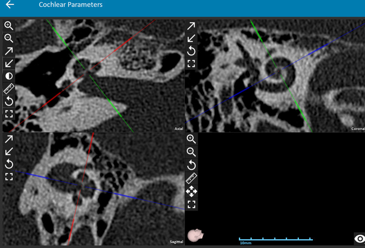
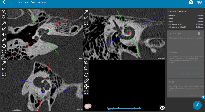
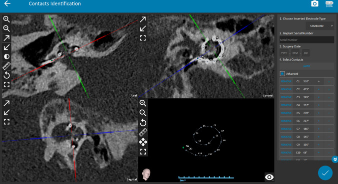
The steps of OTOPLAN analysis are as follows: First, DICOM files of the patient are imported into OTOPLAN software. Second,the implanted ear is selected for analysis. Third, after optimal cochlear view is aligned on axial, coronal and sagittal views, (see Fig. 1), cochlear parameters are measured. Fourth, the cochlear parameters are defined; the diameter of the basal turn of the cochlea, height of the cochlea, and the width of the cochlea are marked using the crosshairs on the screen. The diameter of the basal turn is marked from the RW centre to the lateral wall, which is known as the ‘A” value (see Fig. 2). The unit for measurement for cochlear parameters was millimetres (mm). Fifth, the cochlear duct length (CDLLW), the linear length of the cochlear duct, is defined as the length of the cochlear duct from the centre of the RW to the apical tip of the cochlea. CDLLW is calculated by the software from the parameters—diameter, width, and height of the cochlea. OTOPLAN version 3.0 uses the Elliptic-Circular Approximation (ECA) equation to calculate the CDL. The ECA approach [9] uses both diameter (A) and width (B) of the cochlear basal turn. ECA is a 2-step approach that was developed to compute CDLLW (θ). Sixth, individual electrode contact identification is done through the auto contact identification tool and the position marked by the tool is further finetuned for accuracy. Seventh, the AID is measured in degrees for each electrode contact by the software based on the postoperative CT image (see Fig. 3).
Fig. 1.
Figure demonstrating the “Cochlear View” on the Otoplan which helps in clearly delineating all the cochlear parameters
Fig. 2.
Figure demonstrating the calculation of cochlear parameters, a Diameter of basal turn & b Width of cochlea, using the cross hair in the Otoplan software system
Fig. 3.
Otoplan image showing post operative electrode contact points. Angle of insertion (AID) is calculated using this
Statistical Analysis
Pearson’s Correlation test was used on the AID data obtained from OTOPLAN analysis, to understand the correlation between the two methods of electrode insertion.
Results
This retrospective study included 26 subjects. The data obtained from 26 CT scan DICOM files of the subjects were analysed using the OTOPLAN software.
All 26 subjects had normal cochlear anatomy, and cochlear turns were visible in all three planes (axial, coronal, and sagittal) on the cochlear view function of the OTOPLAN software. The scala tympani was the site of placement in all 26 subjects as visually confirmed by the OTOPLAN software.
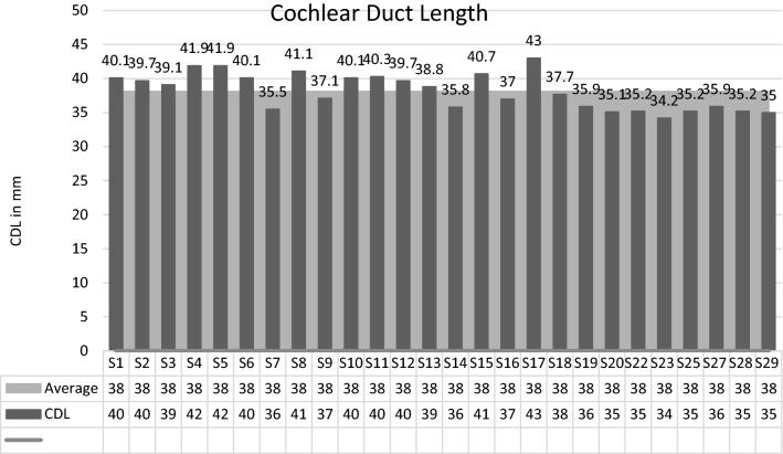
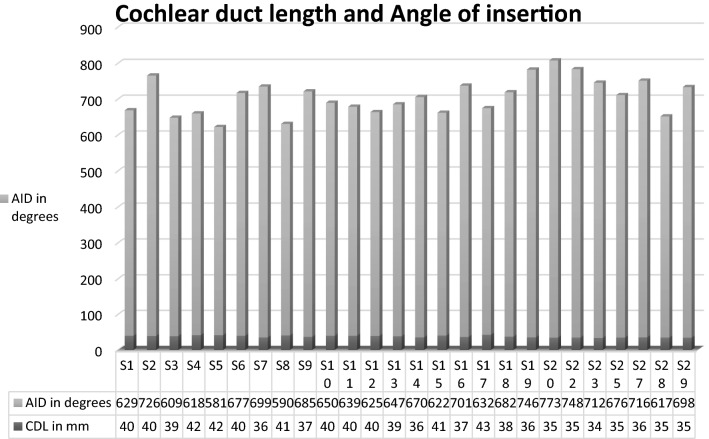
All 26 subjects were implanted through the Veria technique. 16 of them had the electrode array insertion through RW while 10 subjects had electrode array insertion through anteroinferior CS. The CDL measured in the 26 subjects ranged from a minimum of 34.2 mm to a maximum of 43 mm. The average CDL was 38.12 mm (Table 3). This data correlates with findings from other CDL studies reported in the literature. [7]
Table 3.
Table depicting the cochlear duct length of each study participant
The electrode array inserted in all subjects was a MED-EL STANDARD electrode array with a length of 31.5 mm. The standard electrode array has a marker ring at the base of the array indicating a full insertion when the marker ring sits at the RW or in the CS.
The average AID in these subjects with the standard electrode array was 667.89 degrees with a minimum AID of 580.8 degrees and a maximum AID of 773 degrees (Table 4). The subjects that had electrode array insertion through RW had an average AID of 666 degrees while the subjects that had electrode array insertion through CS had an average AID of 670 degrees. The low-frequency coverage provided by the standard electrode ranged from 47 to 269 Hz in all 26 subjects, with the average lowest frequency of 115 Hz. The highest frequency covered with the standard electrode array ranged from 9614 to 13,799 Hz, with an average of 12,124 Hz. On statistical analysis, no significant difference was found in AID between subjects with CS and subjects with RW insertion.
Table 4.
Table depicting the cochlear duct length and corresponding angle of insertion of the cochlear electrodes for each study participant
Discussion
The goal of CI surgery, irrespective of the surgical technique, is to help restore the lost function of hearing by inserting an electrode array into the cochlea, which has a delicate framework of neural structures and vasculature. During the insertion, the surgeon must take utmost care to make the surgery as atraumatic as possible to preserve the intracochlear structures, thereby preserving the residual hearing. Factors, such as design and flexibility of the electrode arrays, insertion force, and insertion speed play a major role in the outcomes of cochlear implant surgery. Various authors have also studied the role of the insertion technique—RW insertion, extended RW approach, CS approach—in structure and hearing preservation. Also, administration of drugs such as dexamethasone [10] or prednisolone [11] to preserve the residual hearing has also been reported in the literature. Hearing preservation was demonstrated 12 months post-implantation in subjects with partial deafness through RW insertion [12]. Hearing preservation with deep and full-length electrodes was also studied through RW insertion [13].
CS approach has been traditionally reported by Veria surgeons as their preferred method of electrode insertion as it is direct in the line with the transcanal tunnel. However, the operating surgeon felt that since the middle ear visualisation along with RW exposure was optimum in Veria technique, insertion of electrode through RW was feasible without difficulty. Also, the proponents of hearing preservation techniques follow RW insertions (soft insertions) where drilling is minimal. Keeping this in view, the surgeon who did CS earlier, switched to RW technique and to corroborate these findings, this study was carried out.
In addition to the primary goal of this study, electrode placement issues were assessed when it is being inserted using the RW as the primary route. The results of this study indicate no significant difference in the angle of insertion of the electrode array using RW insertion and CS. Also, through analysis by the OTOPLAN software, irrespective of the insertion technique used, the electrode array was visualised inside the scala tympani in all subjects without any scalar deviations or tip fold-overs.
Successful usage of the OTOPLAN software was first reported in preoperative analysis and reconstruction of the inner ear structures. OTOPLAN was successfully used for preoperative surgical planning in a case in which conventional temporal bone CT surgery was wrongly contraindicated [14]. Also, the utility of this otological planning software was demonstrated in visualisation and preoperative planning for a case of advanced otosclerosis [15]. It was reported in both studies that the OTOPLAN software provided clear and precise visualisation of RW on CT images through cochlear view functionality. In this study, the OTOPLAN software was used to study the postoperative CT images and to analyse the angle of insertion and any deviations from the scala tympani. This OTOPLAN-based study on electrode array position provides evidence that RW insertion is possible with the transcanal technique; there was no displacement/ kinking/ bending of electrodes when the RW was used to insert the electrode array.
The aim of this study has been to assess the post operative placement of electrode in the cochlea when inserted using RW or CS technique. The surgical technique used was Veria. Since the aim was to analyse the post operative data only, preoperative analysis was not discussed in this study. The Transcanal Veria approach is a versatile approach where it is not only possible to perform RW insertions without any difficulty (against the common perception), but it is also easy to operate different kinds of malformations with much ease as the entire middle ear is in view. This study also tries to refute the common misconception that RW insertion of the electrode array is not possible in the Veria approach and that it will lead to electrode displacements, bending or kinking of electrodes. Detailed study using the OTOPLAN software has established that there remains no difference between RW insertion or CS insertion when it comes to electrode array position and placement in the scala tympani.
Limitations and Future Directions of This Study
Postoperative audiological evaluation couldn’t be done due to lack of availability of subjects during the period of study. Comparison of these post- operative levels with pre-operative levels could have further provided information on structure/hearing preservation through Veria technique both through CS and RW electrode insertion.
Supplementary Information
Below is the link to the electronic supplementary material.
Funding
None.
Declarations
Conflict of interest
The authors declare that they have no conflict of interest.
Footnotes
Publisher's Note
Springer Nature remains neutral with regard to jurisdictional claims in published maps and institutional affiliations.
References
- 1.Adunka OF, Dillon MT, Adunka MC, King ER, Pillsbury HC, Buchman CA. Cochleostomy versus round window insertions. Otol Neurotol. 2014;35(4):613–618. doi: 10.1097/mao.0000000000000269. [DOI] [PubMed] [Google Scholar]
- 2.Bhavana K, Bharti B, Vishwakarma R. Round Window insertion in veria technique of cochlear implantation: an essential modification. Indian J Otolaryngol Head Neck Surg. 2019;71(S2):1586–1591. doi: 10.1007/s12070-019-01677-z. [DOI] [PMC free article] [PubMed] [Google Scholar]
- 3.Hans JM, Prasad R. Cochlear implant surgery by the Veria technique: how and why? Experience from 1400 cases. Indian J Otolaryngol Head Neck Surg. 2015;67(2):107–109. doi: 10.1007/s12070-015-0863-2. [DOI] [PMC free article] [PubMed] [Google Scholar]
- 4.House WF. Cochlear implants. Ann Otol Rhinol Laryngol. 1976;85(3_suppl):3. doi: 10.1177/00034894760850s303. [DOI] [PubMed] [Google Scholar]
- 5.Kiratzidis T, Arnold W, Iliades T. Veria operation updated. ORL. 2002;64(6):406–412. doi: 10.1159/000067578. [DOI] [PubMed] [Google Scholar]
- 6.Kuthubutheen J, Joglekar S, Smith L, Friesen L, Smilsky K, Millman T, Ng A, Shipp D, Coates H, Arnoldner C, Nedzelski J, Chen J, Lin V. The role of preoperative steroids for hearing preservation cochlear implantation: results of a randomized controlled trial. Audiol Neurotol. 2017;22(4–5):292–302. doi: 10.1159/000485310. [DOI] [PubMed] [Google Scholar]
- 7.Koch RW, Ladak HM, Elfarnawany M, et al. Measuring Cochlear Duct Length—a historical analysis of methods and results. J Otolaryngol Head Neck Surg. 2017;46:19. doi: 10.1186/s40463-017-0194-2. [DOI] [PMC free article] [PubMed] [Google Scholar]
- 8.Kiratzidis T, Arnold W, Iliades T. Veria Operation Updated. ORL J OTO-Rhino-Laryngol Relat Spec ORL-J OTO-Rhino-Laryngol. 2002;64:406–412. doi: 10.1159/000067578. [DOI] [PubMed] [Google Scholar]
- 9.Lovato A, de Filippis C. Utility of OTOPLAN reconstructed images for surgical planning of cochlear implantation in a case of post-meningitis ossification. Otol Neurotol. 2019;40(1):e60–e61. doi: 10.1097/mao.0000000000002079. [DOI] [PubMed] [Google Scholar]
- 10.Lovato A, Marioni G, Gamberini L, Bonora C, Genovese E, de Filippis C. OTOPLAN in cochlear implantation for far-advanced otosclerosis. Otol Neurotol. 2020;41(8):e1024–e1028. doi: 10.1097/mao.0000000000002722. [DOI] [PubMed] [Google Scholar]
- 11.Roland PS, Wright CG, Isaacson B. Cochlear implant electrode insertion: the round window revisited. Laryngoscope. 2007;117(8):1397–1402. doi: 10.1097/mlg.0b013e318064e891. [DOI] [PubMed] [Google Scholar]
- 12.Schurzig D, Timm ME, Batsoulis C, Salcher R, Sieber D, Jolly C, Lenarz T, Zoka-Assadi M. A novel method for clinical cochlear duct length estimation toward patient-specific cochlear implant selection. OTO Open. 2018;2(4):2473974X1880023. doi: 10.1177/2473974x18800238. [DOI] [PMC free article] [PubMed] [Google Scholar]
- 13.Skarżyński H, Lorens A, D’Haese P, Walkowiak A, Piotrowska A, Śliwa L, Anderson I. Preservation of residual hearing in children and post-lingually deafened adults after cochlear implantation: an initial study. ORL. 2002;64(4):247–253. doi: 10.1159/000064134. [DOI] [PubMed] [Google Scholar]
- 14.Skarzynski H, Lorens A, Piotrowska A, Anderson I. Preservation of low frequency hearing in partial deafness cochlear implantation (PDCI) using the round window surgical approach. Acta Otolaryngol. 2007;127(1):41–48. doi: 10.1080/00016480500488917. [DOI] [PubMed] [Google Scholar]
- 15.Usami S-I, Moteki H, Suzuki N, Fukuoka H, Miyagawa M, Nishio S-Y, Takumi Y, Iwasaki S, Jolly C. Achievement of hearing preservation in the presence of an electrode covering the residual hearing region. Acta Otolaryngol. 2011;131(4):405–412. doi: 10.3109/00016489.2010.539266. [DOI] [PMC free article] [PubMed] [Google Scholar]
Associated Data
This section collects any data citations, data availability statements, or supplementary materials included in this article.


