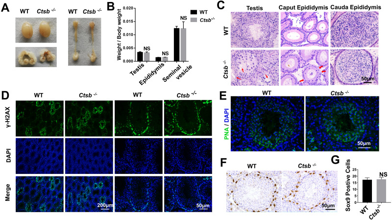
Figure 3. Spermatogenesis of Ctsb-/- mice.
(A) The morphology of the testis, epididymis, and seminal vesicle in WT and Ctsb-/- mice. (B) The weights of the testis, epididymis, and seminal vesicle did not significantly differ between WT and Ctsb-/- mice. (C) H & E-stained sections of the testes, caput, and cauda epididymides from WT and Ctsb-/- mice. (D) Immunofluorescence staining of γH2AX-positive spermatocytes from WT and Ctsb-/- mice. (E) PNA-stained acrosomes in the developmental spermatid of WT and Ctsb-/- mice. (F) Immunohistochemistry staining of SOX9-positive Sertoli cells from WT and Ctsb-/- mice. (G) Sertoli cell counts from WT and Ctsb-/- mice. NS, non-significant. n ≥ 4 in each group.

