Abstract
Clostridial neurotoxins potently and specifically inhibit neurotransmitter release in defined cell types by a mechanism that involves cleavage of specific components of the vesicle docking/fusion complex, the SNARE complex. A derivative of the type A neurotoxin from Clostridium botulinum (termed LHN/A) that retains catalytic activity can be prepared by proteolysis. The LHN/A, however, lacks the putative native binding domain (HC) of the neurotoxin and is thus unable to bind to neurons and effect inhibition of neurotransmitter release. Here we report the chemical conjugation of LHN/A to an alternative cell-binding ligand, wheat germ agglutinin (WGA). When applied to a variety of cell lines, including those that are ordinarily resistant to the effects of neurotoxin, WGA-LHN/A conjugate potently inhibits secretory responses in those cells. Inhibition of release is demonstrated to be ligand mediated and dose dependent and to occur via a mechanism involving endopeptidase-dependent cleavage of the natural botulinum neurotoxin type A substrate. These data confirm that the function of the HC domain of C. botulinum neurotoxin type A is limited to binding to cell surface moieties. The data also demonstrate that the endopeptidase and translocation functions of the neurotoxin are effective in a range of cell types, including those of nonneuronal origin. These observations lead to the conclusion that a clostridial endopeptidase conjugate that can be used to investigate SNARE-mediated processes in a variety of cells has been successfully generated.
The clostridial neurotoxin (CNT) family includes tetanus toxin (TeNT), produced by Clostridium tetani, and the seven antigenically distinct botulinum neurotoxins produced from strains of Clostridium botulinum (BoNTs). These proteins are responsible for the conditions of tetanus and botulism, respectively, that develop as a direct result of inhibition of Ca2+-dependent neurotransmitter release, a mechanism of action common to all the CNTs. In the case of BoNTs, intoxication of the neuromuscular junction is thought to occur in at least three phases: an initial binding phase, an internalization phase, and finally a neurotransmitter blockade phase (24).
All CNTs have a similar structure and consist of a heavy chain (HC: approximately 100 kDa) covalently joined to a light chain (LC: approximately 50 kDa) by a single disulfide bond. Proteolytic cleavage of the HC of C. botulinum neurotoxin type A (BoNT/A) generates two fragments of approximately 50 kDa each. The C-terminal domain (HC) is required for target cell binding, with the N-terminal domain (HN) being proposed to be involved in intracellular membrane translocation (22). Under conditions in which the disulfide bond between the LCs and HCs is maintained, trypsin cleavage results in a 100-kDa species termed LHN/A (originally described as H2L [23]) representing a catalytically active, non-cell-binding derivative of BoNT/A.
The identities of the cellular receptors for CNTs are generally unknown, though it is clear that CNTs are highly specific for the neuromuscular junction. It is proposed that CNTs bind to their target cell by a combination of specific, high-affinity binding events possibly involving more than one component (9). It is thought that gangliosides and membrane-bound glycoproteins may serve as targets for toxin binding. The recent proposal that BoNT/B binds to synaptotagmin and the gangliosides GT1b and GD1a (16) and that BoNT/A and BoNT/E may also bind to synaptotagmin (11) has supported this concept. Having accomplished the first cell intoxication stage of binding, CNTs require mechanisms to facilitate internalization into and intracellular routing within the target cell. The actual mechanism of intracellular routing of CNTs to the cytosol remains unclear, though the role of an acidic compartment has been proposed (10) in common with a number of other bacterial protein toxins (20, 21). Though it is proposed that it is the role of the HN domain to facilitate translocation of the endopeptidase into the cytosol, exclusion of a contribution of the HC domain in intracellular mechanisms has not been possible. One of the aims of this study is to address the issue of HC contribution in the three phases of intoxication. In addition, this study aims to investigate the ability of the HN domain to facilitate translocation of the LC into the cytosol of both neuronal and nonneuronal cells.
Once the CNT (or fragment) has gained access to the cytosol, the proteolytic LCs specifically hydrolyze key components of the soluble NSF accessory protein receptor (SNARE) complex (25) required for synaptic vesicle docking, fusion, and neurotransmitter release. In the case of BoNT/A and BoNT/E, the substrate is synaptosome-associated protein 25 (SNAP-25), whereas BoNT B, D, F, and G cleave the vesicle-associated membrane protein and BoNT/C cleaves syntaxin (2, 14). It has been demonstrated that cleavage of these components of the SNARE complex by CNTs results in inhibition of transmitter release from a variety of neuronal cell systems. It is also known that formation of the SNARE complex is a universal mechanism of vesicle fusion and secretion, not limited to neuronal cell types. Therefore, the range of highly specific endopeptidase activities of CNT serotypes provides an opportunity for the understanding of SNARE-mediated events. Unfortunately, the use of native clostridial toxins for the study of such events is often limited by the availability of the requisite toxin receptor(s) on the target cell of interest. Alternative approaches to internalizing the active endopeptidase have included techniques such as microinjection, permeabilization, and electroporation (3). However, the invasive nature of these techniques makes them less than ideal approaches for the study of complex intracellular processes. To overcome these problems, and to address issues of HN and HC domain functionality, we have sought to identify alternative molecules capable of delivering functional endopeptidase to the cytosol of target cells in vitro. Here we describe the replacement of the native BoNT/A cell-binding domain (HC) with one such alternative targeting ligand, wheat germ agglutinin (WGA), which enables binding to a range of cell types (6).
WGA is a homodimeric lectin molecule of 36 kDa expressed by the plant species Triticum vulgaris. WGA has an affinity for N-acetylglucosamine (GlcNAc) and N-acetyl sialic acid (15) and has been used previously as a neuronal cell marker, for retrograde transport studies, and for analysis of lectin function on a variety of cell processes (12). This profile and the degree of characterization make WGA attractive as a novel cell-binding domain for detoxified CNTs. In this study we report that a WGA-LHN/A conjugate binds to, internalizes into, and inhibits stimulated neurotransmitter release from a range of cultured neuronal cell types. Furthermore, WGA-LHN/A conjugate is demonstrated to inhibit insulin release from a pancreatic B-cell line that is resistant to the effects of neurotoxin. WGA-LHN/A conjugate effects on secretion are shown to be dose dependent, mediated by ligand-dependent binding, and correlated to cleavage of the natural BoNT/A substrate SNAP-25.
MATERIALS AND METHODS
Materials.
PC12 and SH-SY5Y cells were supplied by the European Collection of Cell Cultures, Brighton, United Kingdom. HIT-T15 cells were kindly supplied by I. Green, University of Sussex, United Kingdom. Antibodies to SNAP-25 (SMI-81) were obtained from Sternberger Monoclonals Inc. Peroxidase-conjugated anti-species antibody was from Stratech Scientific Ltd. Western blot detection was performed with enhanced chemiluminescence reagents and Hyperfilm from Amersham. The rat insulin radioimmunoassay kit was obtained from Linco Research Inc. The protein cross-linking reagent, N-succinimidyl-3-(2-pyridyldithio)propionate (SPDP), was obtained from Pierce & Warriner Ltd. Sodium dodecyl sulfate-polyacrylamide gel electrophoresis (SDS-PAGE) materials were obtained from Novex. Triton X-114 was obtained from Fisons. WGA and all other buffers, reagents, and cell culture media were obtained from Sigma, Poole, United Kingdom.
Conjugation and purification of WGA-LHN/A.
WGA-LHN/A conjugate was synthesized by conjugating WGA and LHN/A components that had previously been derivatized to introduce reactive cross-linking groups. Briefly, WGA (10 mg/ml in phosphate-buffered saline [PBS]) was reacted with an equal concentration of SPDP (10 mM in dimethyl sulfoxide [DMSO]) for 1 h at ambient temperature. Reaction by-products were removed by desalting into PBS over a PD-10 column (Pharmacia) prior to reduction of the cross-linker with dithiothreitol (DTT; 5 mM for 30 min) to generate a reactive sulfhydryl (SH) group. The thiopyridone and DTT were then removed by desalting into PBS over a PD-10 column to result in derivatized WGA (dWGA) with 1 mol of SH incorporated per mol of WGA.
LHN/A at a concentration of 5 mg/ml in PBSE (PBS containing 1 mM EDTA) was reacted with a fourfold molar excess of SPDP (10 mM in DMSO). After 3 h at ambient temperature, the reaction was terminated by desalting over a PD-10 column into PBSE. To determine the degree of derivatization, an aliquot of the derivatized LHN/A (dLHN/A) was removed from the solution, reduced with DTT (5 mM, 30 min), and analyzed spectrophotometrically at 280 and 343 nm.
The dWGA and the dLHN/A were mixed in a ≥3:1 molar ratio. After 16 h at 4°C, the mixture was centrifuged to clear any precipitate that had developed. The supernatant was concentrated by ultrafiltration (Millipore Biomax Ultrafree-4; 10,000 molecular weight exclusion limit) before application to a Superose 12 column on an FPLC chromatography system (Pharmacia). The column was eluted with PBS, and the elution profile was monitored at 280 nm. Fractions containing high-molecular-weight conjugate material (separated from free dWGA) were pooled and applied to PBS-washed GlcNAc-agarose. WGA-LHN/A conjugate bound to the GlcNAc-agarose and was eluted from the column by the addition of 0.3 M GlcNAc in PBS. The elution profile was monitored at 280 nm, and fractions containing conjugate were pooled, dialyzed against PBS, and stored at 4°C until use.
Preparation and maintenance of cell cultures.
PC12 cells were seeded at a density of 4 × 10⁵ cells/well onto 24-well (Matrigel coated) plates (Nunc) from stocks grown in suspension. The cells were cultured for 1 week prior to use in RPMI-1640–10% horse serum–5% fetal bovine serum–1% l-glutamine. SH-SY5Y cells were seeded at a density of 5 × 10⁵ cells/well onto 24-well plates (Falcon). The cells were cultured in Ham's F-12 medium–minimal essential medium (MEM) (1:1, vol/vol) containing 15% fetal bovine serum, 1% MEM nonessential amino acids, and 2 mM l-glutamine for 1 week prior to use. Embryonic spinal cord (eSC) neurons were prepared from spinal cords dissected from 14- to 15-day-old fetal Sprague-Dawley rats and used after 21 days in culture in a modification of a previously described method (5, 18). HIT-T15 cells were seeded at a density of 4 × 10⁵ cells/well onto 12-well plates (Falcon). The cells were cultured in RPMI-1640–5% fetal bovine serum–2 mM l-glutamine for 5 days prior to use.
Inhibition of stimulated secretion.
PC12 or SH-SY5Y cells were washed with a balanced salt solution (BSS: 137 mM NaCl, 5 mM KCl, 2 mM CaCl2, 4.2 mM NaHCO3, 1.2 mM MgCl2, 0.44 mM KH2PO4, 5 mM glucose, 20 mM HEPES [pH 7.4]) and loaded for 1 h with [3H]noradrenaline (NA; 2 μCi/ml, 0.5 ml/well) in BSS containing 0.2 mM ascorbic acid and 0.2 mM pargyline. Cells were washed four times (at 15-min intervals for 1 h), and then basal release was determined by a 5-min incubation with BSS–5 mM K+. Cells were then depolarized with 100 mM K+ (BSS with Na+ reduced accordingly) for 5 min to determine stimulated release. Superfusate (0.5 ml) was removed to tubes on ice and briefly centrifuged to pellet any detached cells. Adherent cells were solubilized in 2 M acetic acid–0.1% trifluoroacetic acid (250 μl/well). The quantities of released and nonreleased radiolabel were determined by liquid scintillation counting of cleared superfusates and cell lysates, respectively. Total uptake was calculated by addition of released and retained radioactivity, and the percentage release was as calculated (released counts/total uptake counts) × 100.
eSC neurons were loaded with [3H]glycine for 30 min prior to determination of basal and potassium-stimulated release of transmitter (essentially as described before [29]). A sample (0.2 M) of NaOH-lysed cells was used to determine total counts, from which percent release could be calculated. Insulin release from HIT-T15 cells was stimulated by incubation with high-potassium Krebs-Ringer bicarbonate buffer (KRB: 103.8 mM NaCl, 5 mM NaHCO3, 30 mM KCl, 1.2 mM KH2PO4, 1.0 mM CaCl2, 1.2 mM MgSO4, 2.8 mM glucose, 10 mM HEPES [pH 7.4]) for 30 min. Basal release was determined by identical simultaneous incubation of cells for 30 min with low-potassium KRB (4.8 mM KCl, 129 mM NaCl). Released insulin was determined by radioimmunoassay exactly as instructed by the manufacturer.
In vitro SNAP-25 cleavage.
The in vitro cleavage of SNAP-25 by WGA-LHN/A and other endopeptidase samples was determined essentially as described before (8).
SDS-PAGE and Western blot analysis.
SDS-PAGE and Western blot analyses were performed by standard protocols (Novex). SNAP-25 proteins were extracted from 2 M acetic acid–0.1% trifluoroacetic acid-lysed cells using Triton X-114 in a method described previously (3). The extracted proteins were resolved on a Tris–glycine–4 to 20% polyacrylamide gel (Novex) and subsequently transferred to a nitrocellulose membrane. The membranes were probed with a monoclonal antibody (SMI-81) that recognizes cleaved and intact SNAP-25. Specific binding was visualized with peroxidase-conjugated secondary antibodies and a chemiluminescent detection system essentially as described previously (17). Cleavage of SNAP-25 was quantified by scanning densitometry (Molecular Dynamics Personal SI, ImageQuant data analysis software). Percent SNAP-25 cleavage was calculated by the formula [cleaved SNAP-25/(cleaved + intact SNAP-25)] × 100.
RESULTS
Conjugation and purification of WGA-LHN/A.
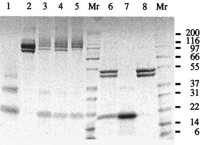
Derivatization with the heterobifunctional cross-linker SPDP has been applied successfully to both WGA and LHN/A. The derivatization ratio of WGA was maintained at 1 introduced —SH per lectin dimer in order to minimize the proportion of very high molecular weight protein aggregates. Conversely, the derivatization ratio of LHN/A was established (observed ratio, 3.53 ± 0.59 mol of SPDP per mol of LHN/A) in order to successfully conjugate at least one WGA per LHN/A. Conjugation of dLHN/A with dWGA generates a heterogenous mixture of high-molecular-weight species as determined by nonreducing SDS-PAGE analysis (Fig. 1). However, when the disulfide bond of the chemical linkage between LHN/A and WGA is reduced in the presence of a thiol (5 mM DTT), the heterogenous mixture is shown to consist of LHN/A and WGA only (Fig. 1, lane 6).
FIG. 1.
SDS-PAGE analysis of WGA-LHN/A purification scheme. Protein fractions were subjected to SDS–4 to 20% PAGE prior to staining with Coomassie blue. Lanes 6 to 8 were run in the presence of 0.1 M DTT. Lanes 1 and 7 and lanes 2 and 8 represent dWGA and dLHN/A, respectively. Lanes 3 to 5 represent the conjugation mixture after Superose-12 chromatography and after GlcNAc-affinity chromatography, respectively. Lane 6 represents a sample of reduced final material. Approximate molecular masses (in kilodaltons) are indicated. Lanes Mr, size markers.
It was also established that the catalytic activity of the LHN/A was not compromised by derivatization. Using a sensitive in vitro assay that is specific for the endopeptidase activity of BoNT/A (8), it was observed that the SPDP derivatization reagent did not significantly decrease the endopeptidase activity of the LHN/A. The concentrations of sample required to achieve 50% substrate cleavage for BoNT/A, dLHN/A, and LHN/A were determined to be of the same order (9.0, 5.1, and 5.9 pM, respectively). Similar data (not shown) were obtained for the WGA-LHN/A conjugate, although since the assay is necessarily performed in the presence of a reducing agent, these data indicate that the catalytic activity of LHN/A has not been compromised by conjugation and subsequent reduction. Therefore, it would be predicted that the specific activity of the LHN/A component would be similar to that of BoNT/A when introduced into the cell.
Purification of WGA-LHN/A was achieved by a two-step strategy. First, size exclusion chromatography was used to remove unconjugated WGA from the mixture, and second, an affinity chromatography step was used to isolate and concentrate species that bound GlcNAc. In this way, the proportion of unconjugated WGA and LHN/A components was kept to a minimum. WGA visualized in the purified conjugate after SDS-PAGE analysis was proposed to be due to the dissociation of the noncovalent homodimeric WGA by SDS.
Inhibition of [3H]NA release from PC12 and SH-SY5Y cells.
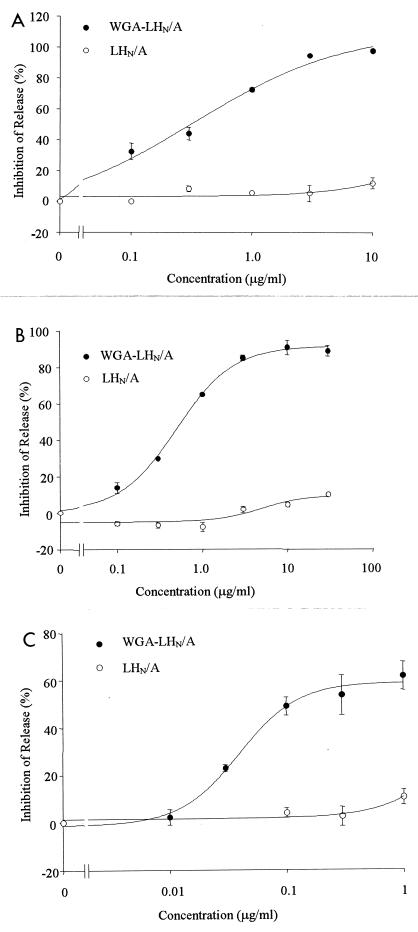
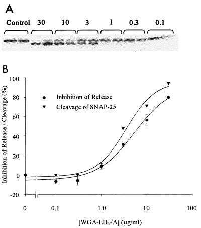
WGA-LHN/A applied to PC12 and SH-SY5Y cells was demonstrated to result in significant inhibition of neurotransmitter release relative to LHN/A alone. Figure 2 illustrates the neurotransmitter release data obtained following application of WGA-LHN/A and LHN/A to PC12 and SH-SY5Y cells for 3 days. The concentrations that inhibited activity by 50% (IC50) for WGA-LHN/A on SH-SY5Y cells were 1.60 ± 0.06 μg/ml (mean ± standard error of the mean, n = 3) after 3 days and 12.63 ± 3.72 μg/ml (n = 4) after 16 h of exposure. Similar data were obtained for PC12 cells (0.63 ± 0.15 μg/ml [n = 3] after 3 days and 4.96 ± 1.13 μg/ml [n = 3] after 16 h). In all cases the inhibitory effects of LHN/A alone were extremely low, and IC50 data were not calculable. To demonstrate that the observed inhibition of release was mediated by SNAP-25 cleavage, hydrophobic proteins were isolated from WGA-LHN/A conjugate-treated cells. The hydrophobic proteins, including SNAP-25, were Western blotted and subsequently probed with a primary antibody that recognizes both cleaved and uncleaved SNAP-25. Figure 3 illustrates the cleavage pattern obtained from one such experiment following treatment of SH-SY5Y cells with WGA-LHN/A for 16 h. Quantitation of the cleavage of SNAP-25 by scanning densitometry indicated that the cleavage of SNAP-25 correlated to the inhibition of release (Fig. 3).
FIG. 2.
Inhibition of neurotransmitter release from cultured neuronal cells. PC12 cells (A), SH-SY5Y cells (B), and eSC neurons (C) exposed for 3 days to a range of concentrations of WGA-LHN/A (solid symbols) and LHN/A (open symbols) were assessed for stimulated [3H]NA release (SH-SY5Y and PC12 cells) or [3H]glycine release (eSC) capability. Results are expressed as percent inhibition compared with untreated controls. Each concentration was assessed in triplicate. For each cell type, the dose-response curve is representative of at least three experiments. Each point shown is the mean of at least three determinations ± the standard error of the mean.
FIG. 3.
Correlation of SNAP-25 cleavage with inhibition of release. Hydrophobic proteins were extracted and probed with antibody SMI-81 for the presence of SNAP-25 (both cleaved and uncleaved forms). (A) Cleavage data for SH-SY5Y cells exposed to a range of concentrations of WGA-LHN/A for 16 h. The shift to a faster-migrating SNAP-25 species with increasing concentrations of conjugate is clearly seen. (B) Cleavage data correlated with inhibition of release of [3H]NA from SH-SY5Y exposed to a range of concentrations of WGA-LHN/A for 16 h.
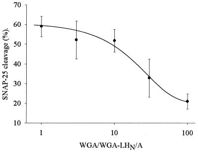
The retargeting of the LHN/A was further characterized by demonstrating that the observed cell effects were mediated by ligand-dependent targeting. SH-SY5Y cells were exposed to WGA-LHN/A on ice (4°C) for 4 h in the presence of various excesses of WGA, washed, and incubated for 16 h at 37°C prior to the determination of SNAP-25 cleavage. Figure 4 clearly shows that at increased WGA concentrations, the cleavage of SNAP-25 is decreased, and therefore the observed endopeptidase effects are a result of WGA-mediated cell entry. These data also confirmed that SNAP-25 cleavage was not a consequence of a WGA effect on cell function.
FIG. 4.
Competition for binding of WGA-LHN/A to SH-SY5Y cells by WGA. Cells were exposed to WGA-LHN/A (10 μg/ml) on ice for 4 h in the presence of various excesses of WGA. The cells were washed and incubated for 16 h at 37°C prior to the determination of SNAP-25 cleavage. SNAP-25 cleavage in the absence of competing WGA was determined to be 63.9 ± 3.9% (n = 3). Each point shown is the mean of at least three determinations ± the standard error of the mean.
Inhibition of [3H]glycine release from eSC neurons.
Primary cultures of eSC neurons are representative of in vivo neuronal cells and are therefore relevant models for a number of in vivo secretory processes. Since eSC neurons have been demonstrated to be sensitive to BoNT/A-dependent inhibition of secretory processes (29), they serve as a useful test model for WGA targeting. In this study we labeled cells with [3H]glycine, an inhibitory neurotransmitter, and therefore the stimulated release observed represented release from an inhibitory population of neurons in these heterogeneous cultures. Comparison of the inhibition profiles of WGA-LHN/A and untargeted LHN/A demonstrates significant enhancement in potency (Fig. 2), with IC50s of 0.06 ± 0.01 μg/ml (n = 3) and 10.6 ± 4.9 μg/ml (n = 3) for WGA-LHN/A and LHN/A, respectively. However, although significant potency was observed, the inhibition of release of glycine reached a plateau at approximately 60%. The failure to achieve complete inhibition probably relates to the substantial portion of the K+-induced release of [3H]glycine from these cells, which has been reported to be Ca2+ independent (28). Ca2+ independence is not a characteristic of synaptic release, and so it is not surprising that clostridial endopeptidases were unable to block this component.
Comparison of inhibition of neurotransmitter release with BoNT/A.
In order to extend this investigation into the properties of WGA-LHN/A, we chose to include a comparison with native neurotoxin and thus determined the IC50 of BoNT/A in the cultured neuronal cell models. The IC50 for inhibition of [3H]NA release from SH-SY5Y and PC12 cells were determined to be of a similar order for both the WGA-LHN/A conjugate and BoNT/A (Table 1). However, in the case of eSC neurons, inhibition of [3H]glycine release was significantly reduced in WGA-LHN/A-treated cells compared with that in BoNT/A-treated cells (Table 1). It is clear that the WGA-LHN/A conjugate and BoNT/A do not represent proteins with identical properties of cell binding and intracellular routing.
TABLE 1.
Comparison of IC50 data obtained from WGA-LHN/A and BoNT/A-treated cellsa
| Cells | IC50 (mean ± SEM)
|
|
|---|---|---|
| WGA-LHN/A | BoNT/A | |
| SH-SY5Y | 9.57 ± 0.36 | 5.56 ± 2.37 nM |
| PC12 | 3.77 ± 0.90 | 4.00 ± 1.30 nM |
| eSC | 0.36 ± 0.08 | 0.03 ± 0.01 pM |
Data are means from a minimum of three independent experiments. Representative examples of the dose-response curves used to generate the IC50s for WGA-LHN/A are shown in Fig. 2. SH-SY5Y cells, PC12 cells, and eSC neurons were treated with a range of concentrations of WGA-LHN/A and BoNT/A for 3 days. In all cases, the IC50s were calculated from dose-response curves that generated both maximum and minimum inhibitory effects.
Inhibition of insulin release from HIT-T15 cells.
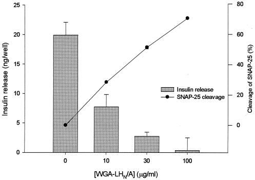
The hamster pancreatic B-cell line HIT-T15 is known to be resistant to the effects of BoNT/A (1, 13). Previous attempts to investigate SNARE-dependent processes in HIT-T15 cells have required permeabilization of the cell membrane to allow the CNT endopeptidase access to the substrate (3, 19). We have used the WGA ligand to demonstrate that a clostridial endopeptidase can be retargeted into the cytosol of HIT-T15 without resorting to physical disruption of the cell membrane. HIT-T15 cells were incubated for 4 h on ice with WGA-LHN/A and washed to remove unbound conjugate, and insulin release was assessed by radioimmunoassay 16 h later. A significant dose-dependent inhibition of stimulated insulin release was observed that correlated with increasing cleavage of SNAP-25 (Fig. 5). At a concentration of 100 μg/ml, WGA-LHN/A inhibition of insulin release was calculated to be 81.6 ± 15.7% (n = 3), which is in good agreement with previously reported neurotoxin-dependent inhibition of insulin release of ∼90% (3).
FIG. 5.
WGA-LHN/A cleaves SNAP-25 and inhibits insulin release from HIT-T15 cells. Cells were exposed to various concentrations of WGA-LHN/A on ice for 4 h. The cells were washed and incubated for 16 h at 37°C prior to the determination of insulin release and SNAP-25 cleavage. Each point shown is the mean of at least three determinations ± standard error (release) or of two determinations (cleavage).
DISCUSSION
This study has demonstrated that LHN/A, BoNT/A which has been enzymatically treated to remove its native cell receptor binding domain, can be chemically derivatized without loss of endopeptidase activity, conjugated to a second protein (WGA) to form a stable, soluble conjugate, and delivered to a variety of cells in vitro by a ligand-dependent process to inhibit secretion in a dose-dependent manner via a mechanism involving endopeptidase-dependent cleavage of the natural BoNT/A substrate. These data clearly demonstrate that a hybrid protein has been created by chemically conjugating WGA and LHN/A and that this conjugate is biologically active. Replacement of the cell-binding moiety of a number of toxins has been reported previously (4, 27), but this work represents the first reported replacement of the BoNT cell-binding domain to result in the creation of a functional hybrid.
We have demonstrated internalization of functional endopeptidase into three different neuronal cell culture models. PC12 and SH-SY5Y are well established in vitro cell lines and neuronal models, derived from rat adrenal chromaffin cells and human sympathetic neurons, respectively. Both cell lines have retained the ability to release NA in a Ca2+-dependent manner (7, 26). eSC neurons, which are more closely representative of in vivo neuronal cells, are exquisitely sensitive to BoNT/A (29) and contain a population of inhibitory glycine-releasing neurons.
It is interesting that in the PC12 and SH-SY5Y cell lines, WGA-LHN/A and BoNT/A had equivalent potency in the NA release assays. For intracellular activity of a targeted endopeptidase, be it native neurotoxin or a hybrid conjugate as reported here, the fundamental elements of cell surface binding, internalization, membrane translocation, substrate localization, and endopeptidase activity must be addressed. When comparing the potencies of native neurotoxin with hybrid conjugates, it is likely that the efficiencies of at least one of these elements may differ. However, the data obtained from PC12 and SH-SY5Y cells suggest that the overall functionality of the conjugated endopeptidase is similar to that exhibited by the native toxin endopeptidase. Significantly, the inhibition-of-neurotransmitter-release data are supported by the in vitro data obtained from the synthetic SNAP-25 peptide cleavage assays, which indicated that the derivatization and conjugation procedures had not adversely affected the LHN/A endopeptidase. Like CNT, it would be assumed that conjugated LHN/A is required to traverse at least one intracellular membrane to facilitate substrate cleavage. Therefore, given the similar overall potencies of the conjugate and BoNT/A in the PC12 and SH-SY5Y cell lines and the equivalent catalytic activity of the LHN/A endopeptidase, it would appear that the membrane translocation functions of the LHN/A have not been significantly compromised by chemical manipulation. Indeed, these observations suggest that the HN domain is fully effective at transporting LC into the cytosol, even though the receptor-mediated mode of entry is different from that encountered during neurointoxication. Thus, it is concluded that the HN domain has the ability to facilitate translocation of the LC in cell types not associated with the neuromuscular junction. In addition, the lack of potency of LHN/A in all three cell types confirms previous data (22) that described the removal of cell-binding ability from BoNT/A by proteolytic removal of the HC domain by trypsin treatment. Therefore, the data obtained with novel conjugates demonstrate that the function of the HC domain of BoNT/A is limited to cell-binding events and exclude a significant role in the intracellular mechanisms of intoxication. The use of a novel binding domain to deliver the LHN fragment to a neuronal cell is one of the few ways that this proposed domain functionality could be experimentally verified.
Furthermore, we have demonstrated internalization of functional endopeptidase into an endocrine cell line, HIT-T15. Inhibition of Ca2+-dependent insulin release was correlated to the cleavage of SNAP-25, and the inhibition of insulin release attained following application of WGA-LHN/A was similar to that previously reported for BoNT/A-treated cells (3), in which permeabilization of the cell membrane was required for toxin entry. It is interesting that the concentration of WGA-LHN/A conjugate (approximately 500 nM) applied to the cells in this study to achieve >80% inhibition was similar to the concentration of BoNT/A used in the previous study (3). These data indicate that the WGA moiety leads to internalization of the endopeptidase component of the conjugate into an intracellular compartment from which the endopeptidase can efficiently translocate. The effective functioning of the LC in the HIT-T15 cytosol, as evidenced by the cleavage of SNAP-25 and inhibition of insulin secretion, also demonstrates that the HN domain is able to function in a nonneuronal environment, enabling translocation of the LC. This is the first demonstration of such an HN function in a nonneuronal cell. The ability to inhibit SNARE-dependent release from a cell that is resistant to the actions of surface-applied neurotoxin is a significant step forward in the design of a tool for investigation of SNARE-mediated processes. In addition, these data are instrumental in defining the specific binding characteristics of the HC domain as being the major factor in susceptibility of cells to intoxication by BoNT.
This study has also indicated differences in potency between the WGA-LHN/A conjugate and intact BoNT/A. The susceptibility of the established cell lines and eSC neuron cells to BoNT/A-dependent inhibition of transmitter release varies markedly (Table 1), with eSC neurons being greater than 105-fold more sensitive than PC12 and SH-SY5Y cells. This difference could possibly be due to the prevalence of the requisite receptor for BoNT/A on the plasma membrane and/or the concentration of SNAP-25 near the point of internalization. As described above, WGA-LHN/A exhibited potency similar to BoNT/A when applied to PC12 and SH-SY5Y cells. However, WGA-LHN/A did not result in inhibition of neurotransmitter release in eSC neurons similar to that determined for BoNT/A and was shown to have an IC50 at least 13,000-fold greater than BoNT/A. Therefore, these data indicate that, although a conjugate of significant potency has been engineered, the binding, and possibly the intracellular routing, characteristics of the conjugate do not represent regeneration of the parental neurotoxin. This is further evidenced by the activity of the conjugate in HIT-T15 cells, in which BoNT/A is ineffective.
The documented cell-binding and internalization characteristics of WGA (30) were instrumental in its choice as a suitable ligand for LHN/A targeting. We have demonstrated the functionality of a conjugate prepared between this broad-specificity targeting ligand and a clostridial endopeptidase. We have demonstrated that WGA can serve as an efficient targeting vehicle, thereby expanding the repertoire of cells that can be studied with native BoNT/A to those that lack the native BoNT/A receptor(s), including, for the first time, nonneuronal cells. The novel cell-binding abilities of such hybrid targeted endopeptidases provide a powerful tool for the investigation of secretory and vesicle fusion mechanisms in a variety of cell systems and membrane fusion pathways. We have also demonstrated that the HN and HC domains of the BoNT/A HC do have defined roles in translocation and membrane binding, respectively, and that the exquisite potency of CNTs is, in major part, due to specific binding to the target cell. The observation that the HN domain is fully capable of effecting translocation of LC in a variety of cells indicates that HC is a very effective targeting moiety for a very effective toxin. The challenge of toxin research is to use our greater understanding of toxin biology to lead to a greater understanding of cellular events.
ACKNOWLEDGMENT
We are grateful to F. Alexander for supplying purified BoNT/A.
REFERENCES
- 1.Black J D, Dolly J O. Selective localisation of acceptors for botulinum neurotoxin A in the central and peripheral nervous systems. Neuroscience. 1987;23:767–779. doi: 10.1016/0306-4522(87)90094-7. [DOI] [PubMed] [Google Scholar]
- 2.Blasi J, Chapman E R, Link E, Binz T, Yamasaki S, De Camilli P, Sudhof T C, Niemann H, Jahn R. Botulinum neurotoxin A selectively cleaves the synaptic protein SNAP-25. Nature. 1993;365:160–163. doi: 10.1038/365160a0. [DOI] [PubMed] [Google Scholar]
- 3.Boyd R S, Duggan M J, Shone C C, Foster K A. The effect of botulinum neurotoxins on the release of insulin from the insulinoma cell lines HIT-15 and RINm5F. J Biol Chem. 1995;270:18216–18218. doi: 10.1074/jbc.270.31.18216. [DOI] [PubMed] [Google Scholar]
- 4.Fitzgerald D, Chaudhary V K, Kreitman R J, Siegall C B, Pastan I. Generation of chimeric toxins. Targeted Diagn Ther. 1992;7:447–462. [PubMed] [Google Scholar]
- 5.Fitzgerald S C. A dissection and tissue culture manual of the nervous system. New York: Alan R. Liss, Inc.; 1989. [Google Scholar]
- 6.Gabor F, Stangl M, Wirth M. Lectin-mediated bioadhesion: binding characteristics of plant lectins on the enterocyte-like cell lines Caco-2, HT-29 and HCT-8. J Controlled Release. 1998;55:131–142. doi: 10.1016/s0168-3659(98)00043-1. [DOI] [PubMed] [Google Scholar]
- 7.Greene L A, Tischler A S. PC12 pheochromocytoma cultures in neurobiological research. In: Federoff S, Hertz L, editors. Advances in cellular neurobiology. Vol. 3. New York: Academic Press; 1982. pp. 373–414. [Google Scholar]
- 8.Hallis B, James B A F, Shone C C. Development of novel assays for botulinum type A and B neurotoxins based on their endopeptidase activities. J Clin Microbiol. 1996;34:1934–1938. doi: 10.1128/jcm.34.8.1934-1938.1996. [DOI] [PMC free article] [PubMed] [Google Scholar]
- 9.Halpern J L, Neale E A. Neurospecific binding, internalization, and retrograde axonal transport. Curr Top Microbiol Immunol. 1995;195:221–241. doi: 10.1007/978-3-642-85173-5_10. [DOI] [PubMed] [Google Scholar]
- 10.Hoch D H, Romero M M, Ehrlich B E, Finkelstein A, DasGupta B R, Simpson L L. Channels formed by botulinum, tetanus, and diphtheria toxins in planar lipid bilayers: relevance to translocation of proteins across membranes. Proc Natl Acad Sci USA. 1985;82:1692–1696. doi: 10.1073/pnas.82.6.1692. [DOI] [PMC free article] [PubMed] [Google Scholar]
- 11.Li L, Singh B R. Isolation of synaptotagmin as a receptor for types A and E botulinum neurotoxin and analysis of their comparative binding using a new microtiter plate assay. J Nat Toxins. 1998;7:215–226. [PubMed] [Google Scholar]
- 12.Lis H, Sharon N. Lectins as molecules and as tools. Annu Rev Biochem. 1986;55:35–67. doi: 10.1146/annurev.bi.55.070186.000343. [DOI] [PubMed] [Google Scholar]
- 13.Montecucco C. How do tetanus and botulinum toxins bind to neuronal membranes? Trends Biochem Sci. 1986;11:314–317. [Google Scholar]
- 14.Montecucco C, Schiavo G. Mechanism of action of tetanus and botulinum neurotoxins. Mol Microbiol. 1994;13:1–8. doi: 10.1111/j.1365-2958.1994.tb00396.x. [DOI] [PubMed] [Google Scholar]
- 15.Nagata Y, Burger M M. Wheat germ agglutinin: molecular characteristics and specificity for sugar binding. J Biol Chem. 1974;249:3116–3122. [PubMed] [Google Scholar]
- 16.Nishiki T, Kamata Y, Nemoto Y, Omori A, Ito T, Takahashi M, Kozaki S. Identification of protein receptor for Clostridium botulinum type B neurotoxin in rat brain synaptosomes. J Biol Chem. 1994;269:10498–10503. [PubMed] [Google Scholar]
- 17.Oyler G A, Higgins G A, Hart R A, Battenberg E, Billingsley M, Bloom F E, Wilson M C. The identification of a novel synaptosomal-associated protein, SNAP-25, differentially expressed by neuronal subpopulations. J Cell Biol. 1989;109:3039–3052. doi: 10.1083/jcb.109.6.3039. [DOI] [PMC free article] [PubMed] [Google Scholar]
- 18.Ransom B R, Neale E, Henkart M, Bullock P N, Nelson P G. Mouse spinal cord in cell culture. I. Morphology and intrinsic neuronal electrophysiologic properties. J Neurophysiol. 1977;40:1132–1150. doi: 10.1152/jn.1977.40.5.1132. [DOI] [PubMed] [Google Scholar]
- 19.Sadoul K, Lang J, Montecucco C, Weller U, Regazzi R, Catsicas S, Wollheim C B, Halban P A. SNAP-25 is expressed in islets of Langerhans and is involved in insulin release. J Cell Biol. 1995;128:1019–1028. doi: 10.1083/jcb.128.6.1019. [DOI] [PMC free article] [PubMed] [Google Scholar]
- 20.Saleh M T, Gariepy J. Local conformational change in the B-subunit of Shiga-like toxin 1 at endosomal pH. Biochemistry. 1993;32:918–922. doi: 10.1021/bi00054a024. [DOI] [PubMed] [Google Scholar]
- 21.Sandvig K, Olsnes S. Diphtheria toxin entry into cells is facilitated by low pH. J Cell Biol. 1980;87:828–832. doi: 10.1083/jcb.87.3.828. [DOI] [PMC free article] [PubMed] [Google Scholar]
- 22.Shone C C, Hambleton P, Melling J. A 50-kDa fragment from the NH2-terminus of the heavy subunit of Clostridium botulinum type A neurotoxin forms channels in lipid vesicles. Eur J Biochem. 1987;167:175–180. doi: 10.1111/j.1432-1033.1987.tb13320.x. [DOI] [PubMed] [Google Scholar]
- 23.Shone C C, Hambleton P, Melling J. Inactivation of Clostridium botulinum type A neurotoxin by trypsin and purification of two tryptic fragments: proteolytic action near the COOH-terminus of the heavy subunit destroys toxin-binding activity. Eur J Biochem. 1985;151:75–82. doi: 10.1111/j.1432-1033.1985.tb09070.x. [DOI] [PubMed] [Google Scholar]
- 24.Simpson L L. The origin, structure, and pharmacological activity of botulinum toxin. Pharmacol Rev. 1981;33:155–188. [PubMed] [Google Scholar]
- 25.Sollner T, Whiteheart S W, Brunner M, Erdjument Bromaga H, Geromanos S, Tempst P, Rothman J E. SNAP receptors implicated in vesicle targeting and fusion. Nature. 1993;362:318–324. doi: 10.1038/362318a0. [DOI] [PubMed] [Google Scholar]
- 26.Vaughan P F T, Peers C, Walker J H. The use of the human neuroblastoma SH-SY5Y to study the effect of second messengers on noradrenaline release. Gen Pharmacol. 1995;26:1191–1201. doi: 10.1016/0306-3623(94)00312-b. [DOI] [PubMed] [Google Scholar]
- 27.Williams D P, Parker K, Bacha P, Bishai W, Borowski M, Genbauffe F, Strom T B, Murphy J R. Diphtheria toxin receptor binding domain substitution with interleukin-2: genetic construction and properties of a diphtheria toxin-related interleukin-2 fusion protein. Protein Eng. 1987;1:493–498. doi: 10.1093/protein/1.6.493. [DOI] [PubMed] [Google Scholar]
- 28.Williamson L, Fitzgerald S C, Neale E A. Differential effects of tetanus toxin on inhibitory and excitatory neurotransmitter release from mammalian spinal cord cells in culture. J Neurochem. 1992;59:2148–2157. doi: 10.1111/j.1471-4159.1992.tb10106.x. [DOI] [PubMed] [Google Scholar]
- 29.Williamson L C, Halpern J L, Montecucco C, Brown J E, Neale E A. Clostridial neurotoxins and substrate proteolysis in intact neurons. J Biol Chem. 1996;271:7694–7699. doi: 10.1074/jbc.271.13.7694. [DOI] [PubMed] [Google Scholar]
- 30.Wirth M, Fuchs A, Wolf M, Ertl B, Gabor F. Lectin-mediated drug targeting: preparation, binding characteristics, and antiproliferative activity of wheat germ agglutinin conjugated doxorubicin on Caco-2 cells. Pharm Res. 1998;15:1031–1037. doi: 10.1023/a:1011926026653. [DOI] [PubMed] [Google Scholar]


